Here are your comprehensive MBBS notes on the Lymphatic System and Hematopoietic & Immune System Organs, compiled from Cellular and Molecular Immunology, Guyton & Hall Medical Physiology, Robbins Pathology, Henry's Clinical Diagnosis, and Harrison's Principles of Internal Medicine.
LYMPHATIC SYSTEM, HEMATOPOIETIC & IMMUNE SYSTEM ORGANS
Comprehensive MBBS Notes - Basic to Advanced
PART 1: THE LYMPHATIC SYSTEM
1.1 Overview and Definition
The lymphatic system consists of:
- Specialized vessels (lymphatics) that drain fluid from tissues
- Lymph nodes interspersed along those vessels
- Secondary lymphoid organs (spleen, tonsils, MALT)
- Primary lymphoid organs (bone marrow, thymus)
Two essential roles:
- Fluid homeostasis - prevents tissue edema by returning interstitial fluid to the bloodstream
- Immune surveillance - transports antigens and immune cells to lymph nodes where adaptive immune responses are initiated
"The lymphatic system represents an accessory route through which fluid can flow from the interstitial spaces into the blood. Most importantly, lymphatics can carry away proteins and large particulate matter that cannot be removed by absorption directly into blood capillaries - without which we would die within about 24 hours."
- Guyton & Hall Medical Physiology
1.2 Components of the Lymphatic System
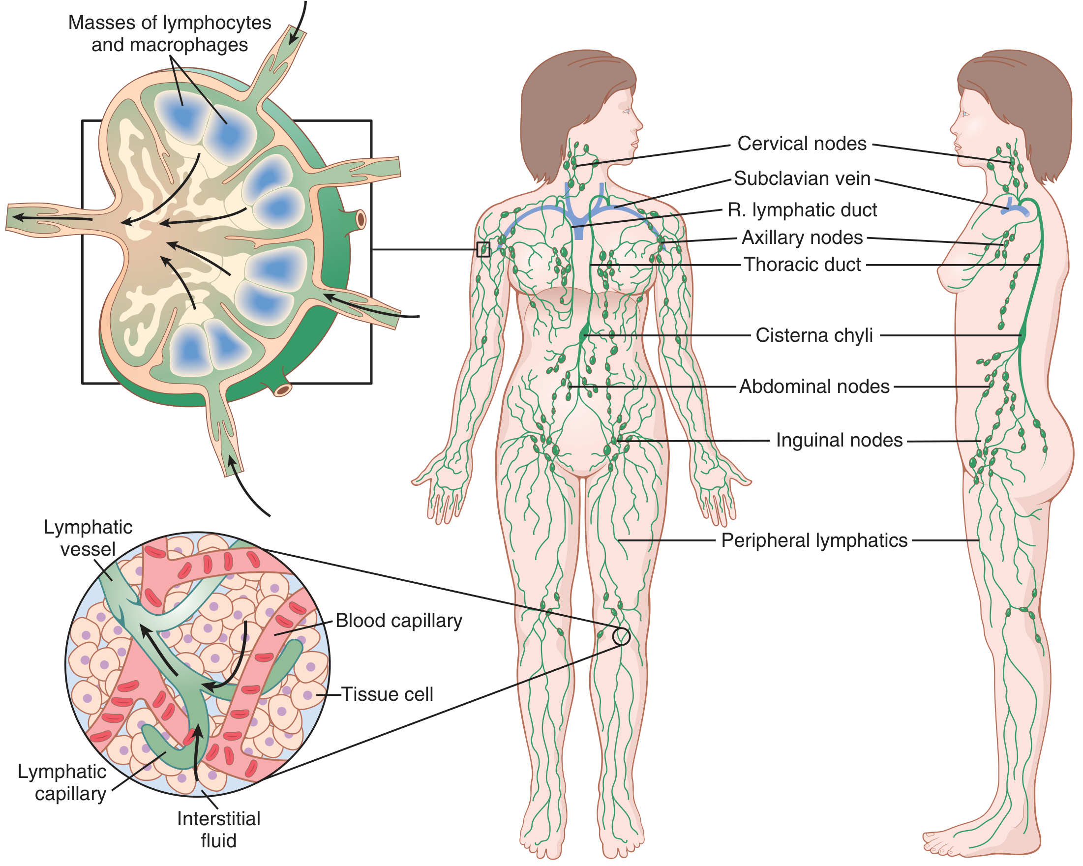
Full body map showing major lymphatic structures:
Figure: The lymphatic system (Guyton & Hall). Major lymphatic vessels drain into the superior vena cava. Inset (upper left) shows a lymph node with afferent/efferent vessels. Inset (lower left) shows the relationship between lymphatic capillaries, blood capillaries, and tissue cells.
1.3 Lymphatic Vessels - Four Types
The lymphatic vascular system is subdivided into four regions based on histological wall structure:
| Type | Diameter | Special Features |
|---|
| Lymphatic capillaries | Smallest | Blind-ended; overlapping endothelial cells; no tight junctions; no continuous basement membrane |
| Precollectors | Small | Contain valve cusps; reinforced connective tissue wall |
| Collectors | 150-600 μm | Valves + venous-type wall (intima/media/adventitia); smooth muscle generates rhythmic contractions (10-12/min) |
| Lymphatic trunks | Largest | Lead to thoracic duct or right lymphatic duct |
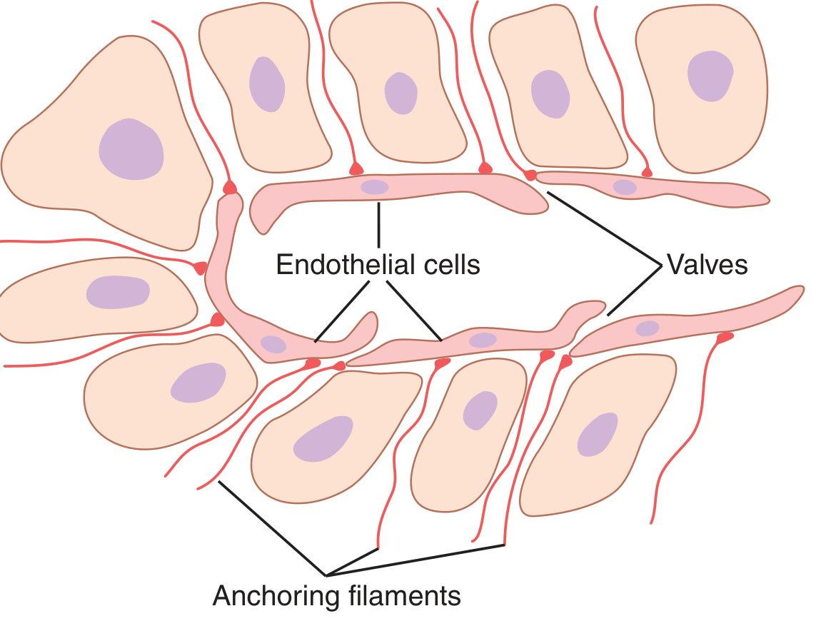
Key structural features of lymphatic capillaries:
Figure: Special structure of lymphatic capillaries (Guyton & Hall). Endothelial cells are attached to surrounding connective tissue via anchoring filaments. Overlapping cell edges act as flap valves - interstitial fluid pushes them open to enter; backflow closes them. This permits uptake of high-molecular-weight proteins, bacteria, and particles.
Mechanism of lymph propulsion:
- Rhythmic smooth muscle contractions in collector segments (peristalsis-like, 10-12 contractions/min)
- Musculoskeletal movement (external compression)
- Arterial pulsations transmitted to adjacent lymphatic vessels
- One-way valves prevent backflow at every level
1.4 Drainage Routes - MUST KNOW
Thoracic Duct (larger) drains:
- Left side of head and neck
- Left arm
- Both legs
- Abdominal viscera (via cisterna chyli)
- Left thorax
→ Empties into junction of left subclavian vein and left internal jugular vein
Right Lymphatic Duct (smaller) drains:
- Right side of head and neck
- Right arm
- Right thorax
→ Empties into junction of right subclavian vein and right internal jugular vein
Cisterna chyli = dilated lymphatic sac at L1-L2 level; receives lymph from intestines (rich in chylomicrons after a fatty meal) and both lower limbs
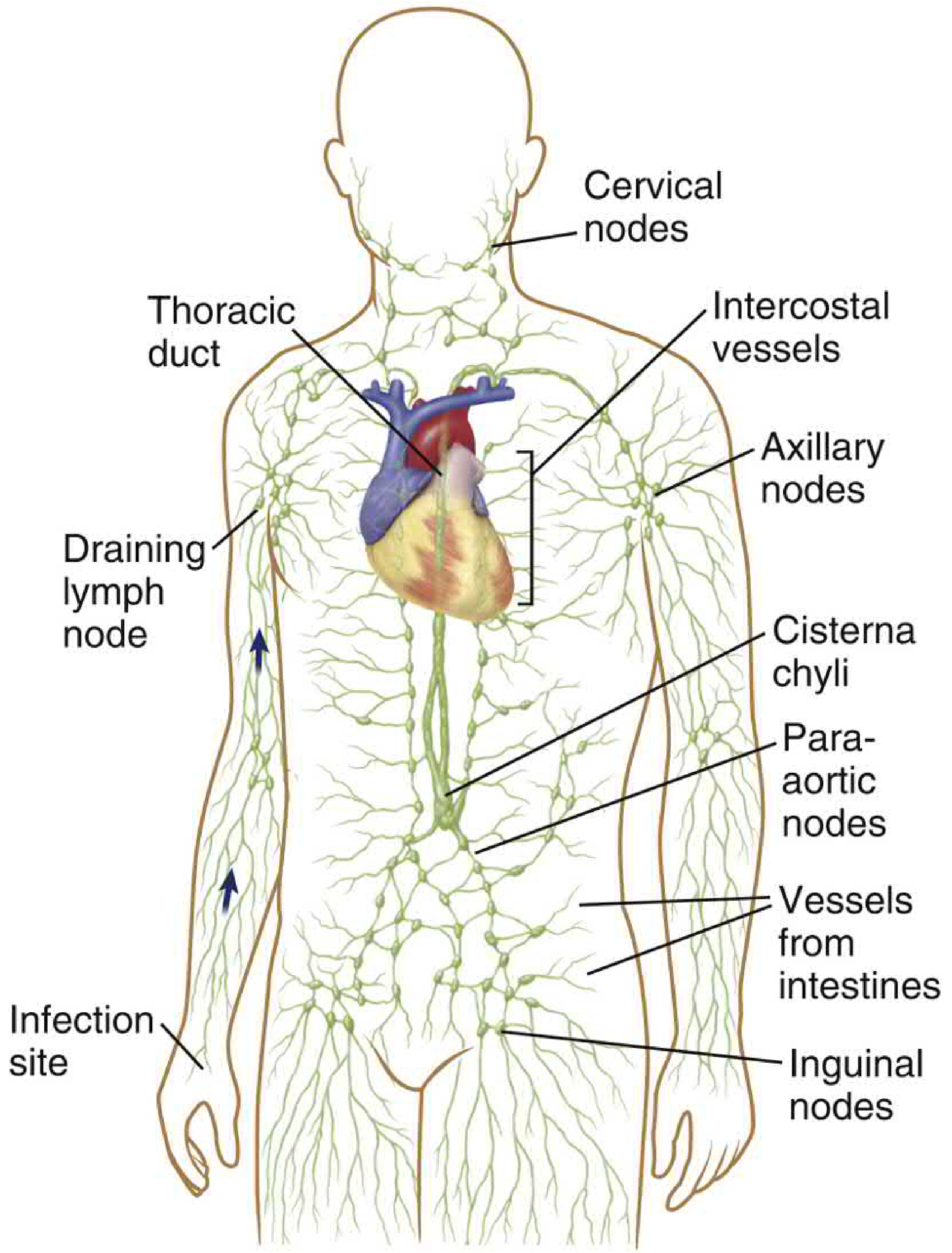
Drainage route overview:
Figure: The lymphatic system (Cellular & Molecular Immunology). Note the thoracic duct, cisterna chyli, para-aortic nodes, inguinal nodes, axillary nodes, and cervical nodes. Antigens from infection sites travel via lymphatics to draining lymph nodes where adaptive immune responses are initiated.
1.5 Formation of Lymph
- Approximately 10% of capillary filtrate enters lymphatics (the other 90% is reabsorbed by venous capillaries)
- Total lymph returned to circulation: 2-3 L/day
- Protein concentration of lymph: ~2 g/dL in most tissues; up to 6 g/dL in liver lymph (liver is very protein-leaky)
- Intestinal lymph: 3-4 g/dL protein; after fatty meal, contains 1-2% fat as chylomicrons
- Thoracic duct lymph (mixture of all): 3-5 g/dL protein
Clinical correlation - Lymphedema: If lymphatics are blocked (by tumor, filariasis, surgery), proteins accumulate in interstitial space, raising oncotic pressure there, causing severe chronic edema.
1.6 The Glymphatic System (Advanced)
- In the CNS, true lymphatic vessels exist only in the meninges
- Deeper brain tissue uses the glymphatic (glial-lymphatic) system:
- CSF flows into perivascular spaces formed by astrocytic endfeet around cerebral arteries
- Flows into brain parenchyma via aquaporin-4 water channels (AQP4) on astrocytes
- Exits via perivenous spaces, carrying waste products (including amyloid-beta) into meningeal lymphatics
- Clinical relevance: Disruption of the glymphatic system is implicated in Alzheimer disease and other neurodegenerative conditions
PART 2: LYMPH NODES
2.1 General Structure
Lymph nodes are encapsulated, vascularized secondary lymphoid organs. The human body contains ~500 lymph nodes.
Architectural layers (outside to inside):
| Layer | Contents | Function |
|---|
| Fibrous capsule | Dense connective tissue | Protection |
| Subcapsular sinus | Macrophages, reticular cells | First-line phagocytosis of organisms |
| Cortex (outer) | B cell follicles (primary and secondary) | B cell activation, germinal center reactions |
| Paracortex (deep cortex) | T lymphocytes, DCs, HEVs | T cell activation; lymphocyte homing |
| Medulla | Medullary cords (plasma cells), medullary sinuses | Antibody secretion; lymph drains out |
| Hilum | Efferent lymphatic, artery, vein exit | Output |
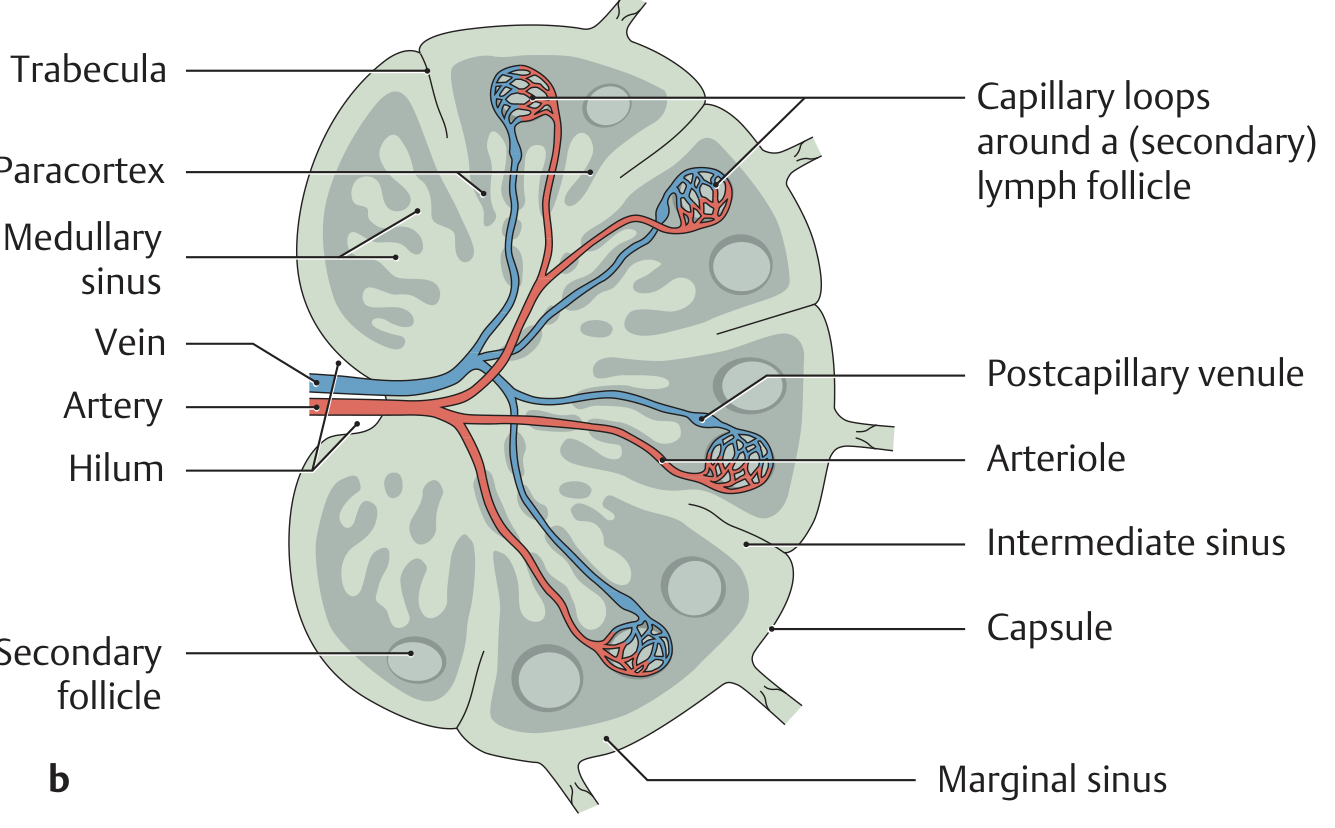
Lymph node cross-section with blood supply:
Figure: Blood supply and structure of a lymph node (Thieme Atlas of Anatomy). The hilum carries the artery in and vein + efferent lymphatic out. High endothelial postcapillary venules (HEVs) in the paracortex allow lymphocyte extravasation from blood. Secondary follicles with germinal centers are visible in the cortex.
2.2 Follicles - Primary vs Secondary
| Feature | Primary Follicle | Secondary Follicle |
|---|
| Germinal center | Absent | Present |
| Cells | Naive, resting B cells | Activated B cells (centroblasts + centrocytes) |
| Significance | No recent antigen exposure | Active immune response ongoing |
Germinal center zones:
- Dark zone - packed with proliferating centroblasts (hypermutation occurring)
- Light zone - centrocytes selected by FDCs for high-affinity antibody; low-affinity cells undergo apoptosis
- Output - memory B cells + long-lived plasma cells (migrate to bone marrow)
2.3 T Cell Zone (Paracortex)
- Populated mainly by T lymphocytes and dendritic cells (DCs)
- Contains High Endothelial Venules (HEVs) - specialized postcapillary venules allowing lymphocyte trafficking from blood into the node
- Naive T cells express CCR7 receptor → binds CCL19/CCL21 produced by paracortical FRCs → directs naive T cells into T cell zone
- Naive B cells express CXCR5 → binds CXCL13 from B zone FRCs → directs B cells into follicles
- FRC conduits (0.2-3 μm diameter tubes of ECM) act as tracks along which T cells and DCs migrate
Practical point: Paracortical expansion (deep cortex hyperplasia) is seen in viral infections and delayed-type hypersensitivity reactions. Follicular hyperplasia indicates B cell stimulation (bacterial infections, autoimmune disease).
2.4 Antigen Transport Through Lymph Nodes
- DCs in skin/mucosa capture antigen → enter afferent lymphatics
- Arrive at subcapsular sinus of draining lymph node
- High-molecular-weight antigens → taken up by sinus macrophages → passed to FDCs in follicles
- Soluble small antigens → travel via FRC conduits to T cell zone
- DCs in T zone present antigen to T cells → adaptive immune response begins
PART 3: PRIMARY LYMPHOID ORGANS
3.1 Bone Marrow
The site of hematopoiesis and B lymphocyte maturation. All blood cells originate from hematopoietic stem cells (HSCs) in the bone marrow.
Sites of active hematopoiesis by age:
| Stage | Active Sites |
|---|
| 3rd week of embryo | Yolk sac (transient; embryonic RBCs) |
| 3rd month of embryo | Liver becomes chief site |
| 4th month onward | Bone marrow takes over |
| Birth | All skeletal marrow active |
| After puberty | Restricted to axial skeleton only (vertebrae, sternum, ribs, ilium, proximal long bones) |
| Normal adult | ~50% of marrow space hematopoietically active |
Embryonic note: Yolk sac-derived cells give rise to long-lived tissue macrophages (microglia in brain, Kupffer cells in liver) - these are NOT from definitive HSCs.
3.2 Hematopoiesis - Full Hierarchy
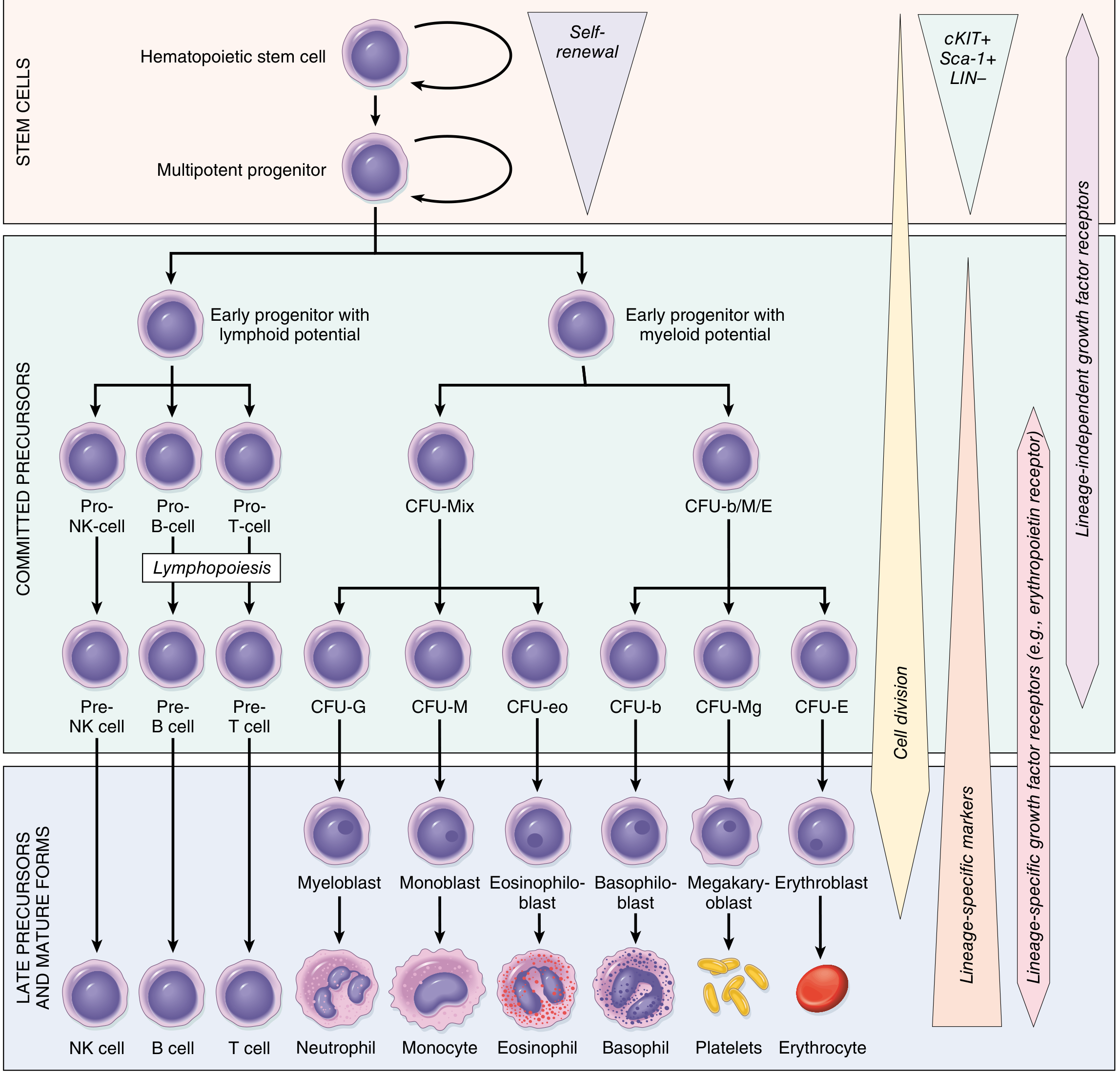
All formed blood elements derive from a single pluripotent Hematopoietic Stem Cell (HSC).
Complete differentiation pathway:
Figure: Differentiation of blood cells (Robbins Pathology). HSCs (cKIT+, Sca-1+, LIN-) self-renew and give rise to multipotent progenitors. These diverge into lymphoid (Pro-NK, Pro-B, Pro-T) and myeloid (CFU-Mix, CFU-b/M/E) early progenitors. Mature cells: NK cells, B cells, T cells, neutrophils, monocytes, eosinophils, basophils, platelets, erythrocytes.
HSC key properties:
- Pluripotency - one HSC generates all mature blood cell types
- Self-renewal - HSCs can divide to produce exact copies of themselves; this maintains the pool throughout life
Estimated adult HSC pool: 50,000-200,000 cells in bone marrow.
Growth factors controlling hematopoiesis (clinically important):
| Growth Factor | Lineage Stimulated | Clinical Use |
|---|
| EPO (Erythropoietin) | Erythroid (CFU-E) | Anemia of chronic kidney disease |
| G-CSF | Granulocytes (CFU-G) | Post-chemotherapy neutropenia |
| GM-CSF | Granulocytes + Monocytes | Stem cell mobilization |
| TPO (Thrombopoietin) | Megakaryocytes/Platelets | Thrombocytopenia |
| SCF (c-KIT ligand) | Multiple lineages | Synergistic with EPO, G-CSF |
| IL-7 | Lymphoid progenitors | T and B cell development |
3.3 B Cell Development in Bone Marrow
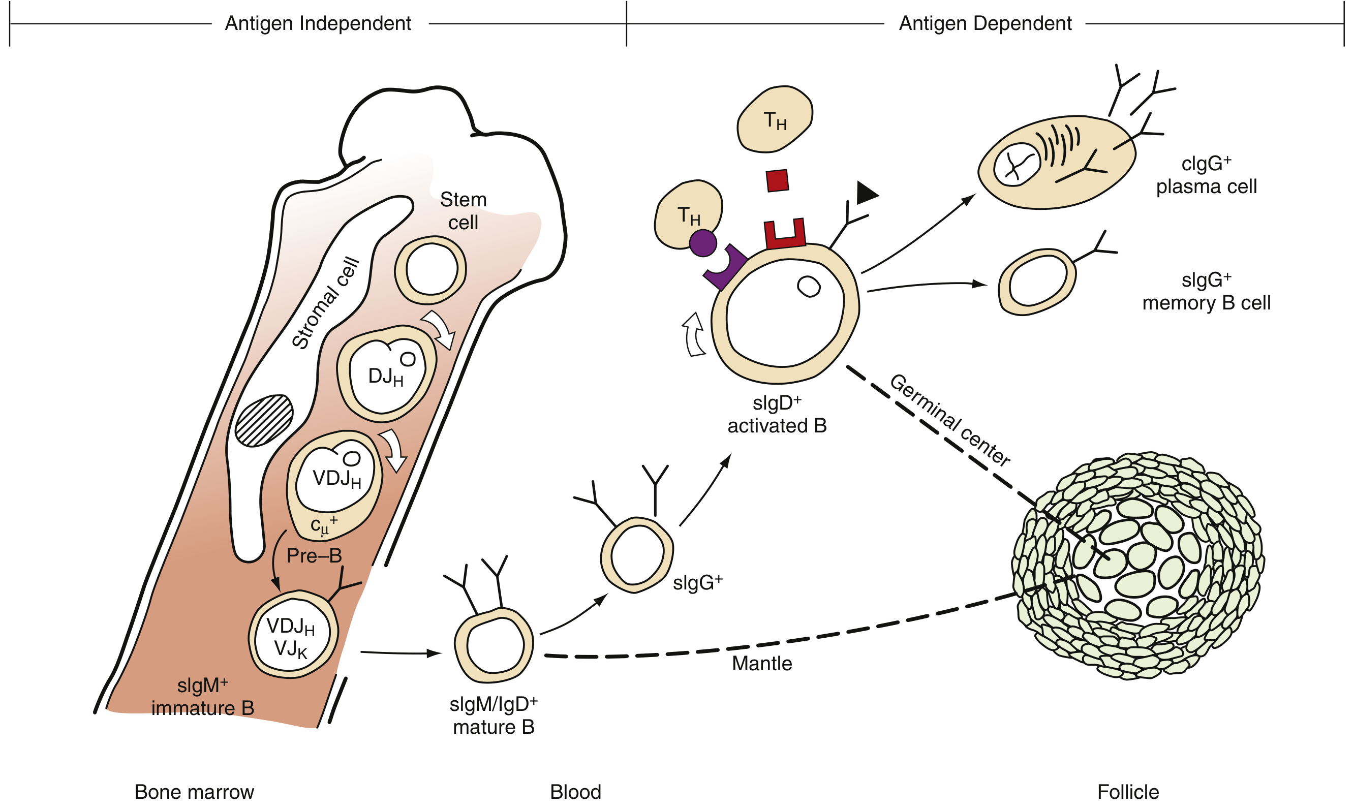
B cells undergo antigen-independent maturation in bone marrow. Key stages:
Figure: B cell differentiation (Henry's Clinical Diagnosis). Left: Antigen-independent maturation in bone marrow - stem cell → Pre-B cell (VDJ rearrangement, cytoplasmic μ chain) → immature B cell (sIgM+) → exits marrow. Right: Antigen-dependent activation in peripheral follicles - mature B (sIgM/IgD+) → activated B → germinal center → plasma cell (secreting IgG) or memory B cell.
Stages summary:
- Pro-B cell - DJ rearrangement begins
- Pre-B cell - VDJ rearrangement complete; cytoplasmic μ heavy chain expressed (cμ+)
- Immature B cell - Complete IgM assembled on surface (sIgM+); leaves bone marrow
- Mature naive B cell - Co-expresses sIgM and sIgD; enters blood
- Activated B cell (antigen-dependent) - encounters antigen + T helper signals → enters germinal center
- Plasma cell - secretes large amounts of antibody
- Memory B cell - long-lived; rapid secondary response
Negative selection: Immature B cells that react against self-antigens in marrow undergo apoptosis (clonal deletion) or receptor editing - this is central tolerance.
3.4 Thymus
The thymus is the primary lymphoid organ where T lymphocytes mature and are selected.
Gross anatomy:
- Bilobed organ in the anterior superior mediastinum
- Each lobe is subdivided into multiple lobules by fibrous trabeculae
- Largest in childhood; begins to involute (fatty atrophy) at puberty
- By adulthood: largely replaced by fat but retains functional tissue
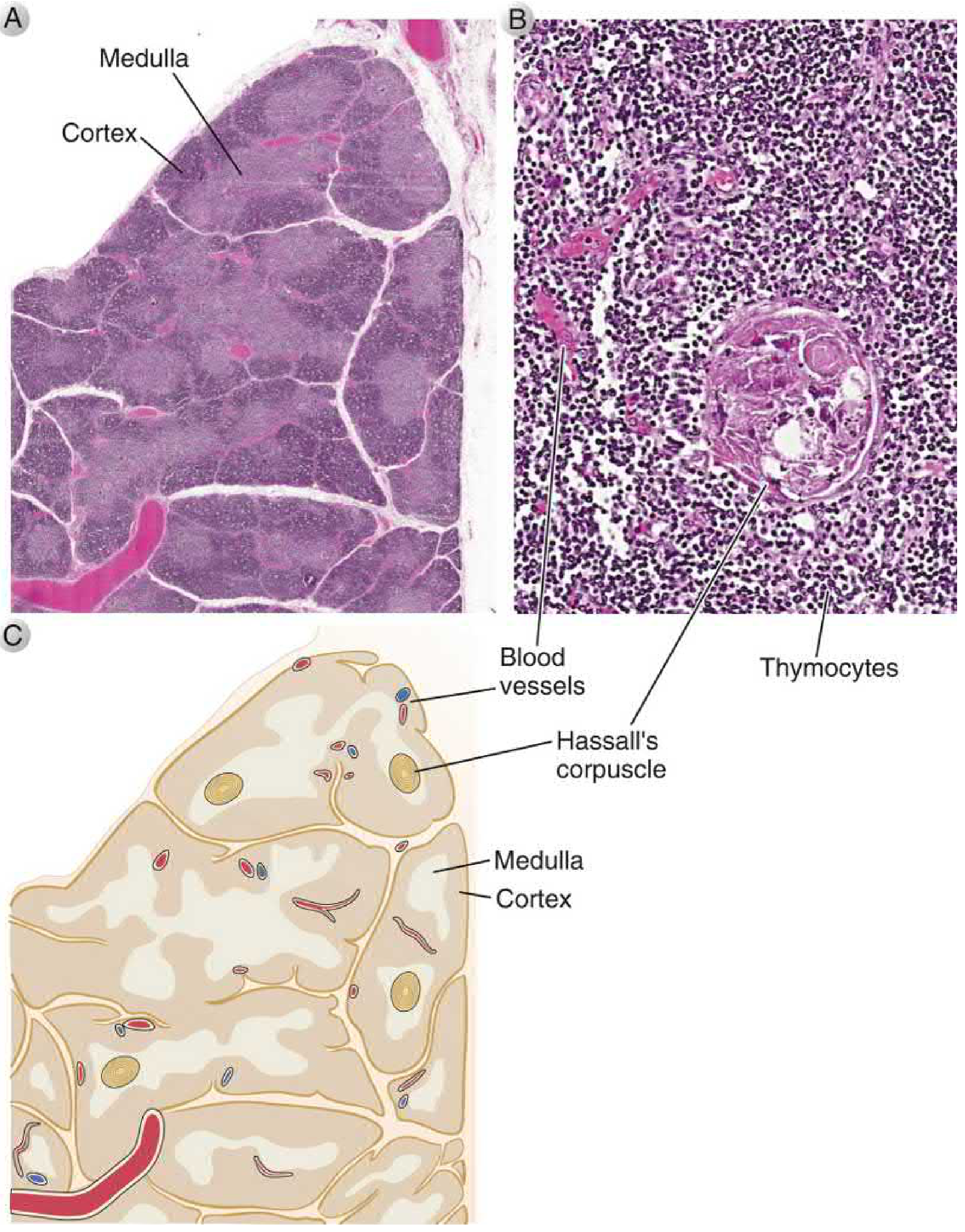
Histology:
Figure: Thymus morphology (Cellular & Molecular Immunology). A: Low power - dark outer cortex (densely packed thymocytes) and paler inner medulla. B: High power medulla - small blue thymocytes + pink Hassall's corpuscle (characteristic of thymic medulla; composed of concentrically arranged keratinized epithelial cells). C: Schematic showing fibrous trabeculae dividing each lobe into lobules.
Thymus compartments:
| Zone | Cells | What Happens Here |
|---|
| Subcapsular cortex | Earliest pro-T cells (double negative: CD4-CD8-) | TCR gene rearrangement begins |
| Inner cortex | Double positive thymocytes (CD4+CD8+) | Positive selection |
| Medulla | Single positive T cells (CD4+ or CD8+) | Negative selection; maturation |
3.5 T Cell Development in the Thymus
Pro-T cells from bone marrow migrate to the thymus via blood. All critical events happen in thymus.
Stages of T cell maturation:
| Stage | CD Markers | Location | Event |
|---|
| Double Negative (DN) | CD4-CD8- | Subcapsular cortex | TCR β-chain gene rearrangement; γδ commitment |
| Double Positive (DP) | CD4+CD8+ | Cortex | TCR α-chain rearrangement; complete TCR formed |
| Positive Selection | CD4+CD8+ → SP | Cortex (cortical epithelium) | Cells that recognize self-MHC survive; others die by neglect |
| Negative Selection | CD4+ or CD8+ | Medulla (AIRE+ medullary epithelium) | Cells reacting too strongly to self-antigens → apoptosis |
| Mature Single Positive | CD4+ or CD8+ | Medulla → Blood | Exit thymus; circulate as naive T cells |
Positive selection: DP thymocytes interact with cortical epithelial cells presenting self-peptide on MHC. If TCR binds MHC-I → keeps CD8; if binds MHC-II → keeps CD4. No binding = death by neglect.
Negative selection: Medullary epithelial cells express the AIRE (autoimmune regulator) gene, driving expression of tissue-specific antigens (thyroid, pancreas, etc.). T cells reacting too strongly against these → clonal deletion. Failure of AIRE = APECED syndrome (autoimmune polyendocrinopathy).
Hassall's corpuscles in the thymic medulla are involved in AIRE-dependent expression of peripheral tissue antigens - their exact functional significance is still being investigated.
PART 4: SECONDARY LYMPHOID ORGANS
4.1 Spleen
The spleen is the largest secondary lymphoid organ. It filters blood (unlike lymph nodes, which filter lymph).
Development: Derived from dorsal mesogastrium at ~5 weeks gestation. ~20% of people have accessory spleens due to incomplete fusion of splenic hillocks.
Gross anatomy:
- Location: Left upper quadrant (LUQ), between 9th-11th ribs
- Attachments: Gastrolienal ligament (to stomach) and lienorenal ligament (to kidney)
- Blood flow: ~150 mL/min through splenic artery; drained by portal circulation
- Weight: ~150 g in adults
Functions (Harrison's):
- Quality control of erythrocytes in red pulp - removal of senescent/defective RBCs
- Synthesis of antibodies in white pulp
- Removal of antibody-coated bacteria and blood cells from circulation
- Storage of monocytes (released during infection/inflammation)
- Site of extramedullary hematopoiesis in certain diseases
4.2 Splenic Microarchitecture
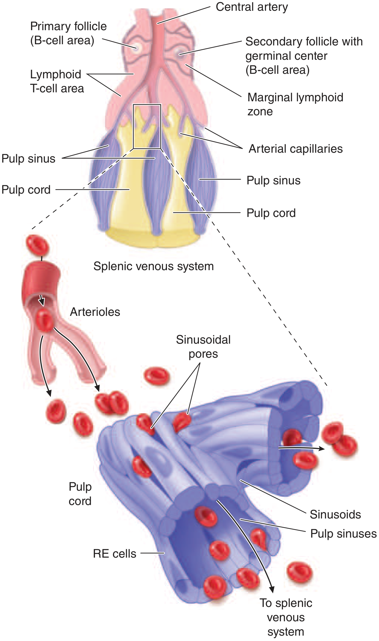
Schematic overview:
Figure: Schematic spleen structure (Harrison's Principles). The splenic artery ramifies into central arterioles around which white pulp is organized. The PALS (T cell zone), B cell follicles, marginal zone, and red pulp (cords + sinusoids) are shown. RBCs must squeeze through sinusoidal slits to re-enter the circulation - old/stiff cells fail this "pitting" process and are destroyed in the cords.
Histology - White Pulp and Red Pulp:
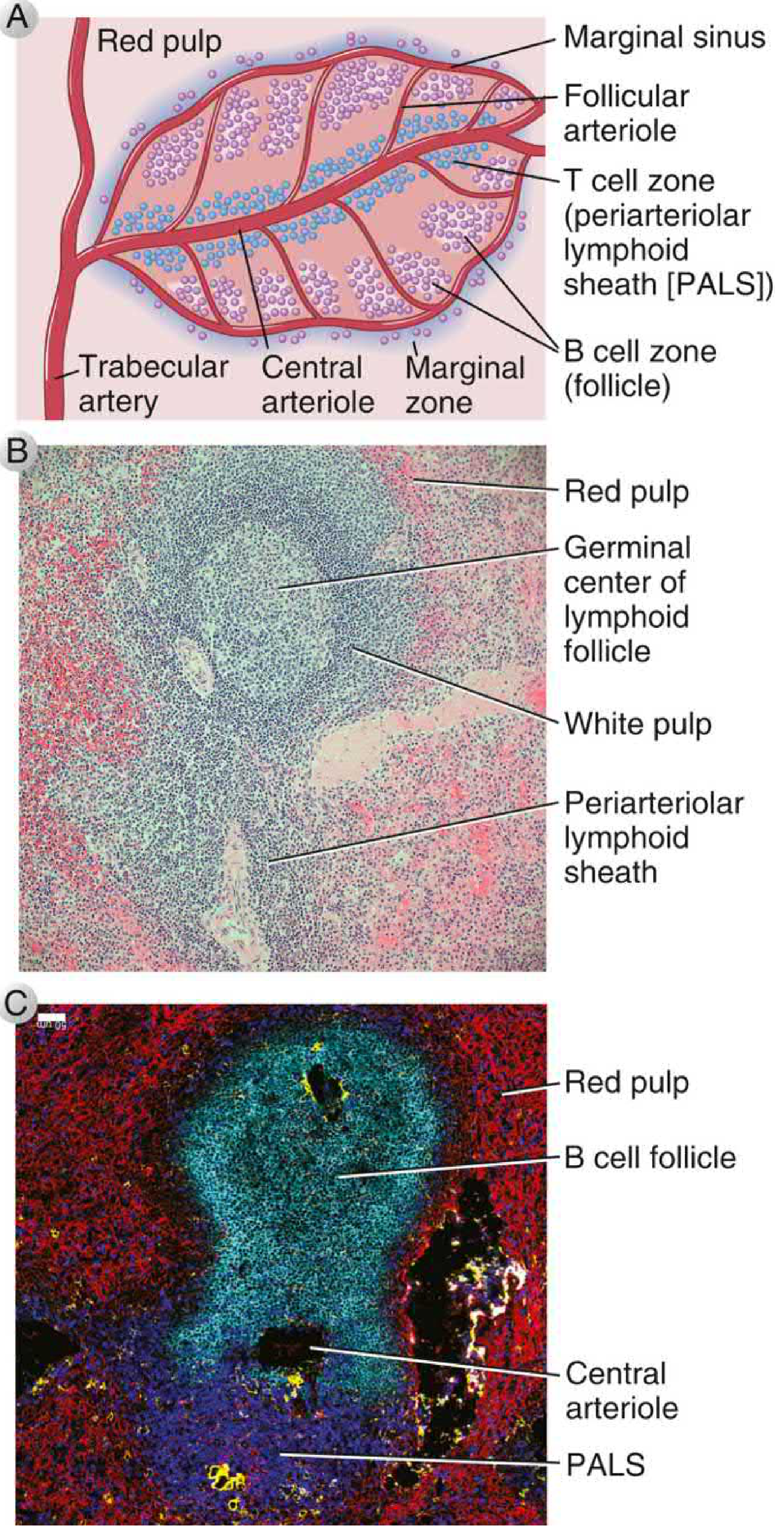
Figure: Morphology of the spleen (Cellular & Molecular Immunology). A: Schematic - T cells surround central arterioles as PALS; B cell follicles are adjacent; marginal zone separates white and red pulp. B: H&E section - germinal center of lymphoid follicle within white pulp, surrounded by red pulp. C: Immunofluorescence - B cells (cyan) in follicle, T cells in PALS (dark), central arteriole (yellow), red pulp (red).
| Compartment | Composition | Function |
|---|
| White Pulp | T zones (PALS) + B zones (follicles) + Marginal zone | Immune response to blood-borne antigens |
| PALS (periarteriolar lymphoid sheath) | T lymphocytes around central arteriole | T cell activation |
| B cell follicles | Primary + secondary follicles adjacent to PALS | B cell activation; germinal center reactions |
| Marginal zone | Macrophages, marginal zone B cells | First-line trap for blood-borne antigens/bacteria |
| Red Pulp | Pulp cords (Billroth's cords) + venous sinusoids | Erythrocyte quality control; phagocytosis |
The RBC filtration mechanism (detailed):
- Blood from central arterioles flows into macrophage-lined sinuses AND cords
- Blood in sinuses re-enters circulation easily (via splenic venules)
- Blood in cords must squeeze through narrow slits (2-3 μm) in the sinus lining
- Young, deformable RBCs pass through → enter sinuses → circulation
- Old, stiff, or damaged RBCs cannot pass → retained in cords → destroyed by macrophages
- Inclusions (Howell-Jolly bodies = nuclear remnants; Heinz bodies = denatured Hb; malaria parasites) are "pitted" out during this squeeze
Clinical correlation - Asplenia/Post-splenectomy:
- Loss of marginal zone macrophages → inability to rapidly clear encapsulated bacteria (Streptococcus pneumoniae, Haemophilus influenzae, Neisseria meningitidis)
- Risk: Overwhelming Post-Splenectomy Infection (OPSI) - mortality up to 50%
- Prevention: Vaccinate against pneumococcus, meningococcus, H. influenzae; lifelong prophylactic penicillin; patient education
- Blood film after splenectomy: Howell-Jolly bodies, target cells, thrombocytosis
4.3 Tonsils and MALT
Mucosa-Associated Lymphoid Tissue (MALT) is a system of non-encapsulated lymphoid structures at epithelial barriers:
| Structure | Location | Protects Against |
|---|
| Palatine tonsils | Oropharynx | Inhaled/ingested pathogens |
| Pharyngeal tonsil (adenoids) | Nasopharynx | Inhaled pathogens |
| Lingual tonsil | Base of tongue | Ingested pathogens |
| Peyer's patches | Small intestine (ileum) submucosa | Intestinal pathogens |
| Appendix | Appendix | Intestinal pathogens |
| BALT | Bronchial submucosa | Inhaled pathogens |
Waldeyer's ring = palatine + pharyngeal + lingual tonsils forming a ring of lymphoid tissue at the oropharyngeal junction - first immune defense against ingested and inhaled antigens.
Peyer's patches contain specialized M (microfold) cells that sample luminal antigens → transport to underlying DCs → initiate IgA responses.
PART 5: CELLS OF THE IMMUNE SYSTEM
5.1 Summary Table of Key Immune Cells
| Cell | Origin | Location | Function | Key Markers |
|---|
| Neutrophil | Bone marrow (CFU-G) | Blood, tissues | First responder; phagocytosis; oxidative burst | CD66, MPO+ |
| Monocyte | Bone marrow (CFU-M) | Blood | Differentiates into macrophages at inflammation sites | CD14, CD16 |
| Macrophage | Monocyte | Tissues | Phagocytosis; antigen presentation; cytokine secretion | CD68, CD163 |
| Dendritic cell | Bone marrow | Skin, mucosa, lymphoid organs | Professional antigen-presenting cell; bridge innate/adaptive | CD11c, MHC-II |
| B lymphocyte | Bone marrow | Blood, lymphoid follicles | Antibody production; antigen presentation | CD19, CD20, sIg |
| T lymphocyte (helper) | Thymus | Blood, T cell zones | Coordinate adaptive immunity; cytokine production | CD4, TCR |
| T lymphocyte (cytotoxic) | Thymus | Blood, tissues | Kill virus-infected/tumor cells | CD8, TCR |
| NK cell | Bone marrow | Blood | Kill infected/tumor cells without prior sensitization | CD56, CD16 |
| Mast cell | Bone marrow | Tissues (especially mucosal) | Allergic responses; antiparasitic | FcεRI, tryptase |
| Eosinophil | Bone marrow (CFU-eo) | Blood, mucosal tissues | Antiparasitic; allergic inflammation | CD16, MBP |
| Basophil | Bone marrow (CFU-b) | Blood | Allergic responses (similar to mast cells) | FcεRI, histamine |
| ILC (innate lymphoid cell) | Bone marrow | Tissues | Innate immunity at epithelial barriers; lymphoid organ development in fetal life | Lineage negative |
5.2 Chemokine-Mediated Cell Homing (Advanced)
The anatomic segregation of T and B cells is controlled by chemokines:
| Receptor | Ligand | Expressed By | Effect |
|---|
| CCR7 | CCL19, CCL21 | Naive T cells; activated DCs | Homing to T cell zone (paracortex) |
| CXCR5 | CXCL13 | Naive B cells; follicular T helper cells | Homing to B cell follicles |
| CCR6 | CCL20 | Plasmablasts, Th17 | Mucosal trafficking |
Knockout experiments confirm function: CXCR5 knockout mice lack B cell follicles; CCR7 knockout mice lack T cell zones.
After antigen activation, lymphocytes switch chemokine receptors - activated B cells downregulate CXCR5 (to exit follicle) and upregulate CCR7; then switch back to CXCR5 when entering germinal centers.
PART 6: REACTIVE CHANGES & CLINICAL CORRELATIONS
6.1 Leukocytosis - Mechanisms
Peripheral blood WBC count is influenced by:
- Size of marrow precursor pool
- Rate of release from marrow storage pools
- Marginal pool (cells adherent to vessel walls)
- Rate of emigration into tissues
| Type | Causes |
|---|
| Neutrophilia | Bacterial infection, tissue necrosis (MI, burns), glucocorticoids, G-CSF |
| Lymphocytosis | Viral infection (EBV, CMV, hepatitis), pertussis, CLL |
| Eosinophilia | Parasitic infection, allergic disease, Addison's disease, malignancy |
| Monocytosis | TB, bacterial endocarditis, chronic inflammation, malaria |
| Basophilia | CML, myeloproliferative neoplasms, hypothyroidism |
6.2 Lymphadenopathy - Pattern Diagnosis
| Pattern | Likely Cause |
|---|
| Follicular hyperplasia (B zone) | Bacterial infection, autoimmune disease (SLE, RA) |
| Paracortical hyperplasia (T zone) | Viral infection, drug reactions, vaccination |
| Sinus histiocytosis | Regional to cancer (reactive); draining lymph node enlargement |
| Granulomatous inflammation | TB, sarcoidosis, fungal infections, Cat-scratch disease |
| Necrotic lymphadenitis | Kikuchi disease, SLE, HSV |
6.3 Splenomegaly - Classification
| Mechanism | Examples |
|---|
| Hyperplasia of immune functions | Infections (EBV, malaria, TB), autoimmune hemolytic anemia |
| Congestion (portal hypertension) | Liver cirrhosis, portal vein thrombosis |
| Infiltration | Leukemia, lymphoma, amyloidosis, Gaucher disease |
| Extramedullary hematopoiesis | Myelofibrosis, thalassemia major |
Massive splenomegaly (crossing midline, >1 kg): CML, myelofibrosis, visceral leishmaniasis (kala-azar), malaria (hyperreactive splenomegaly).
PART 7: EMBRYOLOGY SUMMARY
| Organ | Embryological Origin | Timing |
|---|
| Bone marrow (definitive) | Mesoderm of intraembryonic aorta/gonad/mesonephros region | Week 3-4 of embryo |
| Thymus | 3rd pharyngeal pouch (endoderm) | Week 6 of embryo |
| Spleen | Dorsal mesogastrium (mesoderm) | Week 5 of embryo |
| Lymph nodes | Mesenchyme + lymphoid precursor cells | 2nd trimester |
| Tonsils | 2nd pharyngeal pouch (palatine) | 3rd-5th month |
DiGeorge Syndrome = deletion of chromosome 22q11.2 → failure of 3rd/4th pharyngeal pouch development → thymic aplasia/hypoplasia → profound T cell deficiency → recurrent viral/fungal infections.
QUICK REVISION TABLE - HIGH-YIELD FACTS
| Fact | Detail |
|---|
| Thoracic duct drains | Everything except right head/neck/arm/thorax |
| Right lymphatic duct drains | Right head, neck, arm, thorax |
| Cisterna chyli location | L1-L2 level |
| Lymph volume/day | 2-3 L |
| Lymph protein concentration | 3-5 g/dL (thoracic duct) |
| HSC markers | CD34+, CD38-, Lin-, cKIT+, Sca-1+ |
| Number of lymph nodes | ~500 |
| Thymus compartments | Subcapsular cortex → cortex → medulla |
| Double negative T cells | CD4-CD8- (earliest thymocytes) |
| Double positive T cells | CD4+CD8+ (in cortex; undergo positive selection) |
| AIRE | Gene expressed by medullary thymic epithelium; drives negative selection |
| PALS | Periarteriolar lymphoid sheath = T cell zone of spleen |
| Howell-Jolly bodies | Nuclear remnants in RBCs; indicate asplenia or splenic dysfunction |
| Malpighi's terms | White pulp and red pulp of spleen |
| HEV | High endothelial venule; site of lymphocyte entry into lymph node paracortex |
| Germinal center output | Memory B cells + long-lived plasma cells |
| OPSI | Overwhelming post-splenectomy infection; pneumococcus is #1 pathogen |
| First hematopoietic site | Yolk sac (embryonic) |
| Definitive HSC origin | Aorta-gonad-mesonephros (AGM) region |
| Liver hematopoiesis | 3rd-6th month of fetal life |
| Marrow hematopoiesis begins | 4th month onward; sole site after birth |
*Sources: Cellular and Molecular Immunology (Abbas, Lichtman, Pillai); Guyton & Hall Textbook of Medical Physiology; Robbins, Cotran & Kumar Pathologic Basis of Disease; Henry's Clinical Diagnosis and Management by Laboratory Methods; Harrison's Principles of Internal Medicine 22nd Ed; Gray's Anatomy for Students; Thieme Atlas of General Anatomy.*Your comprehensive MBBS notes are complete. Here's a summary of what's covered:
What These Notes Cover
Part 1 - Lymphatic System: Components, 4 vessel types, drainage routes (thoracic duct vs right lymphatic duct), lymph formation, the glymphatic system, and clinical correlations (lymphedema).
Part 2 - Lymph Nodes: Full architecture (capsule → cortex → paracortex → medulla), primary vs secondary follicles, germinal center reactions, T cell zones, HEVs, antigen transport, and chemokine-mediated cell homing.
Part 3 - Primary Lymphoid Organs (Bone Marrow + Thymus):
- Hematopoiesis hierarchy from HSC to all blood cells (with full diagram)
- B cell maturation stages with VDJ rearrangement
- Thymus histology (cortex/medulla, Hassall's corpuscles)
- T cell development: double negative → double positive → single positive, positive and negative selection, AIRE gene
Part 4 - Secondary Lymphoid Organs: Spleen (white pulp/PALS/red pulp/RBC filtration mechanism), MALT, Waldeyer's ring, Peyer's patches
Part 5 - Immune Cells: Full table of all immune cells with markers
Part 6 - Clinical Correlations: Leukocytosis patterns, lymphadenopathy diagnosis, splenomegaly classification, post-splenectomy infections
Part 7 - Embryology + High-Yield Quick Revision Table
All supported by 6 original textbook images from authoritative sources (Robbins, Cellular & Molecular Immunology, Guyton & Hall, Harrison's, Thieme Atlas).