
Urogenital diaphragm
The urogenital diaphragm is a fibromuscular sheet spanning the pubic arch, forming the deep perineal pouch. It consists of the deep transverse perineal muscle and the external urethral sphincter, enclosed between the superior and inferior fascia of the urogenital diaphragm. It transmits the urethra (and vagina in females) and supports the pelvic floor.
Searching sources...
Reading File
Finding Sources
Searching Images
urogenital diaphragm anatomy diagram perineum
Reading File
Finding Sources
Reading File
Reading File
Reading File
Loading Image
Loading Image
Reading File
Here is a comprehensive overview of the urogenital diaphragm, drawn from multiple authoritative textbooks:
Urogenital Diaphragm
Overview and Terminology
The term "urogenital diaphragm" (UGD) is the traditional name for the fibromuscular structure spanning the anterior pelvic outlet. In modern anatomy, it is increasingly called the perineal membrane, reflecting a better understanding of its three-dimensional (funnel-shaped rather than flat "sandwich-like") structure. It exists only in humans because the human pelvic outlet faces inferiorly, unlike that of quadrupeds. - Mulholland & Greenfield's Surgery, p.3660
Position and Boundaries
The UGD is located in the urogenital triangle of the perineum - the anterior division of the perineal diamond. It is:
- Stretched across the anterior half of the pelvic outlet, between the ischiopubic rami
- Positioned inferior to the pelvic diaphragm (levator ani), reinforcing its anterior gap (urogenital hiatus)
- Bound inferiorly by the perineal membrane (inferior fascia of the UGD)
- Bound superiorly by the superior fascia of the UGD (weaker layer)
The deep perineal compartment (pouch) it forms is bound:
-
Inferiorly - perineal membrane
-
Superiorly - deep fascial layer separating it from the ischiorectal fossa
-
May be directly continuous superiorly with the pelvic cavity
-
Berek & Novak's Gynecology, p.199; Campbell-Walsh Urology
Structural Layers
The pelvic floor is organized in three levels:
| Level | Structure |
|---|---|
| Upper | Pelvic diaphragm (levator ani + coccygeus + fascia) |
| Middle | Deep urogenital muscles (= urogenital diaphragm) |
| Lower | Sphincters and erectile muscles |
- THIEME Atlas of Anatomy: General Anatomy
Muscles and Contents
Deep perineal pouch (within the UGD) contains:
-
Deep transverse perineal muscle
- Originates at the internal aspect of the ischial bone
- Parallels the compressor urethrae
- Attaches to the lateral vaginal wall along the perineal membrane
- Helps fix the perineal body
-
Sphincter urethrae (external urethral sphincter) - a continuous muscle that fans out proximally and distally, comprising:
- External urethral sphincter - surrounds the middle third of the urethra
- Compressor urethrae - arcs across the ventral side of the urethra
- Urethrovaginal sphincter (female) - surrounds the ventral urethra and terminates in the lateral vaginal wall
-
In females: also contains the urethrovaginalis (another name for the female external sphincter) and components related to the vaginal wall
- Berek & Novak's Gynecology, p.198-199; Campbell-Walsh Urology
Superficial perineal pouch (below the perineal membrane) contains:
- Superficial transverse perineal muscle
- Bulbospongiosus
- Ischiocavernosus
- Vestibular bulbs/clitoral crus (in females)
- Bartholin glands (in females)
Structures Passing Through
- Urethra (both sexes)
- Vagina (females) - passing through the urogenital hiatus of the perineal membrane to exit at the vestibule
- In males, the membranous urethra passes through the deep transverse perineal muscle / sphincter urethrae
Two-Region Structure (Female)
The urogenital diaphragm/perineal membrane has two distinct portions:
-
Dorsal portion - bilateral transverse fibrous sheets attaching the lateral vaginal wall and perineal body to the ischiopubic ramus; devoid of striated muscle
-
Ventral portion - a solid three-dimensional tissue mass in which the compressor urethrae and urethrovaginal sphincter are embedded; continuous with the insertion of the arcus tendineus fasciae pelvis; the levator ani muscles connect with its cranial surface; the vestibular bulb and clitoral crus fuse with its caudal surface
-
Berek & Novak's Gynecology, p.198-199
Fascial Relations
- Colles' fascia (superficial perineal fascia) forms the roof of the superficial perineal space; posteriorly it fuses with the inferior fascia of the UGD at the perineal body
- Pubourethral ligaments arise as elevations of the UGD fascia, anchoring the urethra to the pubic symphysis
- The arcus tendineus fasciae pelvis is continuous with the ventral perineal membrane
Clinical Relevance
| Condition | Relevance |
|---|---|
| Urethral injuries | The posterior urethra (above the UGD) vs. anterior urethra (distal to the UGD) distinction governs the Goldman classification of urethral trauma |
| Urinary continence | Skeletal muscle fibers from the UGD intermix with outer urethral smooth muscle at this level, increasing resistance; a smaller striated urogenital sphincter is associated with stress incontinence |
| Episiotomy | Angled laterally to avoid damage to external anal sphincter fibers converging at the perineal body |
| Parturition damage | Can damage the deep transverse perineal and UGD structures, contributing to pelvic floor dysfunction |
Diagrams
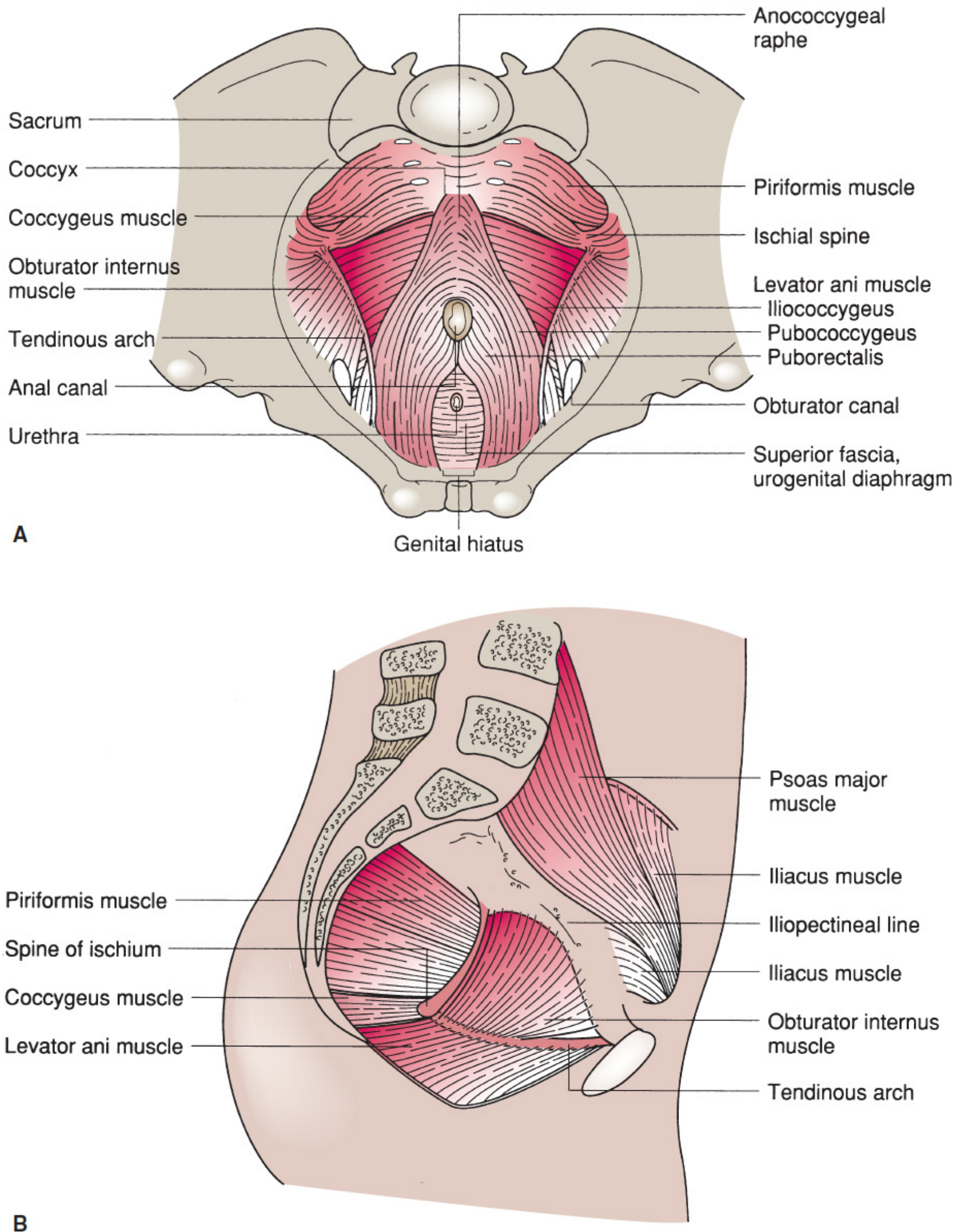
Pelvic diaphragm and urogenital diaphragm viewed from within the pelvis:
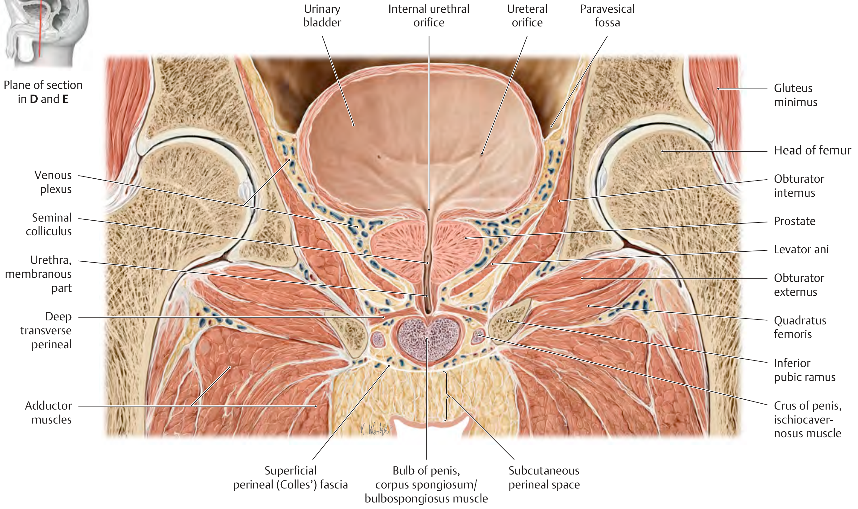
Coronal section through male pelvis showing deep transverse perineal muscle:
Summary: The urogenital diaphragm is the fibromuscular sheet filling the urogenital hiatus of the pelvic diaphragm. Its two key muscles are the deep transverse perineal and the external urethral sphincter (sphincter urethrae). The structure is enclosed between the superior and inferior (perineal membrane) fasciae of the UGD, transmits the urethra (and vagina in females), and plays a primary role in urinary continence and pelvic floor support.
This is a shared conversation. Sign in to Orris to start your own chat.