
Signs and symptoms of migraine and why is it caused
migraine pathophysiology signs symptoms mechanisms 2024
migraine pathophysiology diagram brain
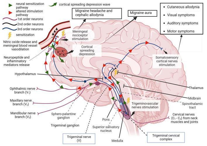
A pathophysiology diagram illustrating the trigeminovascular pathway and its role in migraine development. The diagram depicts a sagittal view of the human brain, highlighting key anatomical structures including the trigeminal ganglion, trigeminal cervical complex (medulla/pons), thalamus, hypothalamus, and somatosensory cortex. It maps the neural hierarchy from 1st-order neurons (trigeminal branches V1, V2, V3 and cervical nerves C1-C3) through the 2nd-order spinothalamic tract to 3rd-order thalamic projections. Visual indicators identify areas of sensitization and altered stimulation pathways. Key educational concepts include the cortical spreading depression wave, meningeal nociceptor stimulation, and neuropeptide release leading to vasodilation. The diagram correlates specific neural activity with clinical symptoms, specifically migraine aura, cephalic and cutaneous allodynia, and visual, auditory, and motor impairments. It further demonstrates the descending pathway via the superior salivatory nucleus involving nitric oxide release in meningeal blood vessels, providing a comprehensive visual of central and peripheral sensitization in migraine pathology.
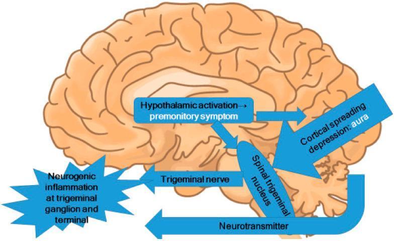
This pathophysiology diagram illustrates the central mechanisms of migraine, overlaid on a sagittal view of the human brain. The illustration highlights the interplay between several key neuroanatomical structures and processes. A central text box indicates 'Hypothalamic activation' leading to 'premonitory symptoms', with arrows showing its influence on the 'Spinal trigeminal nucleus' in the brainstem. Cortical spreading depression, associated with 'aura', is depicted as a large arrow targeting the same spinal trigeminal nucleus. The diagram outlines the involvement of the trigeminal nerve system, showing bidirectional activity: activation of the spinal trigeminal nucleus leads to 'Neurogenic inflammation at the trigeminal ganglion and terminal' (represented by a starburst graphic), while 'Neurotransmitter' release is shown signaling back from the brainstem to the peripheral trigeminal structures. This diagram serves as an educational tool for medical students and clinicians to understand the trigeminovascular system's role in migraine pathogenesis, specifically how hypothalamic and cortical triggers converge on the brainstem and peripheral nerve terminals.
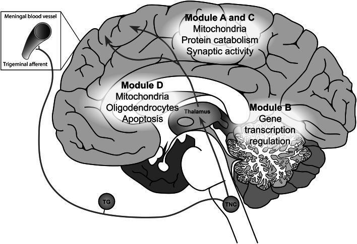
A medical schematic illustrating the pathophysiology of migraine, specifically the trigeminovascular pathway and associated gene co-expression modules in the brain. The diagram depicts a sagittal view of the human brain with key anatomical structures labeled, including the cortex, cerebellum, thalamus, and trigeminal nucleus caudalis (TNC). The trigeminovascular pathway is traced from meningeal blood vessels via trigeminal afferents through the trigeminal ganglion (TG) and TNC, ascending to the thalamus and ultimately the cerebral cortex. Overlaid on the anatomy are localized gene expression modules (A-D) highlighting specific cellular functions and components: Modules A and C in the cortex are associated with mitochondria, protein catabolism, and synaptic activity; Module B in the cerebellum is linked to gene transcription regulation; and Module D in the subcortical white matter and thalamic region is associated with mitochondria, oligodendrocytes, and apoptosis. The illustration serves as an educational tool for understanding the integration of genetic expression patterns with neuroanatomical pain pathways in migraine headache disorders.
migraine pathophysiology trigeminovascular
Migraine: Signs, Symptoms, and Causes
What Is Migraine?
The Four Phases and Their Symptoms
1. Premonitory Phase (Prodrome)
- Fatigue and yawning (very common)
- Mood changes - depression, irritability, or euphoria
- Food cravings (especially for sweet or salty foods)
- Neck stiffness
- Increased thirst and urination
- Difficulty concentrating
- Sensitivity to light and sound (early onset)
2. Aura (in ~25-30% of migraineurs)
| Type | Symptoms |
|---|---|
| Visual (most common) | Zigzag lines (fortification spectra), scotoma (blind spots), flashing lights, blurred vision |
| Sensory | Pins and needles or numbness with a "marching" spread - usually arm or face |
| Motor | Weakness spreading from one area to another (rare) |
| Speech/Language | Mild dysphasia, difficulty finding words |
| Brainstem aura | Vertigo, diplopia, dysarthria, tinnitus, bilateral paresthesias |
3. Headache Phase (The Core Attack)
- Unilateral location
- Pulsating/throbbing quality
- Moderate-to-severe intensity
- Worsened by routine physical activity (walking, climbing stairs)
- Nausea and/or vomiting
- Photophobia (light sensitivity) AND phonophobia (sound sensitivity)
- Osmophobia (smell sensitivity) - highly specific for migraine when present
- Cutaneous allodynia - pain from normally non-painful stimuli (e.g., brushing hair, wearing glasses)
- Pallor and clamminess
- Prostration - patients typically retreat to a dark, quiet room
4. Postdrome ("Migraine Hangover")
- Exhaustion and fatigue
- Cognitive fog ("brain fog")
- Residual neck soreness
- Mood changes - can be either low or unusually elevated
Why Is Migraine Caused? (Pathophysiology)
1. Genetic Predisposition
- Migraine has a strong genetic basis: first-degree relatives of migraine-with-aura patients are ~4x more likely to develop it.
- Familial Hemiplegic Migraine (FHM) provided the clearest genetic clues - three gene mutations are identified:
- FHM1: CACNA1A (chromosome 19p13) - α1 subunit of a P/Q-type voltage-gated calcium channel
- FHM2: ATP1A2 (chromosome 1q23) - α2 subunit of the sodium-potassium ATPase pump
- FHM3: SCN1A (chromosome 2q24) - α subunit of a neuronal voltage-gated sodium channel
- These mutations lead to abnormal ion channel function, increasing neuronal excitability and susceptibility to spreading depolarization.
- Common forms of migraine involve ~44 genetic variants across 38 loci affecting vascular biology, synaptic regulation, and cell-cell interactions.
2. Cortical Spreading Depression (CSD)
- CSD is the mechanism underlying migraine aura and likely also triggers head pain.
- It is a self-propagating wave of near-complete neuronal depolarization followed by suppression, traveling across the cortex at ~3 mm/min.
- CSD activates trigeminal pain fibers around meningeal blood vessels.
- A 2024 University of Rochester study demonstrated for the first time how the spreading wave interacts with brain fluid dynamics (glymphatic system) to trigger headache - identifying new proteins as potential therapeutic targets.
3. The Trigeminovascular System - The Pain Generator
- Pain-sensitive intracranial structures (meningeal blood vessels, dura mater) are innervated by the trigeminal nerve (primarily V1 ophthalmic branch).
- When activated, trigeminal nerve terminals release CGRP (calcitonin gene-related peptide), substance P, and other neuropeptides.
- These neuropeptides cause neurogenic inflammation - vasodilation, plasma protein extravasation, and mast cell degranulation around meningeal vessels.
- Pain signals travel: 1st-order trigeminal neurons → trigeminal cervical complex (brainstem/upper cervical cord) → 2nd-order neurons via spinothalamic tract → thalamus → 3rd-order neurons → somatosensory cortex.
- The convergence of cervical (C1-C3) and trigeminal inputs at the trigeminal cervical complex explains why migraine pain often radiates to the neck and occiput.
4. Central and Peripheral Sensitization
- Repeated activation leads to sensitization of pain pathways.
- Peripheral sensitization: Trigeminal nerve endings become hypersensitive - explaining throbbing pain worsened by movement (pulsatile mechanical stimulation of sensitized vessels).
- Central sensitization: Second- and third-order neurons become sensitized, causing allodynia (pain from non-noxious stimuli like touch or temperature) - a marker of established central sensitization in an attack.
5. Hypothalamic Involvement
- The hypothalamus is activated during the premonitory phase (before headache starts), driving symptoms like food cravings, yawning, thirst, and fatigue.
- It modulates pain processing and acts as a "migraine generator" or pacemaker for attacks.
6. Serotonin and CGRP
- Serotonin (5-HT) levels drop during attacks; serotonin receptors (especially 5-HT1B/1D) on trigeminal terminals and blood vessels are targets for triptans.
- CGRP is the dominant neuropeptide driving neurogenic inflammation and vasodilation. Modern preventive therapies (CGRP monoclonal antibodies like erenumab, fremanezumab) and acute treatments (gepants) target this pathway directly.
Common Triggers
| Category | Examples |
|---|---|
| Hormonal | Estrogen fluctuations, menstruation, oral contraceptives |
| Dietary | Alcohol (esp. red wine), caffeine/caffeine withdrawal, nitrates, aged cheese, chocolate, skipping meals |
| Sleep | Oversleeping or sleep deprivation |
| Sensory | Bright lights, strong odors, loud noise |
| Environmental | Weather/barometric pressure changes |
| Psychological | Stress, anxiety, "let-down" after stress |
Summary
- Bradley and Daroff's Neurology in Clinical Practice - the primary reference for pathophysiology and classification
- Textbook of Family Medicine, 9e - IHS diagnostic criteria
- Recent reviews: Neuroimmune interactions in migraine (PMID 39271369, Trends in Neurosciences, 2024) and Migraine review (Phys Med Rehabil Clin N Am, 2025, PMID 41167851)