
Types of heart blocks with ecg and each component description
heart block ECG types first degree second degree third degree AV block
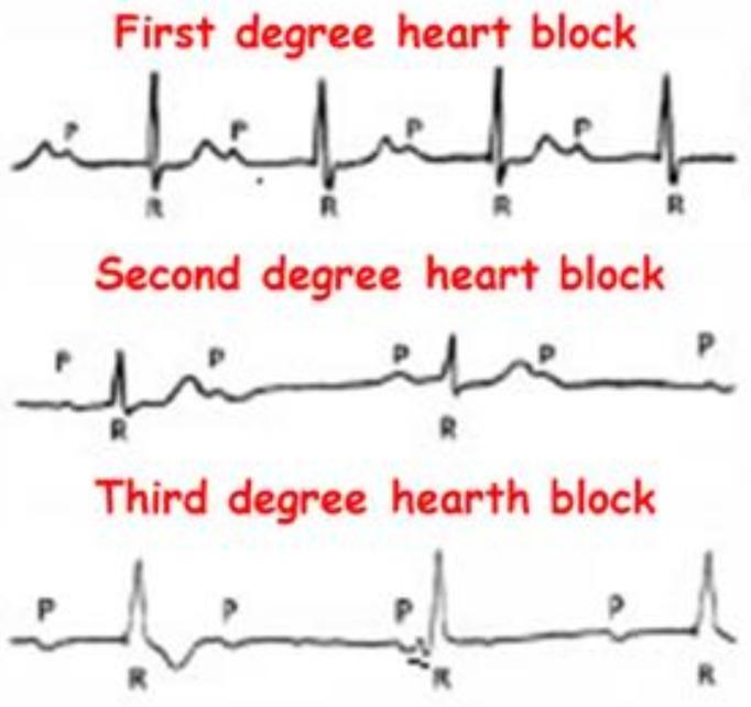
This Comparison Chart illustrates the electrocardiographic (ECG) characteristics of Atrioventricular (AV) blocks. It features three stacked rhythm strips categorized as First, Second, and Third-degree heart blocks, emphasizing the relationship between P waves (atrial depolarization) and QRS complexes (ventricular depolarization). The 'First degree heart block' panel demonstrates a consistent 1:1 P-to-QRS ratio with a fixed, prolonged PR interval. The 'Second degree heart block' panel shows intermittent conduction failure, where specific P waves are not followed by a QRS complex. The 'Third degree heart block' panel depicts complete AV dissociation, where P waves and QRS complexes occur independently and at different rates, indicating a total interruption of impulse transmission and the presence of a ventricular escape rhythm. This diagnostic illustration is designed for cardiovascular education to help students and clinicians distinguish between different stages of cardiac conduction system disease and their associated clinical significance.
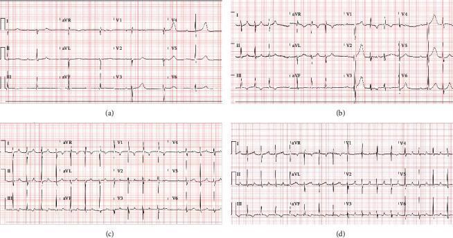
A comparison of four 12-lead electrocardiogram (ECG) tracings illustrating the progression and types of atrioventricular (AV) conduction blocks. (a) Complete heart block (third-degree AV block) demonstrating atrioventricular dissociation with a slow, regular ventricular escape rhythm of 41 bpm and independent atrial activity. (b) Second-degree type 2 AV block (Mobitz II) showing intermittent non-conducted P waves with a constant PR interval in conducted beats. (c) Second-degree Mobitz type 1 AV block (Wenckebach) characterized by progressive lengthening of the PR interval until a QRS complex is dropped. (d) First-degree AV block displaying a 1:1 P-to-QRS ratio with a fixed, prolonged PR interval exceeding 200 ms (specifically >250 ms). The clinical series documents the evolution of conduction system abnormalities in a patient with myocarditis, showing transitions between high-grade blocks and milder conduction delays during treatment with isoproterenol and anti-inflammatory therapies.
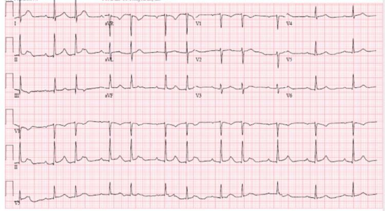
This diagnostic image is a standard 12-lead electrocardiogram (ECG) displayed on a traditional grid background, illustrating a second-degree atrioventricular (AV) block, specifically Mobitz Type I (Wenckebach phenomenon). The tracing shows characteristic patterns across the limb leads (I, II, III, aVR, aVL, aVF) and precordial leads (V1-V6). Key diagnostic features include a progressive lengthening of the PR interval in successive beats until a P wave fails to conduct to the ventricles, resulting in a dropped QRS complex. This cycle then repeats, starting with a shorter PR interval. The QRS complexes are relatively narrow, suggesting the block occurs at the level of the AV node. P-wave morphology is consistent with sinus rhythm, being upright in leads II and V2-V6 and inverted in aVR. This ECG is a critical educational tool for demonstrating non-invasive identification of conduction system pathologies and differentiating between types of second-degree heart blocks.
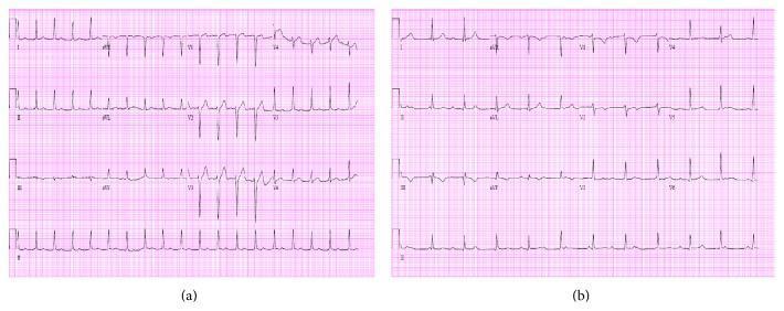
This diagnostic image presents a side-by-side comparison of two 12-lead electrocardiograms (ECGs) demonstrating the progression of conduction disease. The left panel (Day 1) shows a sinus tachycardia with a rate of 111 bpm. Notable features include a consistently prolonged PR interval, indicating a first-degree atrioventricular (AV) block. The right panel (Day 2) displays a significant clinical evolution to a third-degree (complete) heart block. Key diagnostic findings in the second ECG include complete AV dissociation, where P waves and QRS complexes occur independently of each other. The atrial rate is faster than the ventricular rate, which has slowed to 67 bpm with a regular R-R interval, suggesting a stable escape rhythm. This comparison illustrates the dynamic nature of electrical conduction abnormalities in acute clinical settings, such as infective endocarditis where paravalvular abscess formation may compromise the conduction system. The ECGs include standard limb leads (I, II, III), augmented leads (aVR, aVL, aVF), and precordial leads (V1-V6).
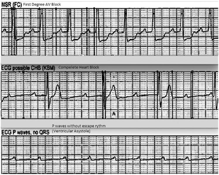
This diagnostic image displays a vertical comparison of three ECG rhythm strips demonstrating the progression and characteristics of different cardiac conduction abnormalities. The top strip, labeled 'NSR (FC) First Degree AV Block,' shows a sinus rhythm with a prolonged PR interval, indicating a delay in conduction between the atria and ventricles. The middle strip, labeled 'ECG possible CHB (KBM) Complete Heart Block,' illustrates third-degree atrioventricular (AV) block characterized by complete AV dissociation; P waves are present but occur independently of the slow, irregular QRS complexes (escape rhythm). The bottom strip, labeled 'ECG P waves, no QRS (Ventricular Asystole),' depicts a high-grade block where atrial activity (P waves) continues, but there is a total failure of ventricular conduction resulting in ventricular asystole. This sequence serves as an educational tool for identifying varying severity levels of heart blocks and the transition from conduction delay to lethal ventricular standstill.
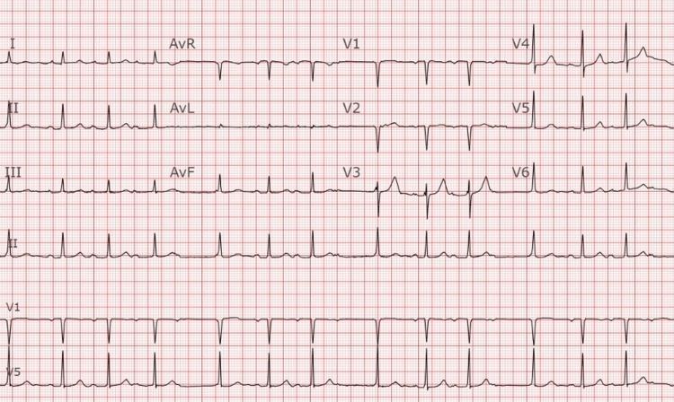
This diagnostic image is a 12-lead electrocardiogram (ECG) printed on standard grid paper, displaying leads I, II, III, aVR, aVL, aVF, and V1-V6, including long rhythm strips for leads II, V1, and V5. The tracing demonstrates classic features of Second-Degree Atrioventricular (AV) Block, Mobitz Type I (Wenckebach phenomenon). The primary diagnostic hallmark visible in lead II is the progressive lengthening of the PR interval over successive cardiac cycles until a P wave fails to conduct to the ventricles, resulting in a 'dropped' QRS complex. Following the non-conducted P wave, the PR interval resets to its shortest duration and the cycle repeats. The QRS complexes are relatively narrow with a normal morphology, and P-wave morphology remains consistent. The ST segments are isoelectric without significant elevation or depression. This ECG is a key educational resource for identifying AV nodal conduction delays and distinguishing between different types of second-degree heart blocks in a clinical cardiology context.
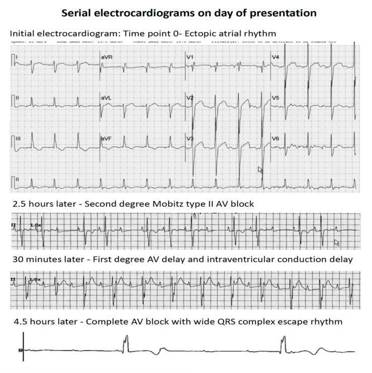
This diagnostic image consists of a series of four electrocardiogram (ECG) rhythm strips demonstrating the rapid clinical progression of atrioventricular (AV) conduction abnormalities over a period of 4.5 hours. The top panel, labeled 'Time point 0', shows an ectopic atrial rhythm with organized P-wave activity and narrow QRS complexes. The second strip, recorded 2.5 hours later, displays a second-degree Mobitz type II AV block, characterized by intermittently dropped QRS complexes without preceding PR interval lengthening. The third strip, 30 minutes later, indicates a first-degree AV delay (prolonged PR interval) and intraventricular conduction delay (widened QRS). The final rhythm strip at 4.5 hours shows third-degree (complete) AV block, characterized by complete AV dissociation and a slow, wide QRS complex ventricular escape rhythm. This progression illustrates acute cardiac conduction system failure, typically associated with myocardial infiltration or severe injury, as seen in patients with leukemic infiltration of the heart.
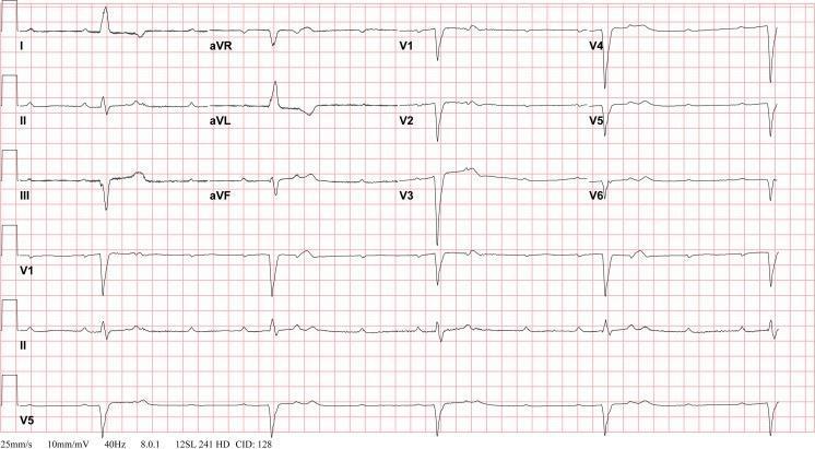
A 12-lead electrocardiogram (ECG) demonstrating a complete atrioventricular (AV) block, also known as third-degree heart block. The diagnostic image shows total AV dissociation characterized by a lack of relationship between P waves and QRS complexes. The atrial rate is significantly faster and independent of the ventricular rate. The ventricular escape rhythm is slow (bradycardic) with wide QRS complexes (duration >120 ms), indicating the escape rhythm originates from a ventricular or infra-Hisian site. Prominent features include deep S waves in the precordial leads (V1-V3) and T-wave inversions in several leads, including I, II, aVL, and V4-V6. This tracing represents a postoperative clinical scenario following tricuspid valve replacement, illustrating a known complication of congenital heart surgery. The ECG is recorded at standard settings of 25 mm/s and 10 mm/mV, serving as a critical educational tool for identifying life-threatening conduction system failures and the necessity for pacing interventions.
Types of Heart Blocks - Complete Guide with ECG Findings
ECG Component Review (Before the Blocks)
| Component | Normal Value | Represents |
|---|---|---|
| P wave | <120 ms, <2.5 mm tall | Atrial depolarization |
| PR interval | 120-200 ms (0.12-0.20 s) | AV nodal + His-Purkinje conduction time |
| QRS complex | <120 ms | Ventricular depolarization |
| QT interval | 350-440 ms (rate-corrected) | Total ventricular activity |
| T wave | Follows QRS in same direction | Ventricular repolarization |
Overview: All Three Degrees
1. First-Degree AV Block
ECG Features:
- PR interval > 0.20 seconds (200 ms) in every beat - this is the hallmark
- PR intervals up to 1.0 second have been recorded
- Regular R-R intervals (every P is followed by a QRS)
- QRS morphology is usually normal width unless concurrent BBB
- P:QRS ratio = 1:1
Clinical Notes:
- Asymptomatic - no treatment required
- Long PR creates a long "a-to-c" jugular venous interval at the bedside
- Diminished intensity of S1 as PR lengthens
- Can progress to Type I second-degree block with vagal stimulation or increased heart rate
Source: Braunwald's Heart Disease, 10th Ed
2. Second-Degree AV Block
2a. Mobitz Type I (Wenckebach)
ECG Features:
- Progressively increasing PR interval in successive beats
- PR interval is shortest right after the dropped beat
- A P wave eventually fails to conduct (no QRS follows it)
- R-R intervals progressively shorten before the pause (because the PR increment gets smaller each cycle)
- The pause (blocked beat) is less than twice the shortest R-R interval
- QRS is narrow (<0.12 sec) - block is at AV node level
- Beat 1: PR = 200 ms (+100 ms increment coming)
- Beat 2: PR = 300 ms (+50 ms increment)
- Beat 3: PR = 350 ms
- Beat 4: P wave BLOCKED (no QRS) → cycle restarts
2b. Mobitz Type II
ECG Features:
- Fixed PR interval in all conducted beats (no progressive lengthening)
- Sudden non-conducted P wave without warning
- QRS may be widened (>0.12 sec) or show bundle branch block pattern - block is infranodal (below His bundle)
- Ratio may be 2:1, 3:1, or higher (e.g., 3:1 = 3 P waves for every 1 QRS)
- More ominous pattern
Distinguishing Type I vs. Type II at a glance:
| Feature | Mobitz I (Wenckebach) | Mobitz II |
|---|---|---|
| PR before dropped beat | Longest | Constant (unchanged) |
| PR after dropped beat | Shortest (resets) | Same as before drop |
| QRS width | Narrow | Wide / BBB pattern |
| Block site | AV node | Infranodal (His-Purkinje) |
| Risk of progression | Low | High |
| Associated MI | Inferior | Anterior |
| Treatment | Observation/atropine | Pacemaker |
Special Case: 2:1 AV Block
3. Third-Degree (Complete) AV Block
ECG Features:
- P waves and QRS complexes are completely independent - no fixed relationship
- Atrial rate > ventricular rate (atria driven by SA node; ventricles by escape pacemaker)
- Regular P-P intervals (atria firing normally)
- Regular R-R intervals (ventricular escape rhythm is regular, but slow)
- QRS morphology depends on escape pacemaker location:
- Junctional escape (His bundle): narrow QRS, rate 40-60 bpm - more stable
- Ventricular escape: wide QRS (>0.12 sec), rate 20-40 bpm - less stable, more dangerous
- Coronary artery disease (inferior MI via RCA - usually reversible; anterior MI - often permanent)
- Drug toxicity (digoxin, beta-blockers, calcium channel blockers)
- Degenerative conduction system disease (Lenègre-Lev disease)
- Infectious: Lyme disease, endocarditis, rheumatic fever
- Congenital (maternal anti-Ro/La antibodies in neonates)
- Post-cardiac surgery
- Syncope (Adams-Stokes attacks)
- Profound bradycardia, hypotension
- Heart failure, angina
- Cannon A waves in jugular venous pulse (atria contracting against closed tricuspid valve)
4. Side-by-Side 12-Lead ECG Comparison
Summary Table
| Type | P:QRS Ratio | PR Interval | QRS Width | Block Site | Treatment |
|---|---|---|---|---|---|
| 1st degree | 1:1 | >200 ms, fixed | Usually narrow | AV node | None |
| 2nd degree Mobitz I | >1:1 (intermittent) | Progressively increases, then drops | Narrow | AV node | Observation / atropine |
| 2nd degree Mobitz II | >1:1 (intermittent) | Fixed (constant) | Wide / BBB | Infranodal | Pacemaker |
| High-grade | 3:1 or more | Variable | Variable | Variable | Pacemaker |
| 3rd degree (complete) | P and QRS independent | No relationship | Narrow (junctional) or wide (ventricular) | Complete AV | Permanent pacemaker |
Generate a clinical summary document on heart block diagnosis and treatment
ls -la /tmp/workspace/ 2>/dev/null || mkdir -p /tmp/workspace && echo "workspace ready"
mkdir -p /tmp/workspace/heart-block-doc && python3 << 'PYEOF' from docx import Document from docx.shared import Pt, Inches, RGBColor, Cm from docx.enum.text import WD_ALIGN_PARAGRAPH from docx.enum.table import WD_TABLE_ALIGNMENT, WD_ALIGN_VERTICAL from docx.oxml.ns import qn from docx.oxml import OxmlElement import datetime doc = Document() # ── Page margins ────────────────────────────────────────────────────────────── for section in doc.sections: section.top_margin = Cm(2.0) section.bottom_margin = Cm(2.0) section.left_margin = Cm(2.5) section.right_margin = Cm(2.5) # ── Default Normal style ────────────────────────────────────────────────────── normal = doc.styles["Normal"] normal.font.name = "Calibri" normal.font.size = Pt(11) # ── Helper: shade a table cell ──────────────────────────────────────────────── def shade_cell(cell, hex_color): tc = cell._tc tcPr = tc.get_or_add_tcPr() shd = OxmlElement("w:shd") shd.set(qn("w:val"), "clear") shd.set(qn("w:color"), "auto") shd.set(qn("w:fill"), hex_color) tcPr.append(shd) def set_cell_border(cell, **kwargs): """Set borders on a cell. kwargs: top, bottom, left, right = (sz, color, val)""" tc = cell._tc tcPr = tc.get_or_add_tcPr() tcBorders = OxmlElement("w:tcBorders") for edge in ("top","left","bottom","right"): if edge in kwargs: sz, color, val = kwargs[edge] border = OxmlElement(f"w:{edge}") border.set(qn("w:val"), val) border.set(qn("w:sz"), str(sz)) border.set(qn("w:color"), color) tcBorders.append(border) tcPr.append(tcBorders) def add_colored_heading(doc, text, level=1, rgb=(0,51,102)): h = doc.add_heading(text, level=level) h.alignment = WD_ALIGN_PARAGRAPH.LEFT for run in h.runs: run.font.color.rgb = RGBColor(*rgb) return h def add_section_box(doc, title, body_lines, title_bg="1F3864", title_fg=(255,255,255), body_bg="EBF3FB"): """Renders a colored box with a title row and content rows.""" tbl = doc.add_table(rows=1+len(body_lines), cols=1) tbl.style = "Table Grid" tbl.alignment = WD_TABLE_ALIGNMENT.CENTER # title row title_cell = tbl.rows[0].cells[0] shade_cell(title_cell, title_bg) p = title_cell.paragraphs[0] p.alignment = WD_ALIGN_PARAGRAPH.CENTER run = p.add_run(title) run.bold = True run.font.color.rgb = RGBColor(*title_fg) run.font.size = Pt(12) # body rows for i, line in enumerate(body_lines): c = tbl.rows[i+1].cells[0] shade_cell(c, body_bg) bp = c.paragraphs[0] bp.alignment = WD_ALIGN_PARAGRAPH.LEFT bp.add_run(line).font.size = Pt(10.5) doc.add_paragraph() # ══════════════════════════════════════════════════════════════════════════════ # COVER HEADER # ══════════════════════════════════════════════════════════════════════════════ p = doc.add_paragraph() p.alignment = WD_ALIGN_PARAGRAPH.CENTER run = p.add_run("CLINICAL SUMMARY") run.bold = True run.font.size = Pt(22) run.font.color.rgb = RGBColor(0, 51, 102) run.font.name = "Calibri" p2 = doc.add_paragraph() p2.alignment = WD_ALIGN_PARAGRAPH.CENTER run2 = p2.add_run("Heart Block: Diagnosis and Treatment") run2.bold = True run2.font.size = Pt(16) run2.font.color.rgb = RGBColor(192, 0, 0) run2.font.name = "Calibri" p3 = doc.add_paragraph() p3.alignment = WD_ALIGN_PARAGRAPH.CENTER meta = p3.add_run(f"Prepared by: Orris Clinical AI | Date: {datetime.date.today().strftime('%B %d, %Y')} | Evidence Based: Braunwald's Heart Disease, Goldman-Cecil Medicine") meta.font.size = Pt(9) meta.font.color.rgb = RGBColor(100, 100, 100) # divider line doc.add_paragraph("─" * 90) doc.add_paragraph() # ══════════════════════════════════════════════════════════════════════════════ # SECTION 1 – DEFINITION & OVERVIEW # ══════════════════════════════════════════════════════════════════════════════ add_colored_heading(doc, "1. Definition and Overview", level=1, rgb=(0,51,102)) p = doc.add_paragraph() p.add_run( "Heart block is a disturbance of impulse conduction along the atrioventricular (AV) " "conduction system that can be permanent or transient depending on the anatomic or " "functional impairment. It is distinct from physiologic interference, which is caused " "by normal refractoriness. The block may occur at the AV node, the His bundle, or the " "bundle branches." ) doc.add_paragraph() # Anatomy box add_section_box(doc, "Cardiac Conduction System - Key Levels", [ " SA Node -> Atria -> AV Node -> Bundle of His -> Bundle Branches (L + R) -> Purkinje Fibers -> Ventricles", " First-degree & Mobitz I blocks: usually at the AV node level (supra-Hisian)", " Mobitz II & complete block: usually infranodal (below His bundle - bundle branches)", ], title_bg="1F3864", body_bg="D6E4F0" ) # ══════════════════════════════════════════════════════════════════════════════ # SECTION 2 – ECG COMPONENTS # ══════════════════════════════════════════════════════════════════════════════ add_colored_heading(doc, "2. Key ECG Components", level=1, rgb=(0,51,102)) ecg_data = [ ("P Wave", "<120 ms; <2.5 mm", "Atrial depolarization (SA node to AV node)"), ("PR Interval", "120-200 ms (0.12-0.20 s)", "AV nodal conduction + His-Purkinje time"), ("QRS Complex", "<120 ms", "Ventricular depolarization"), ("QT Interval", "350-440 ms (rate-corrected)","Total ventricular electrical activity"), ("T Wave", "Concordant with QRS", "Ventricular repolarization"), ] tbl = doc.add_table(rows=1+len(ecg_data), cols=3) tbl.style = "Table Grid" tbl.alignment = WD_TABLE_ALIGNMENT.CENTER headers = ["Component", "Normal Value", "Represents"] for i, h in enumerate(headers): cell = tbl.rows[0].cells[i] shade_cell(cell, "2E5090") run = cell.paragraphs[0].add_run(h) run.bold = True run.font.color.rgb = RGBColor(255,255,255) run.font.size = Pt(10) row_colors = ["FFFFFF","F0F4FA","FFFFFF","F0F4FA","FFFFFF"] for i, (comp, val, rep) in enumerate(ecg_data): row = tbl.rows[i+1] shade_cell(row.cells[0], row_colors[i]) shade_cell(row.cells[1], row_colors[i]) shade_cell(row.cells[2], row_colors[i]) row.cells[0].paragraphs[0].add_run(comp).font.bold = True row.cells[1].paragraphs[0].add_run(val).font.size = Pt(10) row.cells[2].paragraphs[0].add_run(rep).font.size = Pt(10) doc.add_paragraph() # ══════════════════════════════════════════════════════════════════════════════ # SECTION 3 – CLASSIFICATION # ══════════════════════════════════════════════════════════════════════════════ add_colored_heading(doc, "3. Classification of Heart Blocks", level=1, rgb=(0,51,102)) # 3.1 First degree add_colored_heading(doc, "3.1 First-Degree AV Block", level=2, rgb=(31, 73, 125)) p = doc.add_paragraph() p.add_run("Definition: ").bold = True p.add_run("Every atrial impulse is conducted to the ventricles (1:1 ratio) but with prolonged conduction time. The PR interval exceeds 0.20 seconds in every beat.") p = doc.add_paragraph() p.add_run("Site of Block: ").bold = True p.add_run("Usually the AV node (prolonged A-H interval). When QRS shows a bundle branch block pattern, the delay may be within the His-Purkinje system.") doc.add_paragraph() add_colored_heading(doc, "ECG Features", level=3, rgb=(68,114,196)) ecg_1 = doc.add_paragraph(style="List Bullet") ecg_1.add_run("PR interval > 200 ms in every beat (hallmark finding)").bold = True for item in [ "P:QRS ratio = 1:1 (every P wave followed by a QRS)", "Regular R-R intervals", "QRS complex is usually normal width (<120 ms)", "PR intervals as long as 1.0 second have been documented", ]: bp = doc.add_paragraph(style="List Bullet") bp.add_run(item) doc.add_paragraph() add_colored_heading(doc, "Clinical Features & Management", level=3, rgb=(68,114,196)) p = doc.add_paragraph(style="List Bullet"); p.add_run("Asymptomatic - no haemodynamic compromise") p = doc.add_paragraph(style="List Bullet"); p.add_run("Diminished S1 intensity; long a-to-c wave in jugular venous pulse") p = doc.add_paragraph(style="List Bullet"); p.add_run("No treatment required. Monitor for progression") p = doc.add_paragraph(style="List Bullet"); p.add_run("Can progress to Type I second-degree block with vagal stimulation or with increased atrial rate") doc.add_paragraph() # 3.2 Second degree add_colored_heading(doc, "3.2 Second-Degree AV Block", level=2, rgb=(31,73,125)) p = doc.add_paragraph() p.add_run("Definition: ").bold = True p.add_run("Some P waves are not conducted to the ventricles. There are two distinct subtypes with different mechanisms, sites, prognoses, and treatments.") doc.add_paragraph() # Mobitz I add_colored_heading(doc, "Mobitz Type I (Wenckebach)", level=3, rgb=(68,114,196)) p = doc.add_paragraph() p.add_run("Key Hallmark: ").bold = True p.add_run("Progressive PR interval lengthening until a P wave is dropped, then the cycle resets to the shortest PR.") add_colored_heading(doc, "ECG Features", level=3, rgb=(68,114,196)) for item in [ "Progressively increasing PR interval in successive beats", "Longest PR interval immediately before the dropped beat", "Shortest PR interval immediately after the dropped beat (cycle reset)", "R-R intervals progressively shorten before the pause", "Pause (non-conducted beat) is less than twice the shortest R-R interval", "QRS complex is narrow (<0.12 sec) - block is at AV node", "P:QRS ratio e.g. 4:3, 3:2", ]: bp = doc.add_paragraph(style="List Bullet"); bp.add_run(item) add_colored_heading(doc, "Clinical Context", level=3, rgb=(68,114,196)) for item in [ "Associated with inferior MI (RCA territory - right coronary artery supplies AV node)", "May occur during sleep in well-conditioned athletes (vagal tone)", "Rarely progresses to complete heart block", "Treatment: Observation; atropine if symptomatic bradycardia", ]: bp = doc.add_paragraph(style="List Bullet"); bp.add_run(item) doc.add_paragraph() # Mobitz II add_colored_heading(doc, "Mobitz Type II", level=3, rgb=(68,114,196)) p = doc.add_paragraph() p.add_run("Key Hallmark: ").bold = True p.add_run("P waves drop suddenly WITHOUT any preceding PR interval lengthening.") add_colored_heading(doc, "ECG Features", level=3, rgb=(68,114,196)) for item in [ "Fixed, constant PR interval in all conducted beats (no progressive lengthening)", "Sudden non-conducted P wave without warning or preceding PR change", "QRS complex is usually wide (>0.12 sec) or shows bundle branch block pattern", "Block is infranodal (below the His bundle - bundle branch level)", "May show 2:1, 3:1 or higher conduction ratios", ]: bp = doc.add_paragraph(style="List Bullet"); bp.add_run(item) add_colored_heading(doc, "Clinical Context", level=3, rgb=(68,114,196)) for item in [ "Associated with anterior MI (LAD territory - affects bundle branches)", "High risk of progression to complete (third-degree) AV block", "Requires permanent pacemaker implantation", "Emergency transvenous pacing if haemodynamically unstable", ]: bp = doc.add_paragraph(style="List Bullet"); bp.add_run(item) doc.add_paragraph() # 2:1 special note p = doc.add_paragraph() run = p.add_run("Special Case - 2:1 AV Block: ") run.bold = True run.font.color.rgb = RGBColor(192,0,0) p.add_run( "When every alternate P wave is blocked, it is impossible to distinguish Mobitz I from " "Mobitz II from surface ECG alone. Narrow QRS suggests Type I (nodal); wide QRS/BBB " "suggests Type II (infranodal). His bundle electrogram may be required for definitive classification." ) doc.add_paragraph() # High grade add_colored_heading(doc, "High-Grade (Advanced) AV Block", level=3, rgb=(68,114,196)) p = doc.add_paragraph() p.add_run("Definition: ").bold = True p.add_run("Two or more consecutive P waves are blocked (e.g., 3:1, 4:1 conduction). Implies near-complete failure of AV conduction. Indication for pacing.") doc.add_paragraph() # 3.3 Third degree add_colored_heading(doc, "3.3 Third-Degree (Complete) AV Block", level=2, rgb=(31,73,125)) p = doc.add_paragraph() p.add_run("Definition: ").bold = True p.add_run("No atrial impulses are conducted to the ventricles. Complete AV dissociation - atria and ventricles beat independently, each driven by separate pacemakers.") add_colored_heading(doc, "ECG Features", level=3, rgb=(68,114,196)) for item in [ "P waves and QRS complexes are completely independent (no fixed PR relationship)", "Atrial rate is faster than ventricular rate", "Regular P-P intervals (atria firing from SA node normally)", "Regular R-R intervals (escape pacemaker is regular, but slow)", "P wave may appear to fall before a QRS but does NOT conduct it (coincidental timing - PR keeps varying)", "Narrow QRS (40-60 bpm): junctional escape from His bundle - more stable", "Wide QRS (20-40 bpm): ventricular escape from Purkinje/ventricular myocardium - less stable, more dangerous", ]: bp = doc.add_paragraph(style="List Bullet"); bp.add_run(item) doc.add_paragraph() # ══════════════════════════════════════════════════════════════════════════════ # SECTION 4 – COMPARISON TABLE # ══════════════════════════════════════════════════════════════════════════════ add_colored_heading(doc, "4. Summary Comparison Table", level=1, rgb=(0,51,102)) col_headers = ["Feature", "1st Degree", "2nd Deg Mobitz I", "2nd Deg Mobitz II", "3rd Degree (Complete)"] rows_data = [ ("P:QRS Ratio", "1:1", ">1:1 (intermittent)", ">1:1 (intermittent)", "Independent"), ("PR Interval", ">200 ms fixed","Progressive increase","Fixed / constant", "No relationship"), ("QRS Width", "Narrow", "Narrow (<120 ms)", "Wide / BBB", "Narrow (junctional)\nor Wide (ventricular)"), ("Block Site", "AV node", "AV node", "Infranodal", "Complete AV"), ("Associated MI", "—", "Inferior MI", "Anterior MI", "Either"), ("Progression Risk", "Minimal", "Low", "High", "N/A (already complete)"), ("Treatment", "None", "Observe / atropine", "Pacemaker", "Permanent pacemaker"), ] tbl2 = doc.add_table(rows=1+len(rows_data), cols=5) tbl2.style = "Table Grid" tbl2.alignment = WD_TABLE_ALIGNMENT.CENTER # Set column widths col_widths = [Cm(3.2), Cm(2.8), Cm(3.5), Cm(3.5), Cm(4.0)] for i, w in enumerate(col_widths): for row in tbl2.rows: row.cells[i].width = w for i, h in enumerate(col_headers): cell = tbl2.rows[0].cells[i] shade_cell(cell, "1F3864") run = cell.paragraphs[0].add_run(h) run.bold = True run.font.color.rgb = RGBColor(255,255,255) run.font.size = Pt(9) cell.paragraphs[0].alignment = WD_ALIGN_PARAGRAPH.CENTER alt_row_colors = ["FFFFFF","EEF4FB"] highlight_rows = {5: "FFF2CC", 6: "FFE0E0"} # highlight progression & treatment rows for i, row_vals in enumerate(rows_data): row = tbl2.rows[i+1] bg = highlight_rows.get(i, alt_row_colors[i % 2]) for j, val in enumerate(row_vals): cell = row.cells[j] shade_cell(cell, bg) p = cell.paragraphs[0] p.alignment = WD_ALIGN_PARAGRAPH.CENTER if j > 0 else WD_ALIGN_PARAGRAPH.LEFT run = p.add_run(val) run.font.size = Pt(9) if j == 0: run.bold = True doc.add_paragraph() # ══════════════════════════════════════════════════════════════════════════════ # SECTION 5 – AETIOLOGY # ══════════════════════════════════════════════════════════════════════════════ add_colored_heading(doc, "5. Aetiology", level=1, rgb=(0,51,102)) causes = { "Ischaemic": [ "Inferior MI - RCA occlusion (AV node ischaemia): tends to cause 1st/2nd degree, usually reversible", "Anterior MI - LAD occlusion (bundle branch ischaemia): tends to cause Mobitz II / complete block, often permanent", ], "Degenerative": [ "Lenegre disease: fibrosis of the His-Purkinje system (primary idiopathic)", "Lev disease: calcification extending from valves / fibrous skeleton into conduction system", ], "Infectious / Inflammatory": [ "Lyme disease (Borrelia burgdorferi) - most common infectious cause in endemic areas", "Rheumatic fever, endocarditis", "Myocarditis (viral, autoimmune)", "Chagas disease (Trypanosoma cruzi) - common cause in South America", "Sarcoidosis", ], "Drugs": [ "Digoxin toxicity", "Beta-blockers", "Calcium channel blockers (non-dihydropyridine: verapamil, diltiazem)", "Amiodarone", "Antiarrhythmics: Class Ia and Ic agents", ], "Congenital": [ "Isolated congenital complete AV block - most common congenital conduction disorder", "Neonatal lupus: maternal anti-Ro/La (SSA/SSB) antibodies crossing placenta", "Associated with congenital heart disease (e.g., L-TGA, AV canal defect)", ], "Metabolic / Electrolyte": [ "Hyperkalaemia", "Hypothyroidism", "Hypomagnesaemia", ], "Iatrogenic": [ "Post-cardiac surgery (valve replacement, VSD repair)", "Transcatheter aortic valve replacement (TAVR/TAVI)", "Catheter ablation", ], } for category, items in causes.items(): p = doc.add_paragraph() run = p.add_run(category + ":") run.bold = True run.font.color.rgb = RGBColor(0, 51, 102) for item in items: bp = doc.add_paragraph(style="List Bullet") bp.add_run(item).font.size = Pt(10.5) doc.add_paragraph() # ══════════════════════════════════════════════════════════════════════════════ # SECTION 6 – CLINICAL FEATURES # ══════════════════════════════════════════════════════════════════════════════ add_colored_heading(doc, "6. Clinical Features", level=1, rgb=(0,51,102)) p = doc.add_paragraph() p.add_run("Symptoms depend on the degree and haemodynamic impact of the block:").italic = True symptoms_table = [ ("First-Degree", "Asymptomatic", "Soft S1; long a-to-c JVP interval"), ("Second-Degree I", "Usually asymptomatic / mild", "Irregular pulse with pauses; variable S1 intensity"), ("Second-Degree II","Palpitations, presyncope", "Irregular pulse; wide pulse pressure"), ("Third-Degree", "Syncope (Stokes-Adams attacks),\nAngina, dyspnoea, heart failure,\nSudden cardiac death", "Bradycardia, hypotension, cannon A waves\nin JVP, variable S1 intensity"), ] tbl3 = doc.add_table(rows=1+len(symptoms_table), cols=3) tbl3.style = "Table Grid" for i, h in enumerate(["Block Type", "Symptoms", "Signs"]): cell = tbl3.rows[0].cells[i] shade_cell(cell, "2E5090") run = cell.paragraphs[0].add_run(h) run.bold = True; run.font.color.rgb = RGBColor(255,255,255); run.font.size = Pt(10) for i, (btype, symptoms, signs) in enumerate(symptoms_table): row = tbl3.rows[i+1] bg = "FFFFFF" if i % 2 == 0 else "EEF4FB" shade_cell(row.cells[0], bg); shade_cell(row.cells[1], bg); shade_cell(row.cells[2], bg) row.cells[0].paragraphs[0].add_run(btype).font.bold = True row.cells[1].paragraphs[0].add_run(symptoms).font.size = Pt(10) row.cells[2].paragraphs[0].add_run(signs).font.size = Pt(10) doc.add_paragraph() # ══════════════════════════════════════════════════════════════════════════════ # SECTION 7 – DIAGNOSIS # ══════════════════════════════════════════════════════════════════════════════ add_colored_heading(doc, "7. Diagnostic Approach", level=1, rgb=(0,51,102)) diag_steps = [ ("12-Lead ECG (First-line)", "Establishes diagnosis in most cases. Assess: PR interval, P:QRS ratio, PR variation, QRS width, " "P-QRS relationship. Rhythm strip in lead II or V1 is most informative."), ("Ambulatory ECG (Holter Monitor)", "For intermittent/paroxysmal blocks. 24-48 hour recording captures transient episodes not seen on " "resting ECG."), ("Exercise Stress Test", "AV block that worsens with exercise suggests infranodal disease (more serious). AV block that " "improves with exercise (increased sympathetic tone) suggests nodal disease."), ("His Bundle Electrogram (Electrophysiology Study)", "Gold standard for localising the site of block. Measures A-H interval (AV node) and H-V interval " "(His-Purkinje). Required when site cannot be determined from surface ECG (e.g., 2:1 block)."), ("Blood Tests", "FBC, U&E (hyperkalaemia), thyroid function, Lyme serology (endemic areas), ANA/anti-Ro/La " "(autoimmune), troponin (acute MI), digoxin level."), ("Echocardiogram", "Assess underlying structural heart disease, LV function, valve pathology, cardiomyopathy."), ("Cardiac MRI / CT", "If sarcoidosis, myocarditis, or infiltrative disease suspected."), ] for step, detail in diag_steps: p = doc.add_paragraph() run = p.add_run(step + ": ") run.bold = True run.font.color.rgb = RGBColor(31, 73, 125) p.add_run(detail).font.size = Pt(10.5) doc.add_paragraph() # ══════════════════════════════════════════════════════════════════════════════ # SECTION 8 – TREATMENT # ══════════════════════════════════════════════════════════════════════════════ add_colored_heading(doc, "8. Treatment", level=1, rgb=(0,51,102)) add_colored_heading(doc, "8.1 Acute Management", level=2, rgb=(31,73,125)) p = doc.add_paragraph() p.add_run("For haemodynamically unstable or symptomatic high-degree block:").italic = True acute = [ ("Atropine 0.5-1 mg IV", "First-line for symptomatic bradycardia in AV nodal blocks (Type I, vagally mediated). " "Repeat up to 3 mg total. NOT effective in infranodal (Mobitz II / complete) block."), ("Transcutaneous Pacing (TCP)", "Emergency bridge while transvenous pacing is arranged. Use in Mobitz II or complete block " "with haemodynamic compromise."), ("Transvenous Temporary Pacing", "Placed via internal jugular or subclavian vein. Definitive bridge to permanent pacemaker. " "Indicated in complete block, Mobitz II, and high-grade block."), ("Isoprenaline (Isoproterenol) infusion", "Sympathomimetic - increases ventricular escape rate. Used as bridge when pacing unavailable. " "Caution in ischaemic heart disease."), ("Treat reversible causes", "Withdraw offending drugs (digoxin, beta-blocker, CCB). Correct electrolyte abnormalities. " "Treat acute MI with reperfusion (PCI/thrombolysis) - may restore conduction."), ] for drug, detail in acute: p = doc.add_paragraph(style="List Bullet") run = p.add_run(drug + ": ") run.bold = True p.add_run(detail).font.size = Pt(10.5) doc.add_paragraph() add_colored_heading(doc, "8.2 Permanent Pacemaker Implantation", level=2, rgb=(31,73,125)) p = doc.add_paragraph() p.add_run("Indications (ACC/AHA Class I - permanent pacing is indicated):").bold = True ppm_indications = [ "Complete (third-degree) AV block - symptomatic or asymptomatic with ventricular rate <40 bpm or pauses >3 seconds", "Second-degree Mobitz Type II with wide QRS (infranodal block)", "Symptomatic second-degree AV block of any type", "High-grade AV block (>2:1 with symptoms)", "Chronic bifascicular or trifascicular block with intermittent complete heart block", "Post-TAVI complete AV block persisting >24-48 hours", "Congenital complete AV block with symptoms, wide complex escape, or rate <50 bpm", ] for item in ppm_indications: bp = doc.add_paragraph(style="List Bullet"); bp.add_run(item).font.size = Pt(10.5) doc.add_paragraph() p = doc.add_run if False else doc.add_paragraph() p.add_run("Pacemaker Mode Selection:").bold = True mode_table = [ ("DDD(R)", "Dual chamber pacing + sensing + triggered/inhibited response", "Most physiological - preferred for AV block"), ("VVI(R)", "Ventricular pacing + sensing + inhibited response", "For permanent AF with complete AV block"), ("AAI(R)", "Atrial pacing only", "Sinus node dysfunction without AV block only"), ("His-bundle / LBBP pacing", "Physiological pacing of conduction system", "Emerging - preserves normal LV activation, reduces pacing-induced CMP"), ] tbl4 = doc.add_table(rows=1+len(mode_table), cols=3) tbl4.style = "Table Grid" for i, h in enumerate(["Mode", "Description", "Clinical Use"]): cell = tbl4.rows[0].cells[i] shade_cell(cell, "2E5090") run = cell.paragraphs[0].add_run(h) run.bold = True; run.font.color.rgb = RGBColor(255,255,255); run.font.size = Pt(10) for i, (mode, desc, use) in enumerate(mode_table): row = tbl4.rows[i+1] bg = "FFFFFF" if i % 2 == 0 else "EEF4FB" for j, val in enumerate([mode, desc, use]): cell = row.cells[j] shade_cell(cell, bg) run = cell.paragraphs[0].add_run(val) run.font.size = Pt(10) if j == 0: run.bold = True doc.add_paragraph() # ══════════════════════════════════════════════════════════════════════════════ # SECTION 9 – SPECIAL POPULATIONS # ══════════════════════════════════════════════════════════════════════════════ add_colored_heading(doc, "9. Special Populations", level=1, rgb=(0,51,102)) specials = [ ("Congenital AV Block (Neonates/Children)", "Maternal anti-Ro/La antibodies cause most cases in utero/neonatal period. " "Mortality highest in the neonatal period. Heart rate <50 bpm at rest correlates with syncope risk. " "Pacemaker indicated for symptomatic children or those with slow escape rates."), ("Acute MI", "Inferior MI: AV block from ischaemia of AV node - usually reversible with reperfusion. " "Anterior MI: block from septal ischaemia affecting bundle branches - often permanent, higher mortality. " "Temporary pacing is the bridge; assess for recovery of conduction at 5-7 days."), ("Lyme Disease", "AV block occurs in ~1% of Lyme carditis. Usually reversible with antibiotic treatment (doxycycline/amoxicillin). " "Temporary pacing if PR >300 ms, high-degree block, or haemodynamically unstable. " "Permanent pacing only if block persists after antibiotics."), ("Post-TAVR", "Complete AV block occurs in 15-30% of TAVR cases, particularly with self-expanding prostheses " "and pre-existing RBBB. Requires temporary pacing post-procedure; permanent pacemaker if persistent."), ] for title, detail in specials: p = doc.add_paragraph() run = p.add_run(title + ": ") run.bold = True run.font.color.rgb = RGBColor(0, 51, 102) p.add_run(detail).font.size = Pt(10.5) doc.add_paragraph() # ══════════════════════════════════════════════════════════════════════════════ # SECTION 10 – QUICK REFERENCE CLINICAL DECISION BOX # ══════════════════════════════════════════════════════════════════════════════ add_colored_heading(doc, "10. Clinical Decision Quick Reference", level=1, rgb=(0,51,102)) add_section_box(doc, "KEY CLINICAL RULE: Narrow vs. Wide QRS in AV Block", [ " NARROW QRS in AV block -> Block is at AV node level -> Lower risk, atropine may work, may be reversible", " WIDE QRS in AV block -> Block is infranodal (bundle branches) -> Higher risk, atropine INEFFECTIVE, pacemaker needed", " Exercise worsens block -> Infranodal disease (serious) | Exercise improves block -> Nodal disease (benign)", ], title_bg="C00000", title_fg=(255,255,255), body_bg="FFF2CC" ) add_section_box(doc, "ATROPINE: When to Use vs. Avoid", [ " USE atropine: First-degree AV block (if symptomatic), Mobitz Type I (Wenckebach), Inferior MI with AV block", " AVOID atropine: Mobitz Type II, Complete AV block with wide QRS, Post-cardiac transplant", " Dose: 0.5 mg IV; repeat q3-5 min to maximum 3 mg total", ], title_bg="1F3864", title_fg=(255,255,255), body_bg="EBF3FB" ) # ══════════════════════════════════════════════════════════════════════════════ # FOOTER / REFERENCES # ══════════════════════════════════════════════════════════════════════════════ doc.add_paragraph("─" * 90) p = doc.add_paragraph() p.alignment = WD_ALIGN_PARAGRAPH.LEFT run = p.add_run("References & Sources") run.bold = True run.font.color.rgb = RGBColor(0,51,102) run.font.size = Pt(11) refs = [ "1. Zipes DP, et al. Braunwald's Heart Disease: A Textbook of Cardiovascular Medicine, 12th Ed. Elsevier, 2022.", "2. Goldman L, Schafer AI. Goldman-Cecil Medicine, 27th Ed. Elsevier, 2022.", "3. Tierney LM, et al. Symptom to Diagnosis: An Evidence-Based Guide, 4th Ed. McGraw-Hill, 2020.", "4. Epstein AE, et al. ACC/AHA/HRS Guidelines for Device-Based Therapy of Cardiac Rhythm Abnormalities. J Am Coll Cardiol. 2013.", "5. Brignole M, et al. 2021 ESC Guidelines on cardiac pacing and cardiac resynchronization therapy. Eur Heart J. 2021.", ] for ref in refs: p = doc.add_paragraph() p.add_run(ref).font.size = Pt(9) p_footer = doc.add_paragraph() p_footer.alignment = WD_ALIGN_PARAGRAPH.CENTER run_f = p_footer.add_run("This document is intended for educational purposes only. Always apply clinical judgement and refer to current institutional guidelines.") run_f.font.size = Pt(8) run_f.font.italic = True run_f.font.color.rgb = RGBColor(128,128,128) # ── Save ────────────────────────────────────────────────────────────────────── output_path = "/tmp/workspace/heart-block-doc/Heart_Block_Clinical_Summary.docx" doc.save(output_path) print(f"SUCCESS: {output_path}") PYEOF
python3 -c " from docx import Document doc = Document('/tmp/workspace/heart-block-doc/Heart_Block_Clinical_Summary.docx') print(f'Paragraphs: {len(doc.paragraphs)}') print(f'Tables: {len(doc.tables)}') for i, tbl in enumerate(doc.tables): print(f' Table {i+1}: {len(tbl.rows)} rows x {len(tbl.columns)} cols') # print first few headings for p in doc.paragraphs[:30]: if p.style.name.startswith('Heading'): print(f' [{p.style.name}] {p.text}') import os size = os.path.getsize('/tmp/workspace/heart-block-doc/Heart_Block_Clinical_Summary.docx') print(f'File size: {size:,} bytes') "
Heart Block Clinical Summary
Word Document · DOCX
What's inside the document (10 sections, 168 paragraphs, 7 formatted tables):
| Section | Content |
|---|---|
| 1. Definition & Overview | Conduction system anatomy, levels of block (nodal vs. infranodal) |
| 2. ECG Components | Formatted reference table: P wave, PR, QRS, QT, T wave - normal values & meaning |
| 3. Classification | Detailed write-up for 1st degree, Mobitz I, Mobitz II, 2:1 special case, high-grade, and 3rd degree - each with ECG features and clinical context |
| 4. Summary Comparison Table | 5-column colour-coded table comparing all block types across 7 features |
| 5. Aetiology | 7 categories: ischaemic, degenerative, infectious, drugs, congenital, metabolic, iatrogenic |
| 6. Clinical Features | Symptom/sign table by block type including Stokes-Adams attacks, cannon A waves |
| 7. Diagnostic Approach | 7 steps: 12-lead ECG, Holter, stress test, EP study, bloods, echo, cardiac MRI |
| 8. Treatment | Acute management (atropine, TCP, transvenous pacing, isoprenaline) + permanent pacemaker indications + pacemaker mode selection table (DDD, VVI, His-bundle pacing) |
| 9. Special Populations | Congenital block, acute MI, Lyme carditis, post-TAVR block |
| 10. Clinical Decision Boxes | Colour-highlighted: Narrow vs. Wide QRS rule, Atropine use/avoid guidance |