
Explain me this report
cd /tmp/workspace/attachments/9d739b80-0488-47f7-8ada-7f83fed1321f/ && python3 -c " import pdfplumber with pdfplumber.open('20260100193_withHeader.pdf') as pdf: print(f'Total pages: {len(pdf.pages)}') for i, page in enumerate(pdf.pages): text = page.extract_text() print(f'--- PAGE {i+1} ---') print(text) print() "
high grade serous carcinoma ovary histology immunohistochemistry
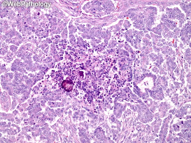
Histology: High-grade serous carcinoma of the ovary is illustrated on hematoxylin and eosin stained paraffin sections prepared for light microscopy. The specimen comprises ovarian/adnexal tissue with intricate papillary and glandular architectures embedded in delicate fibrous stroma. Tumor epithelium demonstrates marked nuclear pleomorphism, conspicuous nucleoli, and high mitotic activity, consistent with high-grade serous morphology. Notably, numerous calcified psammoma bodies are scattered within tumor clusters and in a focal necrotic center, reflecting degenerative change. The papillary fronds display hierarchical branching with complex invaginations into fibrous stroma, and tumor cells line papillary cores, forming psammoma body–laden aggregates. Necrosis is focal rather than widespread, with surrounding viable tumor showing cohesive cells and prominent cytoplasm. Invasion into adjacent ovarian stroma may be present, and cellular solidity varies among fields. The presence of psammoma bodies is characteristic of serous tumors and has potential prognostic implications; when abundant, they are associated with better differentiation and often improved prognosis, though high-grade serous carcinomas remain clinically aggressive. Immunohistochemical staining patterns, if performed, typically reveal WT1 positivity and p53 overexpression with mutant morphology. Overall, the slide highlights classic serous ovarian carcinoma features—papillary architecture, psammoma bodies, necrosis, nuclear atypia, and high proliferative index compatible with an ovarian-origin malignant neoplasm.
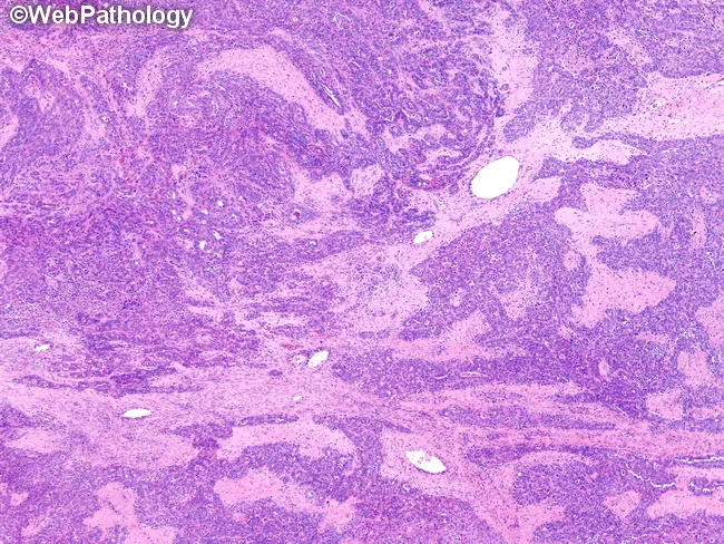
Histopathology slide of an ovarian mass, examined by light microscopy on hematoxylin and eosin (H&E) stained sections. Primary subject: high-grade serous carcinoma of the ovary. Specimen type: ovarian tumor tissue. Imaging perspective: microscopic examination of a formalin-fixed paraffin-embedded section, low to mid magnification. Morphology: tumor shows cohesive solid sheets of malignant epithelial cells with marked nuclear pleomorphism, conspicuous nucleoli, and high mitotic activity. Architectural features include anastomosing trabeculae and glandular/gland-like growth patterns, with occasional slit-like or cribriform glandular arrangements. The neoplasm displays solid, nests, and papillary-like structures embedded in fibrous stroma; tubulocystic differentiation may be evident in focal areas. The overall pattern is characteristic of high-grade serous carcinoma, often accompanied by areas of crush artifact and necrosis. Notable diagnostic features include high-grade cytology, brisk mitotic rate, and complex architectural heterogeneity. Optional ancillary correlation with immunohistochemistry commonly shows p53 abnormal staining and WT1 positivity, supporting serous differentiation. Clinical significance: confirms epithelial ovarian cancer with aggressive phenotype, guiding staging, surgical planning, and platinum-based chemotherapy. Differential considerations include mucinous, endometrioid, clear cell carcinomas, metastatic serous carcinoma, and mixed histology. This image is relevant for educational purposes, pathology teaching, diagnostic practice, and research into ovarian carcinogenesis and targeted therapies.
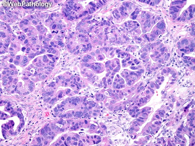
Histopathology image of ovarian high-grade serous carcinoma (HGSC) captured on light microscopy after formalin fixation and hematoxylin and eosin staining. The specimen is ovarian/tumor tissue. The tumor demonstrates complex gland-like spaces lined by markedly atypical, high-grade serous epithelial cells with pleomorphic hyperchromatic nuclei and conspicuous nucleoli. Architectural pattern is predominantly papillary with focal glandular and cribriform areas; other patterns such as cystic, microcystic, and solid components can be present in HGSC and may be observed in the same lesion. The observed glands are densely cellular, with loss of polarity, prominent mitotic activity, and frequent apoptotic figures. The stroma shows desmoplasia and invasion by tumor glands, supporting malignancy. Overall features are typical for high-grade serous carcinoma of the ovary, with aggressive cytology and high-grade nuclei. This histology is critical for differentiating HGSC from lower-grade serous tumors and other ovarian carcinoma subtypes, guiding staging, prognosis, and platinum-based chemotherapy responsiveness. Clinically, HGSCs often present with advanced-stage disease and elevated CA-125. This image provides educational demonstration of serous differentiation, papillary architecture, and invasive gland formation, illustrating key criteria used in diagnostic pathology reports, tumor grading (grade 3), and differential diagnosis including endometrioid, mucinous, and clear cell ovarian carcinomas.
Pathology Report Explained - RAJESHWARI (Age 62, Female)
What Is This Report?
The Final Diagnosis (Bottom Line First)
"IMMUNOHISTOCHEMICAL FINDINGS FAVOUR HIGH GRADE SEROUS CARCINOMA OF OVARY"
Understanding Each Marker Tested
| Marker | Result | What It Means |
|---|---|---|
| ER (Estrogen Receptor) | Positive | Tumour cells express estrogen receptors. This is commonly seen in serous ovarian carcinomas and Mullerian (gynecologic) tumours. |
| WT-1 | Diffusely positive | WT-1 (Wilms Tumour protein 1) is a strong marker for ovarian/Mullerian origin - its diffuse positivity strongly confirms this is an ovarian/gynecologic tumour, not a metastasis from somewhere else. |
| CK7 | Diffusely positive | CK7 (Cytokeratin 7) is a marker for epithelial cells of gynecologic, lung, and breast origin. Diffuse positivity in combination with the others confirms an epithelial ovarian tumour. |
| Vimentin | Negative in tumour cells | Vimentin is a marker typically seen in mesenchymal (connective tissue) tumours. Being negative here rules out sarcoma and confirms this is a pure carcinoma (epithelial cancer). |
| P53 | Strong and diffuse nuclear positivity - "p53 mutant phenotype" | This is the most important marker here. The TP53 gene is mutated in nearly all (~96%) high-grade serous carcinomas. When p53 protein accumulates (strong/diffuse staining), it means the TP53 tumour suppressor gene is mutated and malfunctioning. This is a hallmark of HGSC. - Robbins, Cotran & Kumar Pathologic Basis of Disease, p. 942 |
| P16 | Nuclear and cytoplasmic block positivity | "Block positivity" of p16 (diffuse, strong staining throughout) is another characteristic feature of high-grade serous carcinoma. It reflects widespread cell cycle dysregulation and helps distinguish HGSC from lower-grade or other histological types. |
Why This Combination of Markers Confirms HGSC Ovary
- WT-1 positive + CK7 positive + ER positive = Mullerian/ovarian epithelial origin confirmed
- Vimentin negative = rules out sarcoma or mixed tumour
- p53 strong diffuse (mutant pattern) + p16 block positive = hallmarks of HIGH-GRADE serous carcinoma specifically, not low-grade
What is High-Grade Serous Carcinoma (HGSC)?
- It is the most common type of ovarian cancer (~70% of all ovarian cancers)
- It is aggressive, often diagnosed at an advanced stage (Stage III/IV)
- Most HGSCs are now thought to originate from the fallopian tube fimbriae (the finger-like ends), not the ovary itself, though they present as ovarian masses
- Almost all cases have TP53 mutations (which is what the "p53 mutant phenotype" means in this report)
- They typically respond to platinum-based chemotherapy (carboplatin + paclitaxel) and may benefit from PARP inhibitors (especially if BRCA1/2 mutations are present) - Robbins, Cotran & Kumar Pathologic Basis of Disease, p. 941-942
Who Issued This Report?
Key Takeaway for the Treating Doctor
- Staging (CT scan of chest/abdomen/pelvis, CA-125 levels)
- Surgical consultation (for cytoreductive surgery / debulking)
- Testing for BRCA1/BRCA2 mutations (genetic testing), as this affects eligibility for PARP inhibitor therapy (e.g., olaparib)
- Chemotherapy planning (standard first-line is carboplatin + paclitaxel)
Note: This explanation is for informational purposes. All clinical decisions should be made by the treating oncologist (Dr. Sunil Kumar, Surgical Oncologist, as noted on the report) based on the full clinical picture.
Chemotherapy
"ovarian cancer" AND "first-line chemotherapy" AND "carboplatin"
"ovarian cancer" AND "bevacizumab" AND "PARP inhibitor" AND maintenance
PMID: 40690248
Chemotherapy for High-Grade Serous Ovarian Carcinoma
Step 1: Surgery First (Before or After Chemo)
| Approach | Who it's for |
|---|---|
| Primary cytoreductive surgery → then chemo | If the surgeon can remove all visible tumour (optimal debulking) upfront |
| Neoadjuvant chemo (NACT) → surgery → more chemo | If disease is too widespread for safe upfront surgery, or patient is too unwell |
Step 2: First-Line Chemotherapy (Standard Regimen)
| Drug | Dose | Route | Schedule |
|---|---|---|---|
| Paclitaxel | 175 mg/m² | IV | Day 1, every 3 weeks |
| Carboplatin | AUC 5-6 (Calvert formula) | IV | Day 1, every 3 weeks |
| Total cycles: | 6 cycles (~18 weeks) |
- Carboplatin dose is calculated using AUC (Area Under Curve) based on kidney function (GFR), not just body weight - Berek & Novak's Gynecology, p. 2357
- Carboplatin is preferred over cisplatin because it has less nausea, vomiting, kidney damage, and nerve damage, though it causes more bone marrow suppression
- Replace paclitaxel with Docetaxel 75 mg/m² (less nerve damage, but more neutropenia) - Berek & Novak's Gynecology, p. 2357
- Or single-agent carboplatin alone for elderly/frail patients with comorbidities
Step 3: Add Bevacizumab? (Targeted Anti-Angiogenic Drug)
- Dose: 7.5-15 mg/kg every 3 weeks, given alongside chemotherapy and continued as maintenance for up to 15 months after chemo ends
- Benefit: Improves progression-free survival (PFS) by several months
- Side effects: High blood pressure, delayed wound healing, rare bowel perforation
Step 4: Maintenance Therapy - PARP Inhibitors (Critical for This Patient)
| BRCA Status | Best Maintenance Option | Survival Benefit |
|---|---|---|
| BRCA1/2 mutated | Olaparib (Lynparza) ± bevacizumab | ~70% 5-year survival |
| BRCA wild-type, HRD positive | Niraparib or Olaparib + bevacizumab | Moderate benefit |
| BRCA wild-type, HRD negative | Bevacizumab alone | Modest benefit |
- Olaparib: 300 mg twice daily orally for up to 2 years
- Niraparib: 200-300 mg once daily orally
- PARP inhibitors exploit the "BRCAness" of HGSC - since TP53 is mutated (confirmed in this report) and many HGSCs have defective DNA repair, PARP inhibitors trap cancer cells unable to repair DNA damage
Summary Treatment Pathway for Rajeshwari
Staging (CT scan, CA-125, BRCA testing)
        ↓
Surgery (debulking) - if feasible upfront
        ↓
Carboplatin (AUC 5-6) + Paclitaxel (175 mg/m²) IV
every 3 weeks × 6 cycles
± Bevacizumab 15 mg/kg
        ↓
Response assessment (CT + CA-125)
        ↓
Maintenance therapy:
• If BRCA mutated → Olaparib ± bevacizumab (2 years)
• If BRCA wild-type → Bevacizumab or Niraparib
Side Effects to Monitor (for Rajeshwari, age 62)
| Side Effect | Drug | Management |
|---|---|---|
| Peripheral neuropathy (numbness/tingling in hands/feet) | Paclitaxel | Dose reduction, switch to docetaxel |
| Bone marrow suppression (low WBC, platelets) | Carboplatin | G-CSF injections, dose delays |
| Alopecia (hair loss) | Paclitaxel | Usually reversible after treatment |
| Fatigue | Both | Supportive care |
| Hypertension | Bevacizumab | Anti-hypertensives |
| Nausea/vomiting | Both | Pre-medication with ondansetron + dexamethasone |
What to Do Now (Next Steps for the Treating Team)
- BRCA1/2 germline + somatic testing - this determines eligibility for the most effective maintenance therapy
- Staging CT scan (chest/abdomen/pelvis) and serum CA-125
- Surgical oncology consultation (Dr. Sunil Kumar, as on the report) for debulking feasibility
- Multidisciplinary tumour board discussion
- Performance status assessment (ECOG) - determines if standard vs. modified chemo is appropriate for a 62-year-old
Note: All chemotherapy doses and regimens must be decided and supervised by the treating medical oncologist. This information is for educational context only.
Can this carcinoma is completely cured?
"high grade serous carcinoma" AND prognosis AND survival
PMID: 39919553
Can High-Grade Serous Carcinoma of the Ovary Be Completely Cured?
The Honest Answer
Survival by Stage - The Numbers
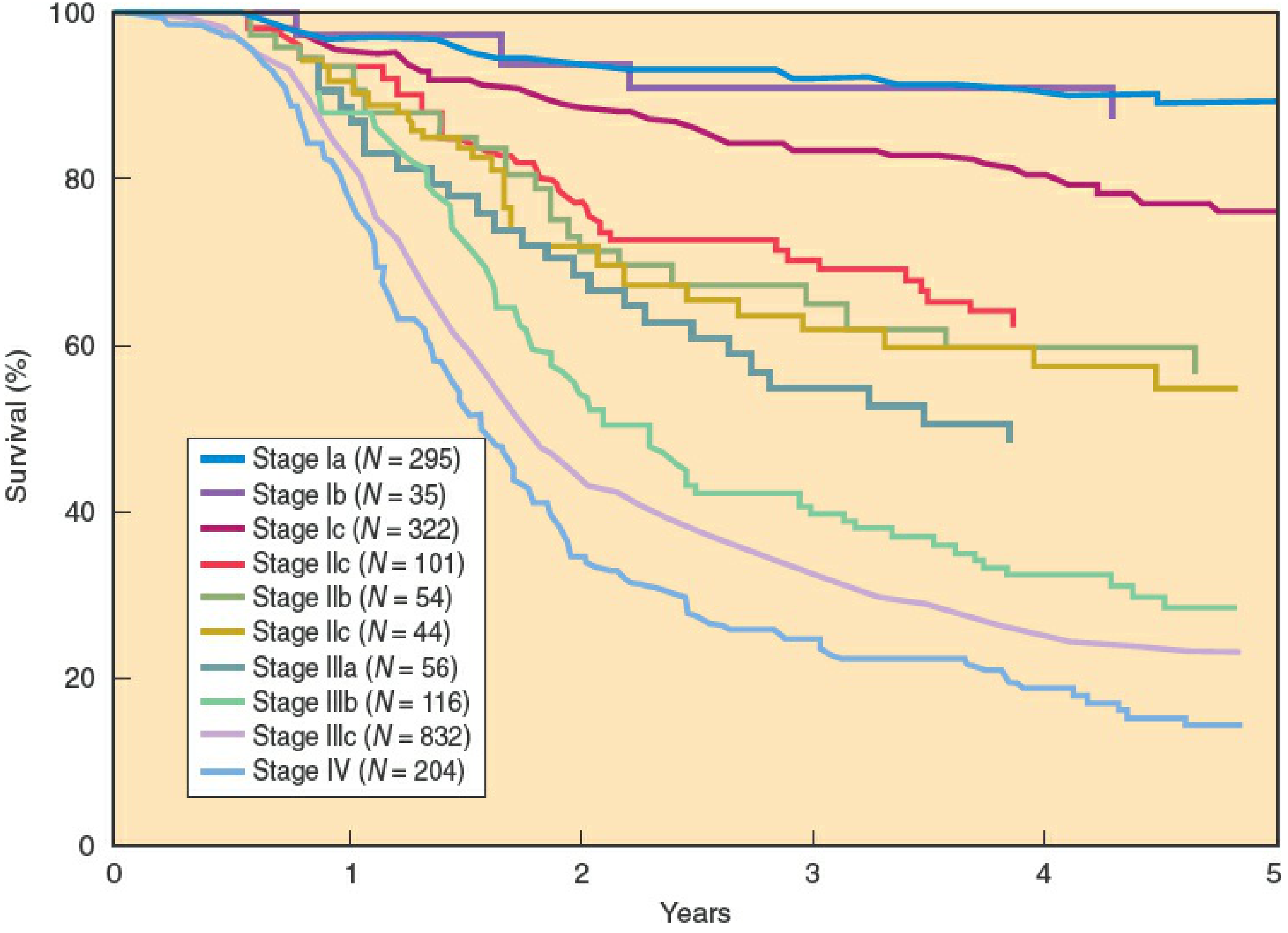
| Stage | Where the cancer is | 5-Year Survival |
|---|---|---|
| Stage I | Confined to the ovary | 93-94% - cure is very likely |
| Stage II | Spread within the pelvis | 70-73% - cure possible |
| Stage IIIa/b | Abdominal spread, small deposits | 29-50% |
| Stage IIIc | Widespread abdominal spread | 23-37% |
| Stage IV | Spread to liver, lungs, distant organs | 11-25% |
Why Doesn't It Cure Easily?
-
Relapse is very common - Even when chemotherapy achieves a "complete remission" (no detectable cancer on scans), approximately 75% of patients with advanced HGSC relapse within 2 years. The cancer hides in microscopic deposits that even scans cannot detect.
-
TP53 mutation (confirmed in Rajeshwari's report) drives aggressive behaviour - high rate of division, rapid spread, and ability to develop resistance to chemotherapy over time.
What Factors Improve the Chances for Rajeshwari?
| Factor | Favourable | Unfavourable |
|---|---|---|
| Age | Under 50 is better | 62 - somewhat less favourable |
| Residual tumour after surgery | None left (R0) | Any visible tumour left behind worsens outlook |
| BRCA mutation status | If BRCA1/2 mutated → PARP inhibitors give ~70% 5-yr survival | BRCA wild-type has lower survival with standard treatment |
| Response to first-line chemo | Complete response → better | Partial/no response → poor outlook |
| Stage | Lower is better | Not yet known for Rajeshwari - CT staging needed |
The Most Hopeful Scenario (With Modern Treatment)
- Is found to be at Stage I or II (cancer confined to pelvis)
- Undergoes complete surgical removal (no residual tumour)
- Responds well to carboplatin + paclitaxel
- Has a BRCA mutation → receives olaparib maintenance
What "Remission" vs "Cure" Means in Ovarian Cancer
- Complete remission: No cancer detectable on scans or blood tests (CA-125 normal) after treatment. This happens in ~80% of patients with advanced HGSC after first-line chemo.
- Cure: No cancer ever returning. This is the outcome for the majority of Stage I patients and a subset of Stage II patients.
- Long-term survivor: Living 5+ years without relapse. Even in Stage III, this is possible with optimal surgery + chemo + maintenance.
Bottom Line
HGSC is a serious cancer, but it is not an automatic death sentence. The single most important factors are: (1) what stage it is found at, and (2) whether the surgeon can remove all visible tumour. Getting the staging CT scan and BRCA test done quickly is the most important next step for Rajeshwari.
They have done hysterectomy
Hysterectomy Has Already Been Done - What This Means
What Surgery Was (Likely) Performed
- Uterus removed (hysterectomy)
- Both ovaries removed (oophorectomy)
- Both fallopian tubes removed (salpingectomy)
- Usually also omentum removed (omentectomy) - the fatty apron of the abdomen where ovarian cancer commonly spreads
Why This Surgery Is So Critical
"Patients whose disease is completely resected to no macroscopic residual disease have the best overall survival. Approximately 30-40% of patients in this category will be free of disease at 5 years."
- Berek & Novak's Gynecology, p. 2339
| Surgical Outcome | Residual Tumour | 5-Year Survival |
|---|---|---|
| Complete resection (R0) | None visible | Best - 30-40% at 5 years for advanced stage; near-cure for early stage |
| Optimal debulking | < 1 cm deposits | Moderate - improved survival over suboptimal |
| Suboptimal debulking | > 1 cm deposits remaining | Poor - ~5% at 5 years |
What Happens After Surgery (The Path Now)
Surgery DONE ✓ (hysterectomy)
        ↓
NEXT: Adjuvant Chemotherapy
Carboplatin + Paclitaxel × 6 cycles
(± Bevacizumab)
        ↓
CA-125 monitoring + CT scan after chemo
        ↓
BRCA test result → decide maintenance therapy
(Olaparib if BRCA mutated)
        ↓
Long-term follow-up every 3 months
This is Actually Good News
- The surgical team considered her fit enough for major surgery - a positive sign
- The primary tumour source has been physically removed
- She is now in the best possible position to benefit from chemotherapy
- If the surgeon achieved no residual disease (R0), her chances of long-term remission are significantly better
Most Important Questions to Ask the Surgeon Now
- Was complete removal achieved? (Was there any visible tumour left behind after surgery?)
- Was the omentum also removed? (Omentectomy is a standard part of the procedure)
- What was the surgical stage? (Stage I, II, III, or IV - based on what was found during surgery)
- Were the lymph nodes checked?
- When can chemotherapy start?