
vaginismus therapy explanation
vaginismus treatment therapy
PMID: 41148166
vaginismus pelvic floor muscles diagram treatment
![Anatomical diagram and infographic depicting the application of magnetic stimulation to the pelvic floor. The upper section displays a stylized anterior view of the human pelvic girdle with the levator ani and associated pelvic floor muscles highlighted in red. The lower section features a line graph titled 'Spatial profile of the magnetic field', showing electromagnetic energy distribution against 'Distance from axis [mm]'. A blue curve exhibits a 'double-dome' distribution, while a red curve shows a singular central peak. Vertical dashed lines correlate the lateral peaks of the blue magnetic field profile with the bilateral anatomical positioning of the pelvic floor muscles. This visual illustrates the biophysical principle of using TOP Flat Magnetic Stimulation (TOP FMS) to achieve uniform, homogeneous stimulation across the pelvic floor area for treating dysfunctions like urinary incontinence and pelvic organ prolapse.](https://cdn.orris.care/cdss_images/pmc_clinical_VQA_fb5836ede8837cfa617a9d75e7eb8b96ff3f9af4e592168737090610e6592a60.jpg)
Anatomical diagram and infographic depicting the application of magnetic stimulation to the pelvic floor. The upper section displays a stylized anterior view of the human pelvic girdle with the levator ani and associated pelvic floor muscles highlighted in red. The lower section features a line graph titled 'Spatial profile of the magnetic field', showing electromagnetic energy distribution against 'Distance from axis [mm]'. A blue curve exhibits a 'double-dome' distribution, while a red curve shows a singular central peak. Vertical dashed lines correlate the lateral peaks of the blue magnetic field profile with the bilateral anatomical positioning of the pelvic floor muscles. This visual illustrates the biophysical principle of using TOP Flat Magnetic Stimulation (TOP FMS) to achieve uniform, homogeneous stimulation across the pelvic floor area for treating dysfunctions like urinary incontinence and pelvic organ prolapse.
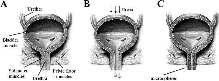
This medical anatomical diagram illustrates the pathophysiology and treatment of stress urinary incontinence (SUI) through three coronal views of the human bladder and urethra (A, B, and C). Diagram A represents normal anatomy and function, showing a healthy bladder muscle, intact ureters, and a closed urethra supported by strong sphincter and pelvic floor muscles. Diagram B depicts the mechanism of stress urinary incontinence, where external 'stress' (indicated by downward arrows) causes involuntary urine leakage through a weakened urethral opening and compromised pelvic floor support. Diagram C demonstrates a therapeutic intervention using urethral bulking agents. It shows the injection of 'microspheres' into the periurethral tissues, which facilitates complete coaptation (closure) of the urethral lumen to prevent leakage. Key labels include the bladder muscle, ureters, urethra, sphincter muscles, and pelvic floor muscles. The illustration is designed for educational use in urology to explain urinary control mechanisms and the clinical application of polymeric microspheres as bulking agents.
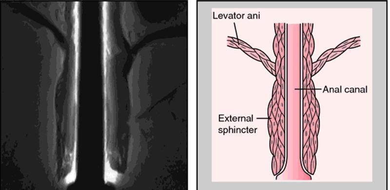
A composite image for medical education comprising a diagnostic radiological image (MRI) on the left and a corresponding anatomical diagram on the right, illustrating the pelvic floor and anal canal. The MRI shows a coronal view of the anorectal region, highlighting the muscular layers of the pelvic outlet. The anatomical diagram provides a schematic representation of the same structures for clarity. It labels the levator ani as a broad, striated muscle group superiorly that forms the pelvic diaphragm and descends to blend with the longitudinal muscle layer of the anal canal. The external sphincter is depicted as a distinct, thicker muscular cuff surrounding the distal portion of the anal canal, located inferior to the levator ani's main attachment points. This visual comparison is designed to teach the spatial relationships between the pelvic floor muscles and the terminal digestive tract, emphasizing the functional anatomy required for fecal continence. The educational focus is on identifying the levator ani and external sphincter during pelvic imaging and understanding their relative morphology.
Vaginismus: Explanation and Therapy
Definition
- Swanson's Family Medicine Review, p. 786
- Campbell-Walsh-Wein Urology, block 41
Classification
| Type | Description |
|---|---|
| Primary | Penetration has never been achieved |
| Secondary | Develops after a period of normal penetration (e.g., after trauma, surgery, childbirth, or menopause) |
| Generalized | Occurs in all situations and with any object |
| Situational | Occurs only in certain contexts (e.g., only with partner, not with dilators) |
Aetiology and Contributing Factors
- Childhood or adolescent exposure to strongly negative religious or cultural messages about sex
- History of sexual trauma or abuse (strong association noted in textbook literature)
- Anxiety about penetration, fear of pain, fear of pregnancy
- Relationship distress or previous painful intercourse creating a conditioned avoidance reflex
-
Infected hymenal remnants or imperforate/thick hymens
-
Episiotomy scars, Bartholin gland infections
-
Vaginitis or cervicitis
-
Vulvodynia / vestibulodynia
-
Postmenopausal vaginal atrophy and reduced lubrication
-
Endometriosis, lichen sclerosis, lichen planus
-
Scarring from pelvic radiation or surgery
-
Testosterone deficiency
-
Kaplan & Sadock's Comprehensive Textbook of Psychiatry, p. 6118-6119
Assessment
- Spasm and rigidity of perineal muscles visible on inspection
- Diagnosis can sometimes be made without speculum insertion
- Gradual progression from inspection to one examiner finger
- Partner should ideally be present throughout the evaluation
Treatment Approaches
1. Vaginal Dilator (Progressive Dilation) Therapy
- Set of plastic dilators in gradually increasing sizes
- The patient inserts these herself, maintaining control
- Begins with the smallest size, progressing only when comfortable and relaxed
- Once the largest dilator is tolerated, the couple may attempt intercourse
- Self-direction is key - the patient controls timing and pace
- Swanson's Family Medicine Review, p. 786
2. Cognitive-Behavioral Therapy (CBT)
- Addresses catastrophic thoughts and fear-avoidance cycles
- Combines psychoeducation, cognitive restructuring, and graded exposure homework
- Often includes systematic desensitization (gradual exposure to anxiety-provoking stimuli while in a relaxed state)
- Homework assignments practiced between sessions
- Pooled success rate ~82% in recent meta-analysis
3. Pelvic Floor Physiotherapy
- Manual therapy targeting the levator ani and pelvic floor muscles
- Myofascial release and soft-tissue massage
- Biofeedback (surface EMG) to teach voluntary relaxation of pelvic floor muscles
- Functional electrical stimulation-biofeedback has also been studied
- Success rate ~85%
4. Sensate Focus
- Developed by Masters and Johnson (LEDO framework)
- Non-demand pleasuring exercises starting with non-genital touch
- Gradually progresses to genital touching, then penetration
- Partners do structured homework exercises together
- Removes performance anxiety and reconnects intimacy with pleasure rather than fear
- Swanson's Family Medicine Review, p. 786
5. Psychodynamic and Adjunct Approaches
| Approach | Role |
|---|---|
| Couples therapy | Addresses relationship dynamics, communication, partner involvement |
| Hypnotherapy | Targets the conditioned anxiety and pain reflex at a deeper level |
| Group therapy | Peer normalization, reduces shame |
| Psychodynamic psychotherapy | Explores unconscious conflicts and past trauma |
| Sex education | Anatomy, physiology, myths - reduces fear through knowledge |
6. Botulinum Toxin (Botox) Injections
- Injection into the pubococcygeus or bulbocavernosus muscles
- Temporarily reduces involuntary spasm chemically
- Especially useful in severe or refractory cases
- Can facilitate dilation therapy by breaking the spasm cycle
- Pooled success ~85%; requires repeat injections in some cases
7. Pharmacological Adjuncts
- Topical anesthetics / lubricants - reduce procedural discomfort during dilator use
- Topical estrogen - for atrophic vaginitis contributing to vaginismus
- Low-dose benzodiazepines - short-term anxiolytic effect to facilitate initial dilation (not first-line, used cautiously)
- Antidepressants - when significant depression, anxiety disorder, or PTSD underlies the condition
Key Principles of Therapy (Any Modality)
- Patient always remains in control - no examination or procedure should be forced
- Partner involvement - ideally present at evaluation, included in all treatment phases
- Rule out organic causes first before purely psychological treatment
- Combine approaches - the data clearly favors multidisciplinary treatment
- Duration is weeks to months - not a quick fix; realistic expectations are important
- Sexual dysfunction almost always involves the couple - treat the dyad, not just the individual
Prognosis
- Swanson's Family Medicine Review, p. 786-787
- Campbell-Walsh-Wein Urology (3-volume set), block 41
- Kaplan & Sadock's Comprehensive Textbook of Psychiatry, p. 6118-6119
- Zulfikaroglu E. "Vaginismus treatment: a systematic review and meta-analysis of contemporary therapeutic approaches." J Sex Med. 2026. (PMID 41148166 - 18 studies, 863 patients)