
Circle of willis
Reading File
Now let me search the medical library for Circle of Willis content in parallel:
Finding Sources
Finding Sources
Good results. Now let me read the most comprehensive sources - the Neuroanatomy textbook and the anatomy section, plus fetch the aneurysm image:
Reading File
Reading File
Reading File
Now let me fetch key images and read a bit more content on aneurysms and clinical significance:
Loading Image
Loading Image
Reading File
Excellent - I have comprehensive content and great diagrams. Let me also fetch the aneurysm image and get the berry aneurysm / clinical pathology content:
Loading Image
Reading File
Finding Sources
Let me fetch the berry aneurysm diagram and the saccular aneurysm image from Robbins:
Loading Image
Loading Image
I now have comprehensive content and rich images to compile a thorough answer. Here is the full response:
Circle of Willis
The Circle of Willis (circulus arteriosus cerebri) is an anastomotic arterial ring at the base of the brain that connects the anterior (carotid) and posterior (vertebrobasilar) circulations, providing collateral supply throughout the cerebral hemispheres.
Anatomy and Components
The ring is formed by the junction of vessels from two sources:
Anterior circulation - supplied by the bilateral internal carotid arteries (ICAs), which originate from the common carotids (the right from the brachiocephalic artery, the left directly from the aorta).
Posterior circulation - supplied by the two vertebral arteries, which arise from the subclavian arteries, ascend through the transverse foramina (foramina transversaria) of cervical vertebrae, enter the foramen magnum, and unite to form the basilar artery.
The ring itself consists of the following vessels (from anterior to posterior):
| Vessel | Connection |
|---|---|
| Anterior communicating artery (AComm) | Joins the two ACAs across the midline |
| Anterior cerebral arteries - A1 segments (bilateral) | Proximal ACAs, between ICA and AComm |
| Internal carotid arteries - supraclinoid segment (bilateral) | Feed both ACA and MCA |
| Posterior communicating arteries - PComms (bilateral) | Link ICAs to PCAs, bridging anterior and posterior circulations |
| Posterior cerebral arteries - P1 segments (bilateral) | Arise from the top of the basilar artery |
The middle cerebral arteries (MCAs) are terminal branches of the ICAs but are not part of the ring itself - they exit laterally.
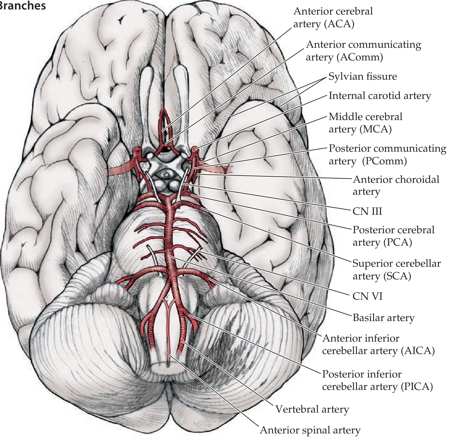
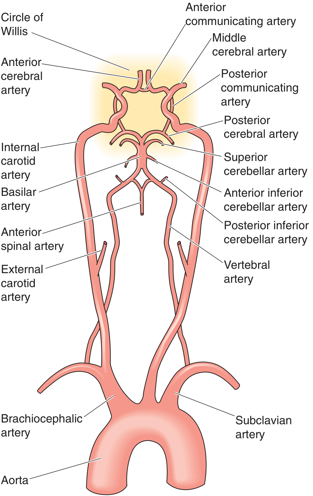
Three Main Cerebral Arteries and Their Territories
| Artery | Origin | Territory Supplied |
|---|---|---|
| Anterior cerebral artery (ACA) | ICA | Medial frontal and parietal lobes, corpus callosum, medial sensorimotor cortex |
| Middle cerebral artery (MCA) | ICA (lateral branch) | Lateral frontal, parietal, temporal lobes; internal capsule (deep perforators); Broca's and Wernicke's areas |
| Posterior cerebral artery (PCA) | Basilar artery apex | Occipital lobes, inferior temporal lobe, thalamus, midbrain |
Deep structures (basal ganglia, thalamus, internal capsule) are supplied by small penetrating (lenticulostriate) arteries arising from the proximal segments of these vessels near the circle.
The OPAAM mnemonic helps recall supraclinoid ICA branches: Ophthalmic, Posterior communicating, Anterior choroidal, Anterior cerebral, Middle cerebral arteries. - Neuroanatomy through Clinical Cases 3rd Edition
Collateral Function
The circle provides abundant opportunity for collateral flow when one vessel is occluded. However, this function is variable:
- A complete, full-caliber ring is present in only approximately 34% of individuals (some sources cite ~40%)
- Common variants include hypoplasia or absence of one A1 segment, hypoplastic PComms, or a fetal PCA (in which the PCA arises from the ICA instead of the basilar artery - occurs in 20-30% of people)
- When the circle is competent, patients with complete carotid occlusion may remain neurologically intact due to cross-filling
"The circle of Willis connects the right and left carotid arteries to each other and each to the vertebrobasilar system. Patients with complete occlusion... may remain neurologically intact." - Schwartz's Principles of Surgery
Clinical Significance
1. Saccular (Berry) Aneurysms
The bifurcation points of the circle are sites of congenital medial weakness and hemodynamic stress, making them the most common locations for berry aneurysms. These develop due to a defect in the tunica media, and their walls are composed of hyalinized fibrous tissue without a normal muscular layer.
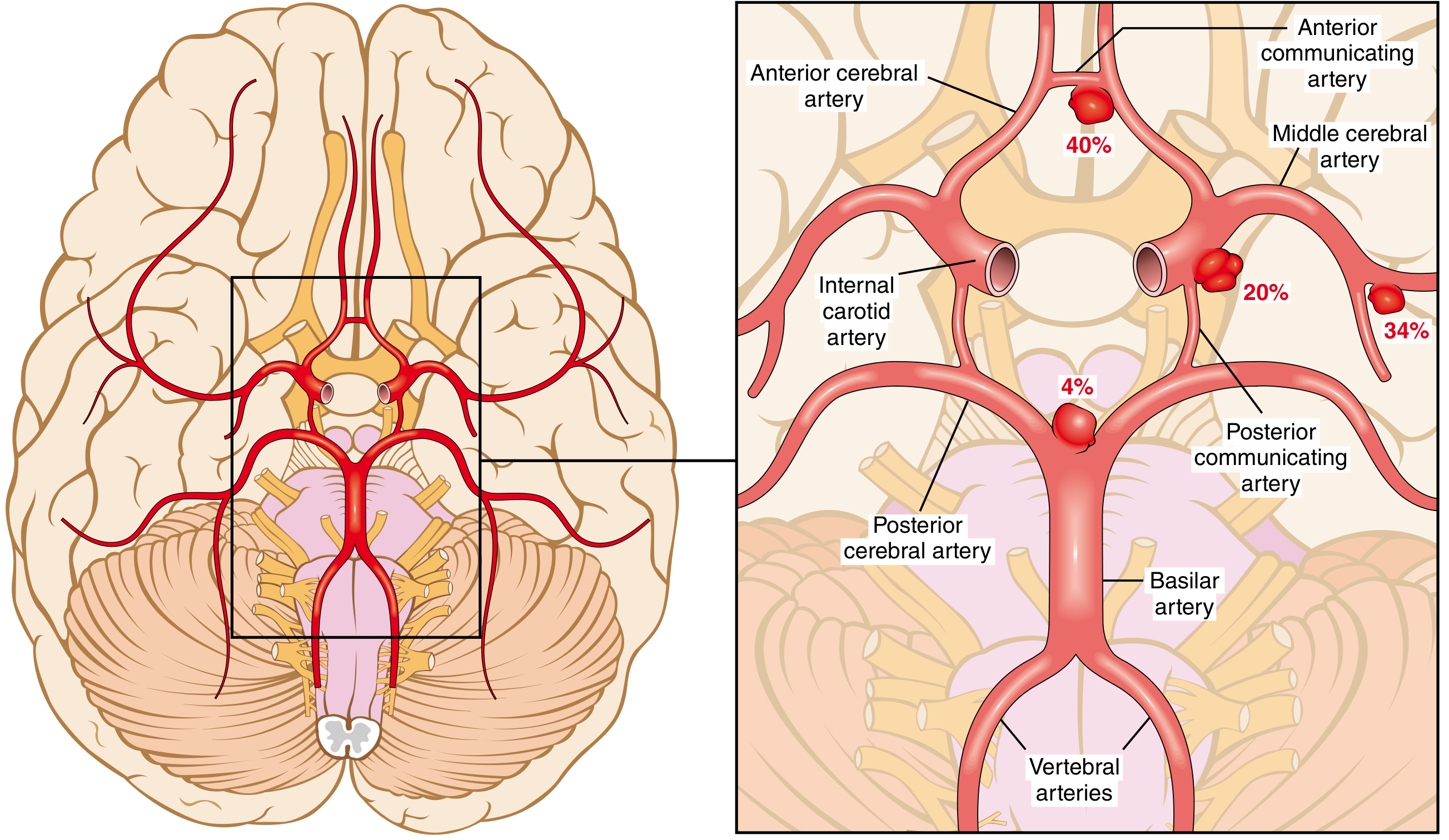
Distribution of berry aneurysms:
- Anterior communicating artery (AComm): ~40% - most common site
- MCA bifurcation: ~34%
- ICA (including PComm junction): ~20%
- Basilar artery bifurcation and posterior circulation: ~4%
Rupture causes subarachnoid hemorrhage (SAH), with the classic "thunderclap headache" (worst headache of life). A large PComm aneurysm can compress CN III (oculomotor nerve), causing a painful, pupil-involving third nerve palsy. Post-SAH vasospasm of circle of Willis vessels is a major cause of secondary ischemic injury, mediated by endothelins, nitric oxide, and arachidonic acid metabolites. - Robbins, Cotran & Kumar
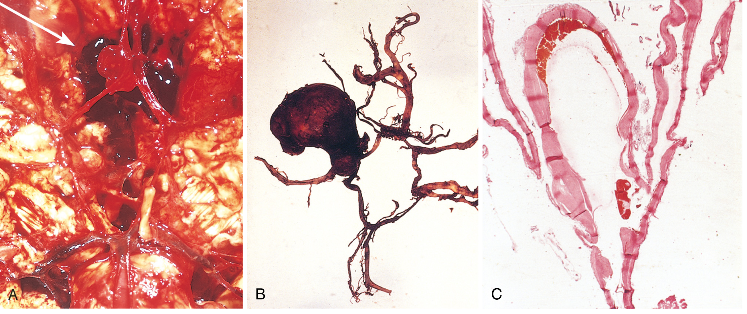
2. Ischemic Stroke
Occlusion of a circle component or its major branches causes territory-specific deficits. Integrity of the circle determines whether collateral flow compensates for the occlusion. This is assessed preoperatively in carotid endarterectomy - if the circle is incomplete, the patient may not tolerate carotid cross-clamping and will need a shunt.
3. Imaging
The circle is best visualized with MR angiography (MRA) or CT angiography (CTA). These modalities reliably demonstrate variants, aneurysms, and stenoses. Digital subtraction angiography (DSA) remains the gold standard for detailed pre-intervention mapping. - Grainger & Allison's Diagnostic Radiology
Summary Table
| Feature | Detail |
|---|---|
| Location | Base of brain, in the subarachnoid space of the interpeduncular cistern |
| Shape | Roughly heptagonal (7-sided) ring |
| Feeding vessels | Bilateral ICAs (anterior) + basilar artery (posterior) |
| Communicating vessels | AComm (anterior), bilateral PComms (posterior) |
| Complete ring prevalence | ~34-40% of individuals |
| Common variant | Fetal PCA (from ICA, not basilar) - 20-30% |
| Main clinical concern | Berry aneurysm - especially at AComm (40%) |
| Collateral role | Compensates for single-vessel occlusion if ring is patent |
This is a shared conversation. Sign in to Orris to start your own chat.