
PPH in detail obstetrics
"postpartum hemorrhage" management
postpartum hemorrhage management uterine atony
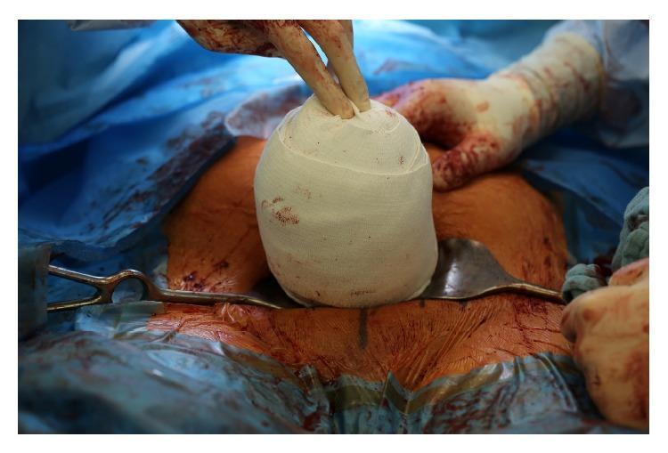
This clinical photograph shows an intraoperative view of an exteriorized uterus during a surgical procedure, specifically illustrating the management of uterine atony. The uterus is tightly wrapped in a white, woven compression bandage, forming a spherical or ovoid shape. This technique, known as the uterine sandwich or a variation of external compression, is used to control postpartum hemorrhage when uterotonics fail. The bandaged organ is positioned centrally within the surgical field, which is draped with blue sterile cloths. Visible landmarks include blood-stained abdominal skin and surgical instruments, including a metal retractor (likely a Doyen or Fritsch retractor) positioned beneath the exteriorized uterus and a surgical clamp on the left. Gloved hands of the surgical team are visible, applying manual pressure to the top of the wrapped uterus. This image serves as an educational resource for obstetricians and surgical residents on mechanical hemostatic techniques during cesarean delivery complications.
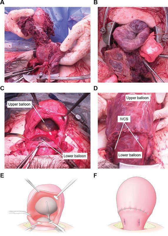
A composite image containing clinical photographs and schematic diagrams (A-F) documenting the management of postpartum hemorrhage (PPH) caused by uterine inversion and severe uterine atony. Panels A and B show the surgical field during cesarean section with an inverted, dark red, congested uterus and adherent placental tissue. Panels C and E (photograph and diagram) demonstrate the intraoperative placement of a double-balloon tamponade (UBT) system through a uterine incision, with an 'upper balloon' positioned in the uterine corpus and a 'lower balloon' in the isthmus. Panels D and F (photograph and diagram) illustrate the postoperative state where Isthmus Vertical Compression Sutures (IVCS) have been applied. These white, vertical sutures compress the lower uterine segment between the two inflated balloons to achieve hemostasis. The sequence highlights a step-by-step minimally invasive technique combining mechanical pressure and surgical compression to treat life-threatening obstetric hemorrhage.
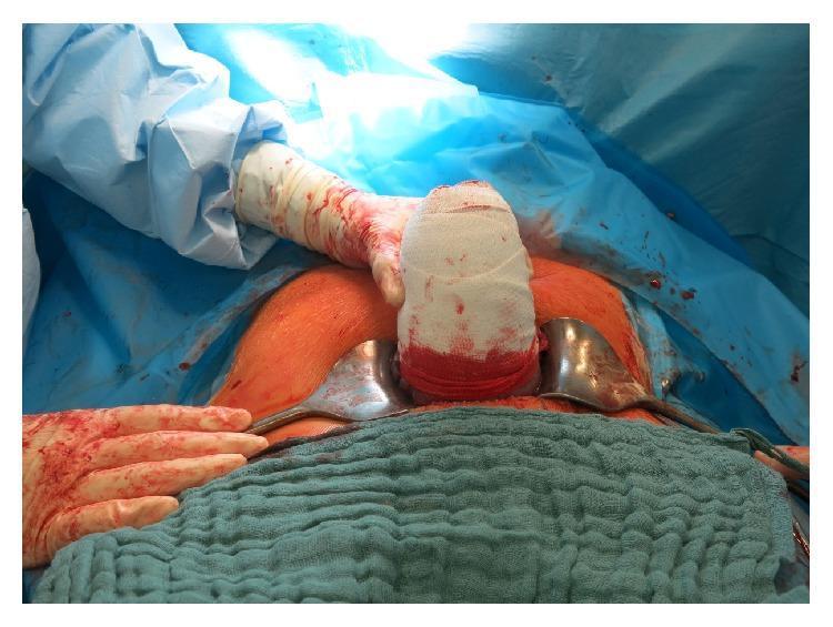
A clinical photograph capturing an intraoperative stage of a cesarean delivery, demonstrating the management of postpartum hemorrhage due to uterine atony. The image shows the exteriorized uterus wrapped tightly in a white elastic compression bandage (uterine bandage technique) to achieve mechanical hemostasis. The bandage shows significant blood staining, particularly at the lower segment. The surgical field is exposed via a transverse abdominal incision held open by two metal retractors. The patient's skin is visible with a reddish-orange hue, likely from antiseptic surgical preparation. The surrounding area is draped with blue sterile cloths and green towels. A surgeon's gloved hand, stained with blood, is visible providing stabilization to the exteriorized organ. This procedure illustrates a conservative surgical intervention used when uterotonic medications fail to induce uterine contraction, aimed at avoiding emergency hysterectomy while promoting uterine tone.
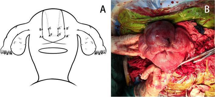
This composite educational material demonstrates the application of modified B-Lynch compression sutures used in the management of postpartum hemorrhage (PPH) due to uterine atony. Figure A is an anatomical diagram of the uterus, ovaries, and fallopian tubes, showing the puncture points (A, B, C, D) and suture pathways designed to compress the uterine corpus. Figure B is an intraoperative clinical photograph showing an exteriorized, atonic uterus during a surgical procedure. The uterus exhibits a reddish-purple, glossy appearance with visible vertical suture loops compressing the fundus and corpus. Surgical instruments, including clamps and suture material, are visible in the operative field alongside blood-stained surgical drapes. The visual illustrates a critical life-saving obstetric intervention, focusing on the mechanical compression of the uterus to achieve hemostasis when conservative measures fail. This content is intended for advanced medical education in obstetrics and gynecology, specifically for surgical training in maternal emergency procedures.
B-Lynch compression suture uterus PPH
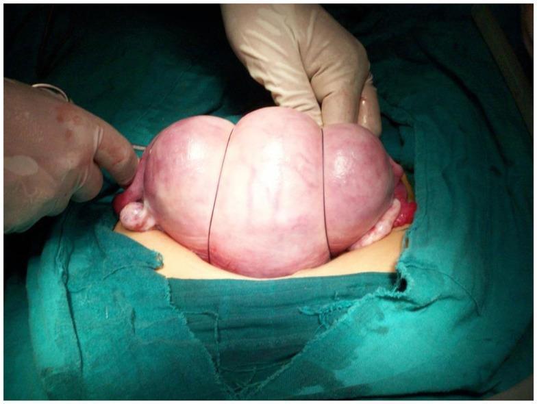
This clinical photograph demonstrates an intraoperative view of a uterine compression suture technique, specifically a B-Lynch or similar brace suture procedure, used to manage postpartum hemorrhage (PPH). The exteriorized uterus is shown as a large, globular, pinkish-red organ. Two dark-colored, non-resorbable sutures are visible encircling the uterine fundus vertically, creating longitudinal grooves and visible tissue compression. These sutures act as braces to provide mechanical compression of the myometrium, facilitating hemostasis by reducing the uterine volume and compressing intramyometrial blood vessels. A gloved hand is shown applying manual pressure to the fundus, assisting in the placement and tightening of the suture. The surgical field is prepared with green sterile drapes. This visual is significant for obstetric education, illustrating a key life-saving surgical intervention when conservative management of uterine atony fails.
This composite educational material demonstrates the application of modified B-Lynch compression sutures used in the management of postpartum hemorrhage (PPH) due to uterine atony. Figure A is an anatomical diagram of the uterus, ovaries, and fallopian tubes, showing the puncture points (A, B, C, D) and suture pathways designed to compress the uterine corpus. Figure B is an intraoperative clinical photograph showing an exteriorized, atonic uterus during a surgical procedure. The uterus exhibits a reddish-purple, glossy appearance with visible vertical suture loops compressing the fundus and corpus. Surgical instruments, including clamps and suture material, are visible in the operative field alongside blood-stained surgical drapes. The visual illustrates a critical life-saving obstetric intervention, focusing on the mechanical compression of the uterus to achieve hemostasis when conservative measures fail. This content is intended for advanced medical education in obstetrics and gynecology, specifically for surgical training in maternal emergency procedures.
Postpartum Hemorrhage (PPH) - Comprehensive Overview
Definition
Important note on pregnancy physiology: Plasma volume increases ~40% and red cell volume ~25% by the end of the third trimester. This can mask hemorrhage - the first sign may be only a mild pulse rate elevation. Up to 30% of total blood volume can be lost before blood pressure drops. - Tintinalli's Emergency Medicine
Incidence & Significance
The 4 Ts - Causes of PPH
| T | Cause | Frequency |
|---|---|---|
| Tone | Uterine atony | ~70-80% of cases |
| Tissue | Retained placental fragments, placenta accreta | ~10% |
| Trauma | Cervical/vaginal/perineal lacerations, uterine rupture, inversion | ~20% |
| Thrombin | Coagulopathy (congenital or acquired, e.g. DIC, HELLP) | Uncommon |
1. TONE - Uterine Atony (Most Common Cause)
Risk Factors for Atony
- Chorioamnionitis / uterine infection
- Prolonged labor
- Oxytocin use during labor (receptor desensitization)
- Multiparity (grand multipara)
- Fetal macrosomia
- Multifetal gestation
- Retained products of conception
- Preeclampsia
- Uterotonic/tocolytic use (volatile anesthetics, magnesium sulfate, terbutaline)
- Uterine structural anomalies
2. TISSUE - Retained Placenta / Placenta Accreta Spectrum
- Retained placental fragments prevent normal uterine contraction
- Placenta accreta (accreta, increta, percreta) - abnormally adherent placenta that does not separate
- Accreta is strongly associated with prior cesarean section and anterior placenta previa
- Real-time ultrasound helps identify retained placenta or residual blood clots
3. TRAUMA - Lacerations, Uterine Rupture, Uterine Inversion
Genital Tract Lacerations
Uterine Rupture
- Rare but carries high maternal and fetal mortality
- Primary risk factor: previous cesarean section (especially single-layer closure)
- Additional risk: augmented labor, fetal weight >3500g
- Signs: breakthrough pain (present in only a minority), fetal heart rate abnormality (most reliable sign)
Uterine Inversion
- Rare but dramatic complication of the third stage of labor
- Mechanism: fundal placental implantation + excessive cord traction or fundal pressure causes the fundus to invaginate
- Complete inversion: inverted fundus protrudes beyond cervix - appears as a beefy-red mass at the introitus
- Incomplete inversion: fundus does not extend beyond external cervical os - suspected when uterine fundus cannot be palpated abdominally
- Treatment: immediate manual replacement (Johnson maneuver); may need tocolysis (magnesium sulfate, nitroglycerin, β-mimetics) or general anesthesia
4. THROMBIN - Coagulopathy
- Hereditary coagulopathies (e.g., von Willebrand disease)
- Acquired: DIC (often complicating placental abruption, AFE, severe preeclampsia/HELLP, sepsis)
- Dilutional coagulopathy from massive transfusion
Secondary PPH (24 hours - 6 weeks)
- Subinvolution of the placental site
- Retained placental tissue
- Genital tract wound infection
- Uterogenital infection (endometritis)
Management of PPH
Immediate Resuscitation
- Frequent vital sign monitoring
- Two large-bore IV access (14-16G)
- Aggressive fluid resuscitation with lactated Ringer's
- Cross-match and prepare for blood transfusion
- Urinary catheter
- Oxygen supplementation
- Call for multidisciplinary help early (obstetrician, anesthesiologist, nursing, blood bank, interventional radiology)
Step-by-Step Management of Uterine Atony
Step 1: Bimanual Uterine Massage
Step 2: Uterotonics (Medical Management)
| Drug | Dose & Route | Notes |
|---|---|---|
| Oxytocin (1st line) | 20-30 units in 1L crystalloid, infusion up to 100 mU/min | Do NOT give IV bolus (risk of hypotension, hyponatremia). WHO: 20 IU in 1L crystalloid after uncomplicated cesarean. |
| Methylergonovine / Ergonovine | 0.2 mg IM | Second-line; contraindicated in hypertension, cardiac disease. Risk of hypertension, CNS vasospasm, coronary artery spasm. IV route forbidden. |
| Carboprost (15-methyl PGF2α) | 250 μg IM, repeat if needed | Contraindicated in cardiovascular disease and asthma. Can cause bronchospasm, pulmonary hypertension, tachycardia. |
| Misoprostol (PGE1 analogue) | 600-800 mcg oral/sublingual/vaginal/rectal | Useful when oxytocin unavailable or desensitization has occurred; also 1000 μg rectal or 800 μg transvaginal intrauterine |
| Tranexamic acid | 1g IV within 3 hours of PPH diagnosis | Antifibrinolytic; ACOG recommends when initial medical therapy fails; WOMAN Trial: RR 0.69 for death from bleeding (P=0.008) when given within 3 hours; administer after cord clamping |
Step 3: Mechanical / Tamponade Methods
- Intrauterine balloon tamponade: Bakri balloon (specially designed), Foley catheter, Rusch balloon, condom catheter, Sengstaken-Blakemore tube. ACOG specifically recommends Bakri balloon. Success rates: 57-100%. The novel Jada System (intrauterine vacuum-induced device) achieved definitive hemorrhage control in >90% of patients at median 3 minutes.
- Uterine cavity packing: Sterile gauze packing - retrospective evidence supports use in atony
- Selective uterine artery embolization (UAE): where facilities and interventional radiology are available; success rates ~85-90%
Step 4: Surgical Management (Laparotomy)
- Identify uterine lacerations - check for occult intra-abdominal sources
- O'Leary sutures - bilateral uterine artery ligation at the level of the internal os
- B-Lynch brace suture - described in 1997; compresses uterine blood supply longitudinally; uterine-conserving. The Hayman technique is a simpler variant.
- Hypogastric (internal iliac) artery ligation - reduces pulse pressure in pelvic vessels by ~85%; collateral circulation preserved (women have had subsequent successful pregnancies after this procedure); transient bladder dysfunction and buttock pain may occur postoperatively
- Peripartum hysterectomy - definitive last resort if all else fails; most commonly performed for uterine atony and placenta accreta
Massive Hemorrhage Protocol (MTP)
- Activate massive transfusion protocol (MTP) early
- Balanced resuscitation: FFP:PRBC ratios (origin from non-obstetric settings; debated in obstetric hemorrhage)
- Cell salvage with leukocyte reduction filter (removes tissue factor, α-fetoprotein, fetal squamous cells) - especially valuable in patients who decline blood products
- For Rh-negative patients: administer anti-D immunoglobulin with Kleihauer-Betke testing when cell salvage is used
- Central venous access / Swan-Ganz catheter in extensive hemorrhage for accurate hemodynamic monitoring
REBOA (Resuscitative Endovascular Balloon Occlusion of the Aorta)
- Occludes Zone III aorta (infra-renal) to reduce pelvic blood flow
- Small (7-Fr) sheaths with ultrasound guidance preferred - lower thrombotic complications
- Note: robust uterine collateral circulation may limit efficacy; additional adjuncts often needed
- Frequent distal pulse monitoring after reperfusion
Prevention of PPH: Active Management of the Third Stage of Labor (AMTSL)
- Uterotonic administration immediately after delivery (oxytocin preferred; equivalent efficacy with methylergonovine or ergometrine)
- Controlled cord traction (Brandt-Andrews maneuver)
- Uterine fundal massage after placental delivery
Summary Algorithm
PPH Detected
    ↓
Bimanual massage + Call for Help + IV Access + Resuscitation
    ↓
Identify Cause (4 Ts): Tone? Tissue? Trauma? Thrombin?
    ↓
Uterine Atony → Uterotonics (Oxytocin → Methylergonovine → Carboprost → Misoprostol)
    + Tranexamic acid (within 3 hours)
    ↓ (if refractory)
Tamponade (Bakri balloon / Jada system / packing)
    ↓ (if refractory)
Interventional Radiology: UAE
    ↓ (if refractory / not available)
Laparotomy: O'Leary sutures → B-Lynch → Hypogastric ligation
    ↓ (last resort)
Peripartum Hysterectomy

Lacerations → Repair directly
Retained Tissue → Manual removal / curettage
Coagulopathy → FFP, cryoprecipitate, platelets, TXA
Uterine Inversion → Manual reinversion ± tocolysis
Key Points Summary
- PPH is the #1 cause of maternal death worldwide
- Uterine atony accounts for >70-80% of PPH
- The 4 Ts (Tone, Tissue, Trauma, Thrombin) guide diagnosis
- Oxytocin is the first-line uterotonic; avoid IV bolus
- Tranexamic acid 1g IV: give within 3 hours of PPH diagnosis (WOMAN Trial evidence; RR 0.69 for death from bleeding)
- Bakri balloon is ACOG-recommended for tamponade
- B-Lynch suture is the key uterine-conserving surgical option
- Peripartum hysterectomy is definitive when all else fails
- Pregnancy physiology masks blood loss - do not wait for BP drop to act