
Diagnosis of bacterial meningitis is paediatrics
"bacterial meningitis" AND diagnosis AND children
bacterial meningitis CSF cerebrospinal fluid analysis diagnosis
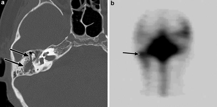
This composite educational image demonstrates a cerebrospinal fluid (CSF) leak into the right temporal bone in a pediatric patient with recurrent bacterial meningitis. Panel A is an axial computed tomography (CT) scan of the temporal bone in a bone window. It shows opacification of the right middle ear and mastoid air cells due to soft tissue density fluid collection (black arrows), contrasting with the normally air-filled spaces. While no acute fracture is visible, the findings suggest a traumatic or congenital pathway for CSF. Panel B displays a radionuclide cisternography scan following lumbar administration of a tracer. A focal area of increased radionuclide accumulation is clearly visible in the region of the right middle ear and mastoid cells (black arrow), providing definitive functional evidence of a CSF leak. The image highlights the diagnostic synergy between anatomical imaging (CT) and functional imaging (nuclear medicine cisternography) in identifying predisposing conditions for meningitis, such as post-traumatic skull base defects.
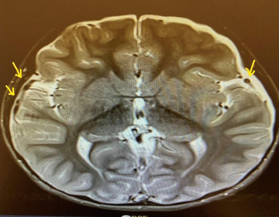
This diagnostic image is an axial MRI of the brain, specifically a T2-weighted sequence where cerebrospinal fluid (CSF) appears bright (hyperintense). The image demonstrates the cerebral hemispheres with visible cortical gyri and sulci, the lateral ventricles, and the third ventricle. Yellow arrows highlight several small, discrete, hypointense (dark) pockets located within the subdural space, particularly adjacent to the frontal and temporal lobes. These localized fluid collections are clinically significant as they represent subdural effusions or empyemas (pockets of pus) in a patient with meningitis. The brain parenchyma shows some architectural crowding, potentially indicative of mild cerebral edema. This image is used to educate clinicians on the neurological complications of bacterial meningitis, such as those caused by Haemophilus influenzae, and the importance of monitoring for intracranial fluid collections that may require neurosurgical intervention.
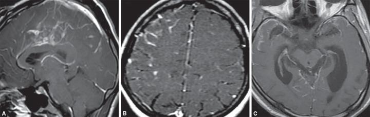
This composite diagnostic image features three panels (A, B, and C) illustrating neuroimaging findings associated with bacterial meningitis and its complications. Figures A and B are contrast-enhanced MRI scans in sagittal and axial planes, respectively, demonstrating diffuse leptomeningeal and pachymeningeal enhancement. This pathological enhancement is particularly prominent along the cerebral convexities and within the basal cisterns, signifying active meningeal inflammation and hypervascularity. Figure C is an axial T1-weighted post-contrast MRI scan showing significant bilateral enlargement of the lateral ventricles. The ventricular system appears markedly dilated with blunting of the frontal horns, characteristic of obstructive hydrocephalus secondary to the inflammatory exudates of meningitis interfering with cerebrospinal fluid (CSF) flow. Collectively, the images provide a clinical demonstration of acute intracranial infection manifesting as diffuse meningeal inflammation and subsequent hydrocephalus, useful for teaching the radiological hallmarks of central nervous system infections.
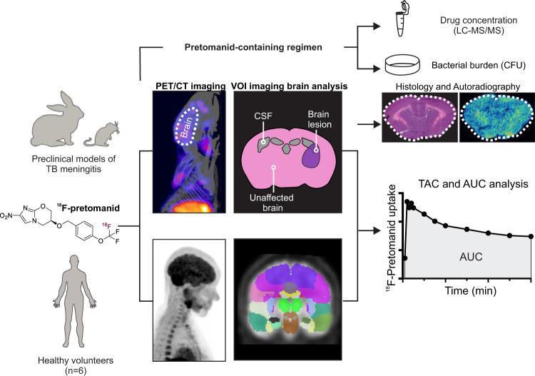
This infographic outlines the experimental design for translating 18F-pretomanid PET/CT imaging from preclinical models to clinical application in humans. The workflow begins with animal models (rabbit and mouse) of TB meningitis and healthy human volunteers (n=6). Both groups undergo dynamic PET/CT imaging to evaluate the biodistribution of 18F-pretomanid, a radiolabeled antitubercular drug. Key analytical steps illustrated include Volume of Interest (VOI) segmentation in the brain to differentiate drug penetration in the cerebrospinal fluid (CSF), brain lesions, and unaffected brain tissue. Pharmacokinetic data is captured through Time-Activity Curves (TAC) and Area Under the Curve (AUC) analysis, demonstrating radiotracer uptake and clearance over time. Preclinical validation is supplemented by direct measurement of drug concentrations using LC-MS/MS, bacterial burden quantification via Colony Forming Units (CFU), and high-resolution spatial mapping through histology and autoradiography. The diagram highlights the drug's ability to cross the blood-brain barrier and its potential as a molecular imaging tool for monitoring treatment regimens in central nervous system tuberculosis.
Diagnosis of Bacterial Meningitis in Paediatrics
1. Common Pathogens by Age Group
| Age | Organisms |
|---|---|
| 0-1 month | Group B Streptococcus, Listeria monocytogenes, S. pneumoniae, E. coli |
| 1-3 months | Group B Streptococcus, Listeria, S. pneumoniae, N. meningitidis, H. influenzae, E. coli |
| 3 months - 18 years | S. pneumoniae, N. meningitidis, H. influenzae |
- Textbook of Family Medicine 9e, p. 1232
2. Clinical Features
Neonates (< 30 days)
- Lethargy, poor feeding, weak sucking
- Irritability, abnormal cry
- Temperature instability (fever or hypothermia)
- Bulging fontanelle (may appear late)
- Vomiting, diarrhoea, apnoea, grunting, respiratory distress
- Seizures (occurs in ~40% of neonates)
Nuchal rigidity and meningismus are not reliable signs in children under 1 year of age.
- Tintinalli's Emergency Medicine, p. 795
Infants (1-3 months)
- Fever or hypothermia, toxic appearance
- Lethargy, mottling, bulging fontanelle
- Abnormal cry, grunting, respiratory distress
- Increased or decreased muscle tone
Older Infants and Children (> 3 months - 18 years)
| Sign / Symptom | Likelihood Ratio |
|---|---|
| Bulging fontanelle (caregiver-reported) | LR 8 (95% CI 2.4-26) |
| Neck stiffness | LR 7.7 (95% CI 3.2-19) |
| Seizures (outside febrile seizure range of 6 months-6 years) | LR 4.4 (95% CI 3.0-6.4) |
| Reduced feeds | LR 2 (95% CI 1.2-3.4) |
- Kernig sign: Patient supine, hip flexed 90°, patient cannot extend knee fully without pain.
- Brudzinski sign: Patient supine, involuntary flexion of legs on passive neck flexion.
- A petechial/purpuric rash on the extremities (beginning as maculopapular) should prompt urgent consideration of meningococcal meningitis.
The WHO Pocket Book of Hospital Care for Children notes that no single clinical feature is sufficient for a "robust diagnosis," but the combination of fever + seizures + meningeal signs + altered consciousness is consistently associated with bacterial meningitis.
- Tintinalli's Emergency Medicine, p. 795
3. Diagnosis: Lumbar Puncture (LP)
Contraindications / Indications to Defer LP:
- Clinical instability / shock / hypoxia
- Focal neurologic signs (do CT first, then LP)
- Papilloedema
- Thrombocytopenia
- Local infection at lumbar site
- Vertebral abnormalities
Children with focal neurologic signs should receive antibiotics immediately without waiting for CT or LP.
CSF Findings: Bacterial vs. Viral Meningitis
| Parameter | Bacterial Meningitis | Viral Meningitis |
|---|---|---|
| Opening pressure (mm H₂O) | > 180 | Often normal |
| WBC count (cells/mm³) | 1000-10,000 (range 100-20,000) | < 300 (range 100-1000) |
| Neutrophils (%) | > 80% | < 20% |
| Glucose (mg/dL) | < 40 (or CSF:serum ratio < 0.4) | Normal |
| Protein (mg/dL) | > 100 (range 100-500) | < 100 (range 50-100) |
| Gram stain | Positive in 60-90% | Negative |
| Culture | Positive in most untreated | Negative |
Note: In infants and children, opening pressures are lower than in adults (normal adult range 80-210 mm H₂O).
- Textbook of Family Medicine 9e, p. 1232; Tintinalli's Emergency Medicine, p. 795
4. The Paediatric Bacterial Meningitis Score
| Criterion | Points |
|---|---|
| Positive CSF Gram stain | 2 |
| CSF protein > 80 mg/dL | 1 |
| Blood absolute neutrophil count ≥ 10,000 cells/mm³ | 1 |
| Seizure at or before presentation | 1 |
| CSF neutrophil count ≥ 1000 cells/mm³ | 1 |
-
0 points - Aseptic meningitis very likely
-
1 point - Aseptic meningitis less likely
-
≥ 2 points - Bacterial meningitis more likely
-
Tintinalli's Emergency Medicine, p. 796 (Nigrovic et al., Pediatr Emerg Care 2009)
5. Blood and Other Investigations
- Blood cultures: Mandatory before antibiotics; positive in many cases
- CBC with differential: Leucocytosis with neutrophilia; ANC ≥ 10,000 cells/mm³ is significant
- CRP: Elevated; can help distinguish bacterial from viral (CRP less reliable than procalcitonin)
- Procalcitonin: Level ≥ 2 ng/mL has ~90% specificity for bacterial infection; < 0.5 ng/mL (sensitivity ~80%, specificity ~70%) suggests low bacterial risk
- Serum glucose: Check bedside glucose in all acutely ill children with altered mental status to calculate CSF:serum glucose ratio
- Gram stain: Identifies organism in 60-90% of untreated cases; specificity > 97%
- Multiplex PCR: Increasingly used - can detect bacterial and viral causes simultaneously; especially valuable when antibiotics have been given prior to LP
- Blood sodium: Assess for syndrome of inappropriate ADH (SIADH), a common complication
6. Neuroimaging
- Immunocompromised state
- History of CNS disease
- New-onset seizure (within 1 week)
- Papilloedema
- Abnormal level of consciousness
- Focal neurologic deficit
7. Differential Diagnosis
In Neonates:
- Viral meningitis (herpes simplex, enteroviruses)
- Fungal meningitis (Candida)
- Cardiac disease, NEC, congenital adrenal hyperplasia
- Inborn errors of metabolism
- Intracranial haemorrhage
In Older Infants / Children:
- Viral (aseptic) meningitis
- Encephalitis (HSV - consider if focal signs + altered sensorium)
- Brain abscess
- Tuberculous meningitis
- Collagen vascular disease
- Malignancy (CNS)
- Drug/toxin-induced
8. Key Principles - Summary
- High clinical suspicion is essential, especially in neonates where signs are non-specific.
- Do not delay antibiotics waiting for LP or CT if the child is unstable or has focal signs.
- LP is the gold standard - CSF analysis with culture, Gram stain, glucose, protein, and cell count.
- The Bacterial Meningitis Score helps stratify risk once CSF results are available.
- Procalcitonin is more useful than CRP for distinguishing bacterial from viral meningitis.
- Multiplex PCR is the emerging standard for pathogen identification, especially post-antibiotic treatment.