
Classification and recent update in renal glomerular lesions/ glomerulonephritis immunofluorescense for md pathology exam
glomerulonephritis immunofluorescence classification update
glomerulonephritis classification pathology update
IgA nephropathy mesangial deposits immunofluorescence
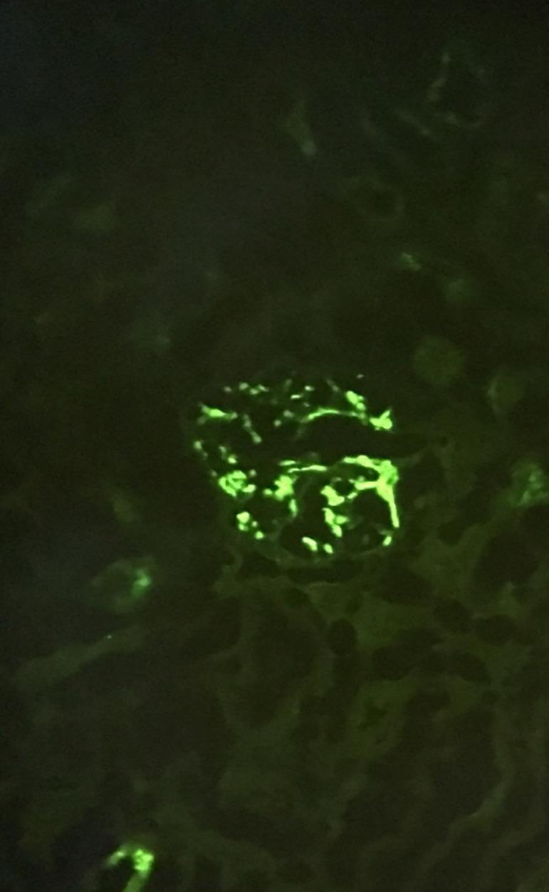
This diagnostic image shows a renal biopsy specimen analyzed via direct immunofluorescence (DIF). The central feature is a glomerulus exhibiting intense, bright green fluorescence against a dark background. The fluorescence displays a characteristic granular pattern, localized predominantly within the mesangium (mesangial distribution). These granular deposits are irregular in size and shape, effectively outlining the branching structures of the glomerular tuft. This specific immunopathological finding is diagnostic for IgA nephropathy, representing the deposition of IgA-containing immune complexes in the renal mesangial areas. The surrounding renal parenchyma shows minimal non-specific background fluorescence. This visual is representative of Haas class III IgA nephropathy, typically associated with hematuria and significant proteinuria.
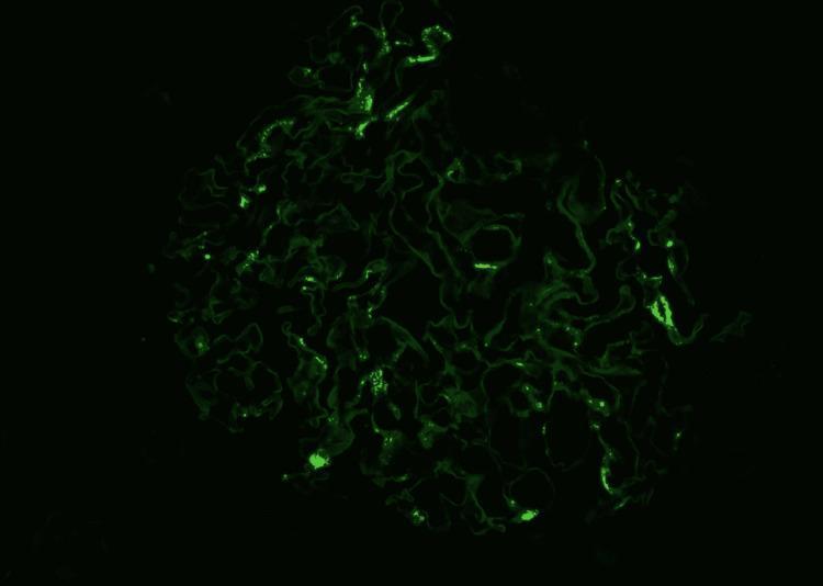
This diagnostic immunofluorescence image demonstrates a renal glomerulus exhibiting a characteristic staining pattern for IgA nephropathy. Against a dark background, a bright green fluorescent signal is localized primarily within the mesangial regions. The signal presents as granular, irregular deposits concentrated in the mesangial matrix, reflecting the entrapment of immunoglobulin A (IgA) immune complexes. The overall structure of the glomerulus shows a complex, lobulated architecture with visible capillary loops. While some areas demonstrate intense, discrete granular fluorescence, others show a more diffuse or attenuated signal, which may correlate with underlying morphological changes such as mesangial hypercellularity or segmental sclerosis. This imaging modality is essential in nephropathology for differentiating between various forms of glomerulonephritis based on the specific distribution (mesangial vs. capillary wall) and nature (granular vs. linear) of immune deposits.
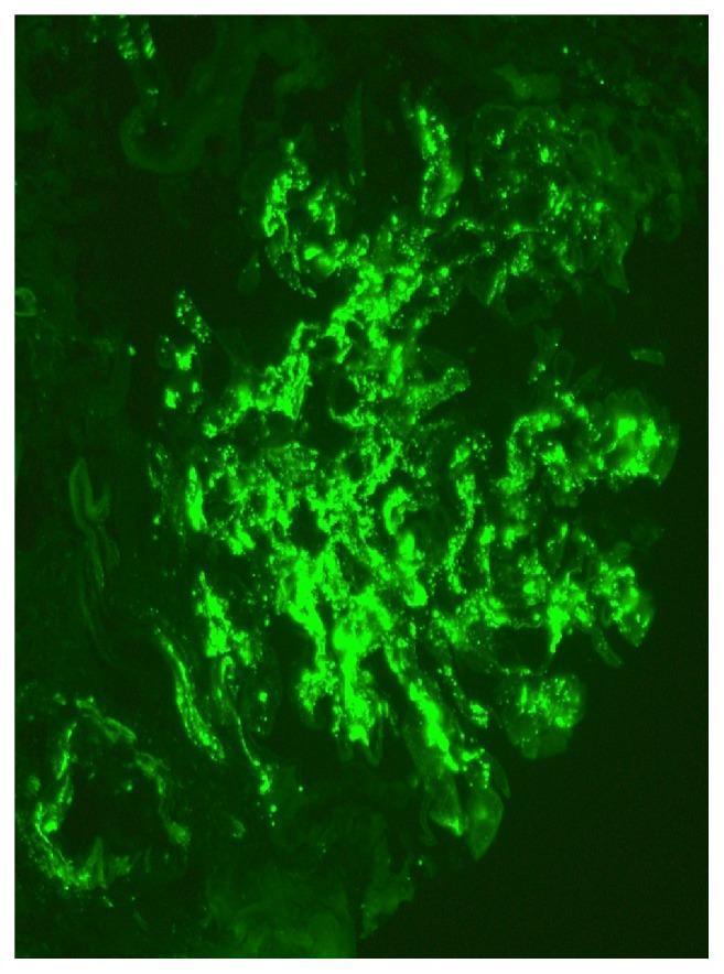
This diagnostic image is a high-magnification (x400) immunofluorescence microscopy of a renal biopsy specimen. The visual field demonstrates a single glomerulus exhibiting a classic pathological pattern of intense, apple-green fluorescent staining. The primary finding is the global, 3+ intensity mesangial deposition of immunoglobulin A (IgA). The deposits are characterized by a granular and confluent distribution primarily restricted to the mesangial matrix and stalk regions of the glomerular tuft, highlighting the branched architecture of the mesangium. There is a notable absence of significant capillary loop or basement membrane staining, which is highly characteristic of IgA nephropathy (Berger disease). The surrounding renal parenchyma is dark, providing high contrast to the localized immunocomplex accumulation within the glomerular architecture. This diagnostic finding is a hallmark educational example used to confirm the diagnosis of primary IgA nephropathy or Henoch-Schönlein purpura nephritis in clinical pathology.
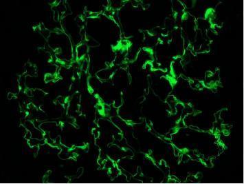
membranous nephropathy granular subepithelial deposits immunofluorescence
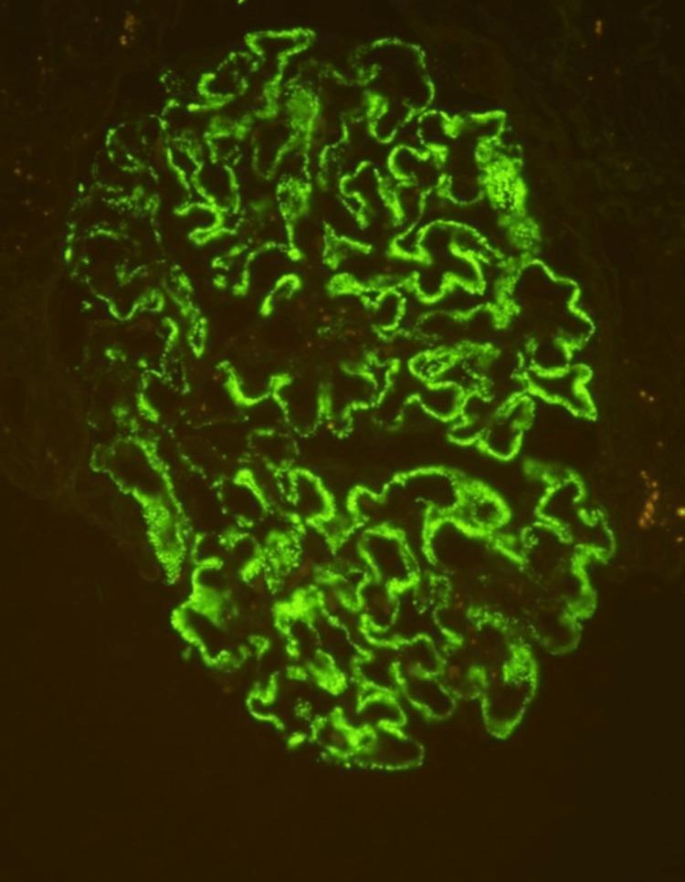
This diagnostic image is a high-power (400x) immunofluorescence micrograph of a human glomerulus from a renal biopsy specimen. The image demonstrates a diffuse, granular staining pattern of 3+ intensity, specifically localized along the glomerular capillary loops. The green fluorescent signal highlights the convoluted, interconnected morphology of the basement membrane area in a 'beaded' fashion, which is characteristic of immune complex deposition. Pathologically, this visual finding represents positive staining for phospholipase A2 receptor (PLA2R) antibodies, a definitive diagnostic marker for primary membranous nephropathy. The absence of mesangial or endocapillary staining emphasizes the subepithelial nature of the deposits. This specimen is critical for differentiating primary autoimmune-mediated glomerular disease from secondary causes in patients presenting with nephrotic syndrome.
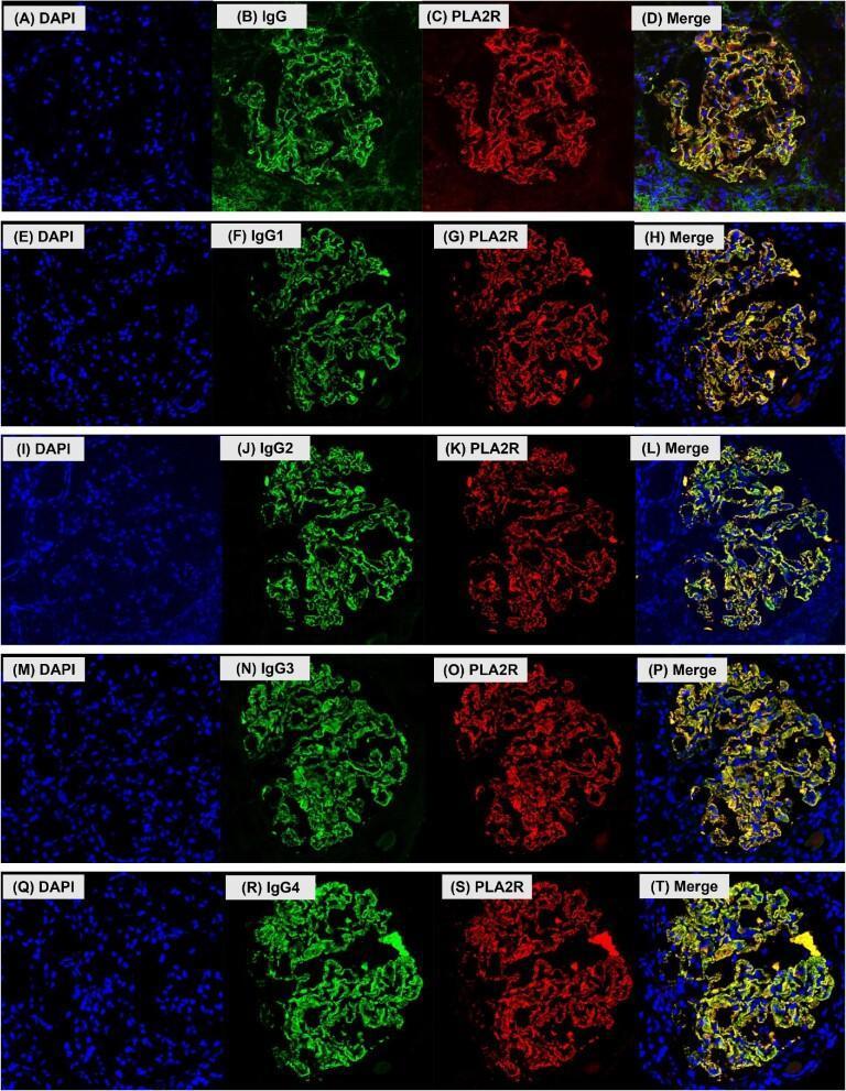
This composite of confocal light microscopy images illustrates immunofluorescence staining patterns in human glomerular tissue, specifically focusing on membranous nephropathy (MN). The 20-panel figure is organized into five rows (A-T), each representing a different IgG target: total IgG, IgG1, IgG2, IgG3, and IgG4. Within each row, four panels show: (1) DAPI nuclear staining in blue, (2) IgG or specific IgG subclasses in green, (3) PLA2R antigen in red, and (4) a merged overlay. The staining reveals a granular, loop-like distribution along the glomerular basement membrane (GBM), characteristic of subepithelial immune complex deposits. In the merged columns (D, H, L, P, T), the overlap of green (IgG) and red (PLA2R) signals produces a yellow color, demonstrating the co-localization of the PLA2R antigen with total IgG and all four IgG subclasses. DAPI staining confirms that these deposits are extracellular and distinct from cellular nuclei. This visual evidence supports the role of PLA2R as a major autoantigen in primary MN and characterizes the subclass composition of the associated immune deposits.
Renal Glomerular Lesions / Glomerulonephritis: Classification & Immunofluorescence
MD Pathology Exam Review
PART 1: CLASSIFICATION OF GLOMERULAR LESIONS
A. Based on Extent of Involvement (Descriptive Terms)
| Term | Definition |
|---|---|
| Diffuse | >50% of all glomeruli affected |
| Focal | <50% of glomeruli affected |
| Global | Entire glomerulus involved |
| Segmental | Only part of a glomerulus involved |
| Proliferative | Increase in number of cells (mesangial, endothelial, epithelial) |
| Membranous | Basement membrane thickening without proliferation |
| Membranoproliferative | Both BM thickening + proliferation |
| Crescentic | Epithelial cell proliferation in Bowman space forming crescents |
| Sclerosing | Collagenous obliteration of glomerular loops |
B. Based on Clinical Presentation
| Syndrome | Hallmarks | Prototypic Disease |
|---|---|---|
| Nephritic | Hematuria, RBC casts, hypertension, oliguria, mild proteinuria | Post-infectious GN, Crescentic GN |
| Nephrotic | Massive proteinuria (>3.5 g/day), hypoalbuminemia, edema, hyperlipidemia, lipiduria | Membranous nephropathy, MCD, FSGS |
| Nephritic-Nephrotic | Overlap features | MPGN, Diffuse Proliferative Lupus GN |
| Asymptomatic hematuria | Microscopic hematuria ± mild proteinuria | IgA nephropathy |
| RPGN | Rapid loss of renal function over weeks-months, crescents | Anti-GBM, ANCA-vasculitis |
C. Primary Glomerulonephritides - Full Classification with IF Patterns
PART 2: IMMUNOFLUORESCENCE (IF) PATTERNS - THE CORE OF MD PATHOLOGY EXAM
Key IF Concept: Three Fundamental Patterns
- Smooth, ribbon-like fluorescence along the entire GBM
- Caused by anti-GBM antibodies binding uniformly distributed intrinsic GBM antigens (NC1 domain of Type IV collagen)
- Diseases: Anti-GBM GN (Goodpasture syndrome)
- Staining: IgG linear + C3 linear along GBM
- Irregular, discontinuous, granular deposits along GBM and/or mesangium
- Caused by immune complex deposition (either circulating or in situ)
- Diseases: Post-infectious GN, Membranous nephropathy, IgA nephropathy, MPGN, Lupus nephritis, Cryoglobulinemia
- Staining varies by disease (see table below)
- Absent or minimal immunoglobulin/complement staining
- Caused by ANCA-mediated neutrophil injury WITHOUT immune complexes
- Diseases: ANCA-associated vasculitis (Granulomatosis with polyangiitis - GPA; Microscopic polyangiitis - MPA; Eosinophilic granulomatosis with polyangiitis - EGPA)
Linear vs. Granular: The Classic Distinction
PART 3: DISEASE-BY-DISEASE IF PATTERN TABLE
| Disease | LM | IF Pattern | IF Immunoreactants | EM |
|---|---|---|---|---|
| Post-infectious GN | Diffuse endocapillary proliferation, "starry sky" | Granular | IgG + C3 in GBM and mesangium; "lumpy-bumpy" | Subepithelial humps (hallmark) |
| Crescentic (RPGN) - Type I (Anti-GBM) | Crescents, fibrinoid necrosis | LINEAR | IgG (linear) + C3 along GBM | No deposits |
| Crescentic (RPGN) - Type II (Immune complex) | Crescents + proliferation | Granular | IgG + C3 ± IgM, IgA | Immune complex deposits at various sites |
| Crescentic (RPGN) - Type III (ANCA) | Crescents, necrosis | Pauci-immune (negative) | No deposits (or trace) | No deposits |
| Membranous Nephropathy | Diffuse GBM thickening; "spike and dome" (silver stain) | Granular capillary wall | IgG + C3 (diffuse granular along capillary loops) | Subepithelial deposits; GBM spikes |
| Minimal Change Disease (MCD) | Normal (lipid in tubules) | Negative or faint IgM | Usually negative; fine granular podocyte IgG (anti-nephrin) in most recent update | Effacement of foot processes; NO deposits |
| FSGS | Focal segmental sclerosis + hyalinosis | IgM + C3 (segmental) | Nonspecific IgM and C3 trapped in sclerosed segments | Foot process effacement; epithelial denudation |
| MPGN Type I | Mesangial proliferation + GBM thickening + "tram-track" (double contour) | Granular | IgG + C3; C1q + C4 (classical pathway activation) | Subendothelial deposits |
| Dense Deposit Disease (C3G / MPGN Type II) | Tram-track; ribbon-like dense deposits | C3 only (no C1q, no C4, no Ig) | C3 dominant - alternative pathway dysregulation | Extremely dense, ribbon-like intramembranous deposits |
| C3 Glomerulonephritis (C3GN) | Variable (mesangial to MPGN pattern) | C3 dominant (C3 ≥ 2 orders more than any Ig) | C3 only or dominant | Mesangial/subendothelial/subepithelial deposits |
| IgA Nephropathy (Berger disease) | Focal mesangial proliferative GN | Mesangial granular | Dominant IgA + C3 in mesangium ± IgG, IgM | Mesangial and paramesangial deposits |
| Lupus Nephritis (Class III/IV) | Proliferative GN; "wire loops" | "Full house" | IgG + IgA + IgM + C3 + C1q (all positive = "full house") | Subendothelial + subepithelial + mesangial deposits; tubuloreticular inclusions |
| Fibrillary GN | Mesangial expansion; MPGN pattern | Granular | IgG (dominant IgG4) + C3; DNAJB9 positive (diagnostic marker) | Fibrils 20 nm diameter (larger than amyloid) |
| Immunotactoid GN | Similar to Fibrillary GN | Granular | IgG (monoclonal) | Microtubules 30-50 nm diameter |
IgA Nephropathy IF Images
Membranous Nephropathy IF
PART 4: RAPID PROGRESSIVE GN (CRESCENTIC GN) - CLASSIFICATION DETAIL
| Type | Mechanism | IF | Serology |
|---|---|---|---|
| Type I - Anti-GBM | Anti-NC1 (Type IV collagen) antibodies | Linear IgG + C3 | Anti-GBM antibody positive |
| Type II - Immune Complex | Circulating/in situ IC deposition | Granular IgG ± C3 | Low complement, variable serology |
| Type III - Pauci-immune (ANCA) | Neutrophil-mediated, no IC | Negative (pauci-immune) | c-ANCA (PR3) or p-ANCA (MPO) positive |
| Type IV | Combined anti-GBM + ANCA | Linear + positive ANCA | Both anti-GBM and ANCA positive |
PART 5: C3 GLOMERULOPATHY - IMPORTANT RECENT UPDATE
- MPGN Type I → remains immune complex-mediated (IC-MPGN)
- MPGN Type II (Dense Deposit Disease) → reclassified as C3 Glomerulopathy - Dense Deposit Disease (DDD)
- C3 Glomerulonephritis (C3GN) → new entity, part of C3 glomerulopathy spectrum
- C3 staining dominance: C3 intensity ≥ 2 full orders of magnitude (2+) above any immunoglobulin
- Reflects alternative complement pathway dysregulation (C3 nephritic factor, Factor H/I mutations)
- No immune complexes (no IgG, IgA, IgM, C1q, C4)
PART 6: LUPUS NEPHRITIS (ISN/RPS Classification 2018 Update)
| Class | Pathology | IF |
|---|---|---|
| I | Minimal mesangial | Normal or minimal mesangial deposits |
| II | Mesangial proliferative | Mesangial IgG/IgA/IgM/C3/C1q |
| III | Focal proliferative (<50% glomeruli) | Segmental "full house" IF |
| IV | Diffuse proliferative (>50% glomeruli) | Diffuse "full house" IF; wire loops |
| V | Membranous (may co-exist with III/IV) | Granular capillary IF ("full house") |
| VI | Advanced sclerosing (>90% global sclerosis) | Absent/minimal IF |
PART 7: NEW ANTIGENS IN MEMBRANOUS NEPHROPATHY (Key Recent Updates)
| Antigen | Association | IF/Lab Finding |
|---|---|---|
| PLA2R (phospholipase A2 receptor) | Primary MN (~70-80%) | IgG4 dominant; serum anti-PLA2R antibody |
| THSD7A (thrombospondin domain 7A) | Primary MN (~3-5%) | IgG4 dominant |
| NELL1 (Neural epidermal growth factor-like 1) | Primary MN (older males, ~15-20%) | IgG1 dominant |
| EXT1/EXT2 (Exostosin 1/2) | Autoimmune-associated (lupus, SLE-associated MN) | IgG1/IgG2 dominant |
| NCAM1 (Neural cell adhesion molecule 1) | Primary MN | IgG1 dominant |
PART 8: MINIMAL CHANGE DISEASE - RECENT UPDATE (2024-2025)
- Diffuse fine granular podocyte IgG associated with nephrin clustering is now recognized on careful IF in most cases
- This explains the podocyte dysfunction and foot process effacement without classic immune complex deposits
PART 9: KEY IF PATTERNS - MEMORY AID
| IF Pattern | Think Of |
|---|---|
| Linear IgG + C3 | Anti-GBM (Goodpasture) |
| Granular IgG + C3 (GBM + mesangium) | Post-infectious GN |
| Granular IgG + C3 (capillary wall only) | Membranous nephropathy |
| Dominant IgA (mesangial) | IgA nephropathy / HSP |
| C3 only (no Ig) | C3 Glomerulopathy (DDD or C3GN) |
| IgG + C3 + C1q + C4 | MPGN Type I (immune complex) |
| Full house (IgG+IgA+IgM+C3+C1q) | Lupus nephritis |
| Negative / Pauci-immune | ANCA vasculitis |
| IgM + C3 (segmental) | FSGS (nonspecific entrapment) |
| Congo red + amyloid P | Amyloidosis (apple-green birefringence) |
| IgG (monoclonal) - microtubules 30-50nm | Immunotactoid GN |
| IgG + DNAJB9 (fibrils 20nm) | Fibrillary GN |
PART 10: ELECTRON MICROSCOPY DEPOSIT LOCATIONS - CORRELATE WITH IF
| Location | Associated Disease |
|---|---|
| Subepithelial humps | Post-infectious GN (characteristic) |
| Subepithelial spikes | Membranous nephropathy |
| Subendothelial deposits | MPGN Type I, Lupus (Class III/IV) |
| Mesangial deposits | IgA nephropathy, Lupus Class II |
| Intramembranous (dense) | Dense Deposit Disease (C3G-DDD) |
| Foot process effacement only (no deposits) | MCD, early FSGS |
| Tubuloreticular inclusions (endothelium) | Lupus nephritis (interferon signature) |
| Fibrils 10-12nm (randomly arranged) | Amyloidosis |
| Fibrils 20nm | Fibrillary GN |
| Microtubules 30-50nm | Immunotactoid GN |
- Robbins, Cotran & Kumar Pathologic Basis of Disease (11th ed.), Table 20.5 - complete glomerulonephritis summary
- Robbins & Kumar Basic Pathology, Ch. 12 - Mechanisms of glomerular injury and disease-specific IF
- Comprehensive Clinical Nephrology, 7th Edition - clinical-pathologic correlation
- CARI Guidelines Commentary on KDIGO GN Guidelines (2025) - PMID 40955731
Morphological spectrum of diabetic nephropathy and next question eosinophilia variants of kidney tumor for md pathology exam 10 marks each
Kimmelstiel-Wilson nodule diabetic nephropathy mesangial sclerosis PAS stain
renal oncocytoma chromophobe renal cell carcinoma eosinophilic histology
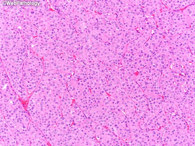
This histopathology photomicrograph displays a renal tumor tissue section stained with Hematoxylin and Eosin (H&E) at high magnification. Tumor cells are arranged in densely packed nests and interlacing trabeculae with a predominantly solid growth pattern. Cells have abundant eosinophilic cytoplasm, distinct cell borders, and minimal cytoplasmic vacuolization. Nuclei vary slightly in size and contour, often showing irregular outlines, occasional binucleation, and conspicuously irregular chromatin. Perinuclear halos—clear rings surrounding nuclei—are evident, a characteristic feature described in chromophobe renal cell carcinoma, particularly its eosinophilic variant. The overall architecture lacks prominent necrosis or conspicuous mitotic activity in this field. Immunohistochemistry would aid definitive subclassification: diffuse CK7 positivity strongly favors chromophobe RCC, whereas oncocytoma typically shows CK7 negativity or only focal staining; CD117 (c-KIT) is commonly expressed in chromophobe tumors as well. The diagnostic significance lies in distinguishing chromophobe RCC from other renal neoplasms due to differences in prognosis and management. Clinically, accurate classification informs surgical planning (partial versus radical nephrectomy), influences risk stratification, and guides consideration of adjuvant therapies. This image serves as an educational example of renal tumor histology and the role of immunophenotyping in differentiating eosinophilic chromophobe RCC from mimickers, such as oncocytoma within diagnostic pathology. This aids decision-making and education.
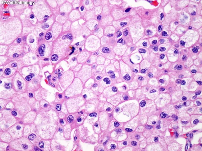
This high-power field represents hematoxylin and eosin stained renal tumor tissue from a chromophobe renal cell carcinoma. The microscope image shows solid/nested growth of polygonal tumor cells with distinct cell borders and ample cytoplasm characterized by a fine reticular eosinophilic pattern. Nuclei are generally central with clumped chromatin; focal binucleation and mild pleomorphism are evident, with small nucleoli. The cytoplasm appears reticular and pale to eosinophilic and is often described as raisinoid in chromophobe RCC, though in this field the reticular cytoplasm is the prominent feature. The cells are arranged in sheets and nests with minimal cytoplasmic clearing and a light, granular background. The combination of well-demarcated cell membranes, perinuclear clearing (if present), and cohesive cell borders are consistent with chromophobe RCC morphology. Clinically, such features aid in distinguishing chromophobe RCC from clear cell RCC and papillary RCC and from oncocytoma, which tends to display more eosinophilic, granular cytoplasm and uniform nuclei. This image is relevant for diagnostic pathology education, differential diagnosis, and for correlating histology with immunohistochemistry (CK7 positive, CD117 positive) and prognosis. Potential clinical applications include histopathologic confirmation after nephrectomy or biopsy and teaching differential diagnostic reasoning in renal tumors.
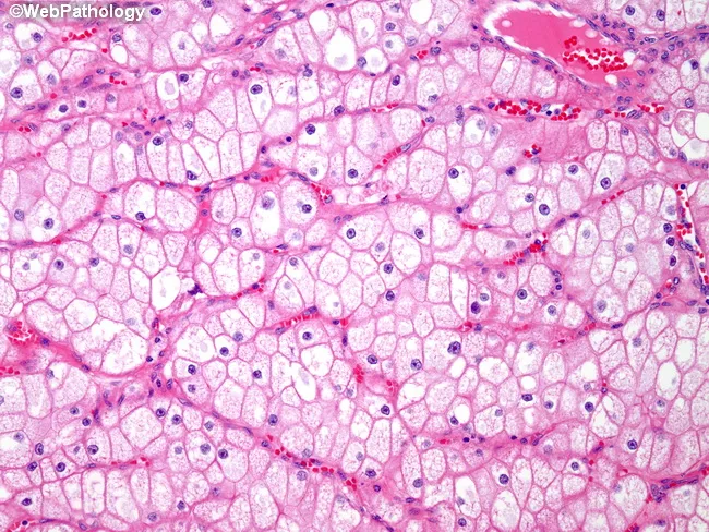
Under brightfield illumination the renal tumor tissue displays an alveolar, organoid pattern consistent with chromophobe renal cell carcinoma. Hematoxylin and eosin stained sections reveal tumor cells with pale pink cytoplasm that is finely reticular rather than optically clear. Cell borders are distinctly thick, giving a plant‑like, boxy outline to each cell, and the cytoplasmic membranes appear prominently delineated. In many fields there are perinuclear halos around nuclei with granular chromatin and relatively small, uniform nuclei, which helps differentiate chromophobe RCC from clear cell RCC, where cytoplasm is typically clear and vacuolated. Vascular networks are elaborate, contributing to the rich intratumoral perfusion seen in this sample. Notably, only a minority of cells exhibit true clear cytoplasm, further arguing against clear cell carcinoma. The combination of reticular cytoplasm, conspicuous cell membranes, and perinuclear halos is characteristic of chromophobe histology, supporting this diagnosis in the appropriate clinical context. From a diagnostic standpoint, these features guide the pathologist toward chromophobe RCC and away from other eosinophilic renal neoplasms such as oncocytoma or papillary RCC. Clinically, recognizing this morphology informs prognosis and potential therapeutic strategies, including surveillance and surgical management. This image exemplifies characteristic chromophobe features and reinforces differential diagnosis in renal neoplasia for education.
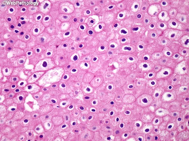
Light microscopy of renal tumor tissue demonstrates chromophobe features with scattered tumor cells showing raisinoid nuclei and prominent nuclear halos. Cells possess very distinct, thick cytoplasmic borders producing a plant-like appearance and pale eosinophilic, reticulated cytoplasm. The architectural pattern is variable, including diffuse and nested arrangements with perinuclear clearing and delicate septae; mitotic activity is usually low. These histologic attributes are characteristic of chromophobe renal cell carcinoma, a renal neoplasm arising from intercalated cells of the collecting ducts. The diagnostic significance lies in the relatively favorable prognosis of chromophobe RCC compared with clear cell RCC when detected early and adequately excised. Immunohistochemical profiling commonly shows CK7 positivity and CD117 positivity, with CAIX negativity, aiding confirmation in challenging cases. Differential considerations include oncocytoma, papillary RCC, and clear cell RCC; correlation with imaging, clinical history, and molecular studies may be necessary. Clinically, the image is relevant for education in renal tumor pathology, histologic differential diagnosis, and pathology-based decision making in urologic oncology. This histology supports surgical planning, staging, and prognostication in patients with renal masses. The visual cues - raisinoid nuclei, nuclear halos, and plant-like cytoplasmic membranes - are critical for recognizing chromophobe RCC in diagnostic practice.
diabetic nephropathy glomerular hyaline cap fibrin cap armanni-ebstein lesion
diabetic nephropathy nodular glomerulosclerosis kidney biopsy
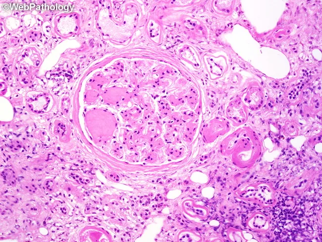
Imaging modality: Light microscopy of a renal biopsy specimen stained with Hematoxylin and Eosin (H&E). The focal region of interest is the glomerulus from the renal cortex, with mid-power to high-power view illustrating nodular mesangial sclerosis typical of diabetic nephropathy. Central to the image are Kimmelstiel–Wilson nodules: spherical, acellular to pale pink nodules within the mesangial matrix that disrupt normal glomerular architecture and compress capillary tufts. Surrounding glomerular capillary loops show mesangial expansion; there are also arterioles adjacent to the glomerulus with arteriolar hyalinosis (hyaline thickening of small vessels) on the external side; this hyaline arteriolosclerosis reflects chronic ischemic injury from hyperglycemia. The periglomerular interstitium exhibits chronic inflammatory infiltrate and mild interstitial fibrosis, consistent with long-standing diabetic kidney disease. The specimen demonstrates features of nodular diabetic glomerulosclerosis with associated vascular and interstitial changes, which collectively contribute to progressive proteinuria and renal insufficiency. Clinically, these histopathologic findings corroborate diabetic nephropathy in patients with long-standing diabetes mellitus, correlating with stage of kidney involvement, guiding management such as optimized glycemic control, renin-angiotensin system blockade, and nephroprotective strategies. Differential considerations include nodular glomerulosclerosis due to other etiologies (e.g., light chain deposition disease) but KW nodules are classic for diabetic nephropathy. These findings guide prognosis.
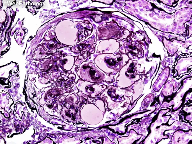
This histopathology image shows a renal glomerulus from a kidney biopsy, examined with bright-field light microscopy after Jones’ methenamine silver staining. The primary subject is nodular glomerulosclerosis associated with diabetic nephropathy. The mesangial matrix is markedly expanded, producing discrete Kimmelstiel–Wilson nodules within the glomerular tuft. These eosinophilic, rounded nodules compress adjacent capillary loops and disrupt normal capillary architecture. The glomerular basement membranes appear thickened, a feature accentuated by the silver stain as dark outlines surrounding the sclerotic nodules. The surrounding mesangial matrix shows increased density, while capillary lumina are variably narrowed. Bowman’s space is variably preserved, and there may be mild hyalinosis of arterioles consistent with diabetic microangiopathy. Overall, the image displays the classic triad of diabetic nephropathy: mesangial expansion, nodular glomerulosclerosis (KW lesions), and GBM thickening. Diagnostic significance is high for long-standing diabetes with renal involvement and proteinuria; the findings contribute to staging and prognosis. Differential considerations include nodular FSGS and nodular amyloidosis, but the appearance of discrete KW nodules is characteristic. Clinically, these histologic changes correlate with reduced GFR and progression to chronic kidney disease; management emphasizes glycemic control and anti-hypertensive therapy. This image is suitable for teaching, case reviews, and pathology QA.
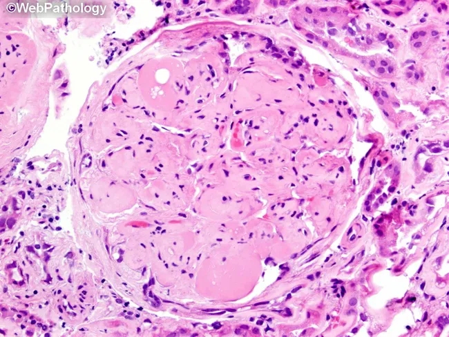
Imaging modality: brightfield histopathology. This slide shows a renal glomerulus from a biopsy specimen of diabetic nephropathy. The mesangial matrix is markedly expanded, producing round to nodular consolidations within the tuft. Several Kimmelstiel‑Wilson nodules are evident as acellular, hyaline nodules that distend the mesangial stalks and encroach on capillary loops. The glomerular basement membranes appear thickened on corresponding PAS‑positive and silver‑stained sections (referenced in adjacent images), consistent with nodular diabetic glomerulosclerosis. The surrounding cortex exhibits mild chronic interstitial changes and arteriolar hyalinosis in keeping with chronic diabetic kidney disease. The glomerulus displays relatively extensive mesangial deposition with focally patent capillary lumina, creating a characteristic nodular pattern. Overall, the morphology is diagnostic for diabetic microangiopathy with progressive nodular sclerosis. Clinically, these findings correlate with long‑standing hyperglycemia, proteinuria, and reduced GFR. Pathological significance: KW nodules indicate advanced disease and higher risk of progression to end‑stage renal disease. Differential considerations include non‑diabetic nodular glomerulosclerosis and other glomerulopathies, but the classic KW nodules and mesangial expansion strongly support diabetic nephropathy. This image is valuable for education, pathology review, and correlating histology with clinical diabetes management. It highlights key features for learners: mesangial expansion, KW nodules, PAS positivity, and altered capillary luminal flow in practice.
renal oncocytoma central scar mahogany brown gross pathology
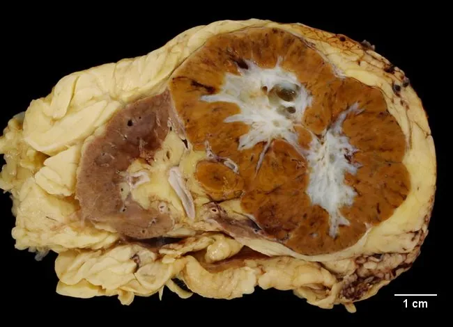
Gross macroscopic image of a kidney nephrectomy specimen displaying a solitary renal mass. The cut surface is mahogany brown to yellow-brown, with a well-circumscribed, but not clearly encapsulated, tumor replacing a portion of the renal cortex. A distinct white, stellate central scar is visible in the majority of the sectioned tumor areas, particularly in larger tumors, and commonly corresponds to fibrous stroma and degenerative changes. The surrounding renal parenchyma appears otherwise unremarkable, with a sharp pseudocapsule and clear delineation between lesion and normal cortex. The lesion demonstrates a heterogeneous color pattern on gross inspection, lacking overt hemorrhage or necrosis. The specimen is imaged in a gross pathology setting, with a ruler indicating scale (approximately 1 cm divisions) to convey tumor dimensions. The overall morphology—well-circumscribed borders, discrete cortical involvement, mahogany coloration, and central scar—aligns with classic features described for renal oncocytoma, a benign epithelial neoplasm arising from collecting ducts (intercalated cells) of the kidney. Clinically, renal oncocytoma accounts for a minority of renal tumors and is frequently discovered incidentally on imaging studies performed for unrelated indications. Although benign, differentiation from renal cell carcinoma remains essential; histology and immunohistochemistry are commonly employed to confirm the diagnosis. This image is intended for education.
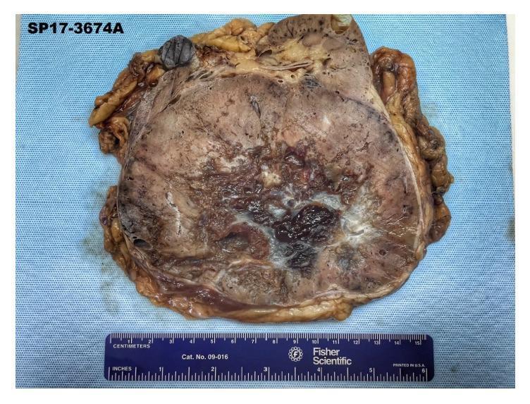
This clinical photograph displays a gross pathology specimen of a bivalved right kidney. The specimen is dominated by a large, well-circumscribed, spherical mass that has almost entirely replaced the renal parenchyma. The cut surface of the tumor exhibits a characteristic mahogany or maroon-colored, homogenous appearance. A prominent, stellate, gray-white fibrous scar is visible in the central region of the mass, radiating outward. These gross findings—a mahogany-brown color and a central stellate scar—are classic diagnostic hallmarks of a renal oncocytoma. The mass is encapsulated, and the surrounding perinephric fat is visible at the margins. A metric ruler at the bottom of the frame provides scale, indicating the significant size of the lesion, which corresponds to the clinical presentation of a large renal mass. This specimen serves as a primary educational example of benign renal neoplasm morphology in urologic pathology.
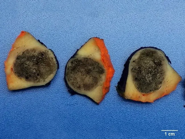
Imaging modality: gross pathology photograph of a resected kidney mass. Anatomical location: kidney, renal parenchyma. The specimen shows a 3.5 cm telangiectatic oncocytoma; a rare vascular variant of renal oncocytoma. The cut surface demonstrates a well-circumscribed, dark-brown hemorrhagic, spongy mass with conspicuous vascular channels throughout the parenchyma and no central stellate scar. The tumor is embedded in normal renal tissue and displays solid to sponge-like consistency. The hemorrhagic cavities reflect telangiectatic vascularity; the capsule is not enlarge? not clearly delineated in this specimen. The color is dark brown to mahogany with reddish-orange rind on the periphery. The specimen lacks a central scar, which can help differentiate from classic oncocytoma that often shows a central scar on imaging or grossly. The vascularity contributes to radiologic misdiagnosis risk. Clinical significance: telangiectatic oncocytoma accounts for about 15% of all renal oncocytomas and is frequently misinterpreted as malignant or suspicious on imaging. Management: surgical excision; prognosis is excellent after complete resection. This image serves as educational reference for gross renal tumor morphology, differential diagnosis vs RCC and angiomyolipoma; it supports correlation with histology showing oncocytic cells with abundant mitochondria, prominent vasculature, and hemorrhagic, sponge-like architecture in telangiectatic variant.
QUESTION 1: Morphological Spectrum of Diabetic Nephropathy (10 Marks)
Introduction
MORPHOLOGICAL SPECTRUM
A. GLOMERULAR LESIONS (Most Important)
1. Glomerular Basement Membrane (GBM) Thickening
- Earliest and most sensitive morphological indicator of diabetes
- Detectable by electron microscopy within 1-2 years of clinical onset
- GBM thickens along its entire length
- Composition altered - loss of heparan sulfate (negatively charged proteoglycans), disrupting the filtration barrier
- Results in increased filtration of plasma proteins (proteinuria)
- Correlates poorly with clinical nephropathy on its own
2. Diffuse Mesangial Sclerosis (Diffuse Diabetic Glomerulosclerosis)
- Most common glomerular lesion of DN
- Increase in mesangial matrix with mesangial cell proliferation + GBM thickening
- Affects all glomeruli diffusely
- Present in most diabetics with >10 years duration
- More common in older patients and those with hypertension
- When severe, leads to nephrotic syndrome (proteinuria, hypoalbuminemia, edema)
- Not specific to diabetes (also in aging, hypertension), but most pronounced in diabetics
3. Nodular Glomerulosclerosis (Kimmelstiel-Wilson Lesion) ★
- Pathognomonic of diabetic nephropathy
- Present in 15-30% of long-standing diabetics
- Gross/LM: Ball-like, ovoid, laminated deposits of matrix material ("nodules") at the periphery of the glomerulus, in the mesangium
- Nodules are acellular, eosinophilic, PAS-positive, silver-positive (methenamine silver)
- Located at the periphery of glomerular lobules, compressing adjacent capillary loops
- The nodules enlarge the mesangial stalks and sit eccentrically
- Differential diagnosis of nodular glomerulosclerosis:
- Light chain deposition disease (LCDD)
- Fibrillary GN
- Nodular FSGS (smoking-related)
- Amyloidosis (Congo red +, unlike KW nodules)
4. Hyaline Lesions (Exudative/Capsular Drop Lesions)
- Hyaline caps ("fibrin caps"): Homogeneous, eosinophilic, acellular cap over glomerular capillary loops - contain plasma proteins (not fibrin despite the name)
- Capsular drop lesions: Accumulation of homogeneous hyaline material between parietal epithelium and Bowman's capsule
- Both are PAS-positive and seen in advanced DN
- Pathogenesis: plasma protein insudation through damaged capillaries
5. Glomerular Hyperfiltration and Hypertrophy
- Earliest functional change - precedes morphological lesions
- Increased GFR and glomerular enlargement
- Due to activation of RAAS, SGLT2 upregulation in proximal tubule → decreased distal Na delivery → further hyperfiltration
B. VASCULAR LESIONS
1. Hyaline Arteriolosclerosis
- Affects both afferent AND efferent arterioles
- Efferent arteriolar hyalinosis is virtually unique to diabetes (in non-diabetics, hyalinosis affects only afferent arterioles)
- Appears as amorphous, eosinophilic, hyaline thickening of arteriolar walls
- Results in ischemia → diffuse cortical scarring (nephrosclerosis)
- Contributes to finely granular cortical surface
2. Atherosclerosis (Macrovascular)
- Accelerated and premature in diabetics
- Affects main renal artery and larger intrarenal arteries
- Contributes to renal ischemia and hypertension
C. TUBULAR LESIONS
1. Armanni-Ebstein Lesion (Glycogen Nephropathy)
- Pathognomonic of poorly controlled diabetes
- Accumulation of glycogen in tubular epithelial cells (especially loop of Henle and collecting duct cells)
- Cells appear clear and vacuolated on H&E
- PAS-positive (glycogen); diastase-labile (distinguishes from lipid vacuoles)
- Reversible with glycemic control
- Associated with glucosuria and osmotic diuresis
2. Tubular Basement Membrane Thickening
- Parallel to GBM thickening
- Less specific than glomerular changes
3. Tubular Atrophy and Interstitial Fibrosis
- Occurs in advanced DN as a result of ischemia and proteinuria
- Correlates better with rate of GFR decline than glomerular stage
D. INTERSTITIAL AND INFECTIOUS LESIONS
1. Chronic Pyelonephritis
- More frequent and severe in diabetics
- Related to immunosuppression, glycosuria (favorable medium for bacteria), and neurogenic bladder
2. Necrotizing Papillitis (Renal Papillary Necrosis)
- Much more prevalent in diabetics than non-diabetics
- Ischemic necrosis of renal papillae due to vascular compromise
- Grossly: grey-white necrotic sloughed papillae
- Can present with acute renal failure and flank pain
- Sloughed papillae can cause ureteral obstruction
E. IMMUNOFLUORESCENCE IN DIABETIC NEPHROPATHY
| Finding | Description |
|---|---|
| Linear IgG along GBM | Nonspecific "trapping" - no immune deposits on EM |
| IgG along tubular BM | Nonspecific |
| C3 | Occasional |
| Electron microscopy | Markedly thickened GBM; subendothelial space expanded; no immune complex deposits |
Summary Table: Morphological Spectrum
| Lesion | Specificity | Main Stain | When Appears |
|---|---|---|---|
| GBM thickening | Non-specific | EM | 1-2 years |
| Diffuse mesangial sclerosis | Non-specific (but severe in DM) | PAS, Silver | >5 years |
| Kimmelstiel-Wilson nodules | Pathognomonic | PAS, Silver | 10-20 years |
| Hyaline caps/capsular drops | Highly suggestive | PAS | Advanced |
| Efferent arteriolar hyalinosis | Virtually unique to DM | H&E | Variable |
| Armanni-Ebstein | Pathognomonic (acute poorly controlled DM) | PAS (diastase labile) | Acute |
| Papillary necrosis | Highly associated with DM | Gross + H&E | Any stage |
QUESTION 2: Eosinophilic Variants of Kidney Tumors (10 Marks)
Introduction
CLASSIFICATION OF EOSINOPHILIC RENAL TUMORS
1. RENAL ONCOCYTOMA (Benign) ★
- 5% of all renal neoplasms
- Male > Female; mean age 60-70 years
- Usually incidental finding on imaging
- Can be bilateral/multifocal in Birt-Hogg-Dubé syndrome
- Tan to mahogany brown (due to abundant mitochondria)
- Relatively homogeneous cut surface
- Characteristic central stellate scar (fibrous stroma, especially in larger tumors)
- Well-circumscribed, NO necrosis, NO hemorrhage (except telangiectatic variant)
- Usually solitary; may reach >10 cm
- Cells with abundant, finely granular, deeply eosinophilic cytoplasm (packed mitochondria on EM)
- Small, round, benign-appearing nuclei with small but conspicuous nucleoli
- NO significant nuclear pleomorphism, NO mitotic activity, NO necrosis
- Architecture: nests and tubules (alveolar pattern) embedded in loose, myxoid stroma
- Cells arranged in solid nests, tubules, or microcysts
- Intercellular edematous stroma
- Cytoplasm packed with abnormal mitochondria (diagnostic feature)
| Marker | Oncocytoma |
|---|---|
| CK7 | Negative or focal (scattered cells only) |
| CD117 (c-KIT) | Positive |
| CAIX | Negative |
| Vimentin | Negative |
| RCC antigen | Variable |
| E-cadherin | Positive |
- Loss of chromosome 1 and Y
- Mitochondrial DNA mutations
2. CHROMOPHOBE RENAL CELL CARCINOMA - EOSINOPHILIC VARIANT ★
- 5% of all renal cell carcinomas
- Shares origin with oncocytoma (hence morphologic overlap)
- Best prognosis among RCC subtypes (better than clear cell and papillary)
- Classic/Chromophobic type - pale, transparent cytoplasm
- Eosinophilic type - granular, pink cytoplasm (the key exam entity)
- Well-circumscribed, homogeneous, tan-brown tumor
- NO central scar (distinguishes from oncocytoma)
- May show focal hemorrhage
- Pale to light tan (not as dark as oncocytoma)
- Cells with pale eosinophilic, finely reticulated cytoplasm
- Prominent, thick cell membranes ("plant cell" or "boxy" appearance) - KEY feature
- Perinuclear halo (clearing around nucleus) - PATHOGNOMONIC
- Nuclei: binucleated or multinucleated; irregular, "raisinoid" (wrinkled) chromatin
- Arranged in solid sheets with concentration of largest cells around blood vessels
- Koilocyte-like cells in some areas
- Hale's colloidal iron stain: Diffuse cytoplasmic positivity (reticular pattern) - diagnostic for chromophobe RCC; negative in oncocytoma
| Marker | Chromophobe RCC |
|---|---|
| CK7 | Diffuse and strong positive (key distinction from oncocytoma) |
| CD117 (c-KIT) | Positive |
| CAIX | Negative |
| Vimentin | Negative |
| Hale's colloidal iron | Diffuse reticular positive |
| CD10 | Negative |
- Multiple chromosome losses (chromosomes 1, 2, 6, 10, 13, 17, 21)
- Extreme hypodiploidy
- TP53, PTEN mutations in some aggressive cases
3. CLEAR CELL RCC WITH EOSINOPHILIC/GRANULAR CHANGE
- 70-80% of all RCCs
- Subset has eosinophilic/granular cytoplasm (less lipid accumulation)
- Polygonal cells with granular eosinophilic cytoplasm instead of the classic clear cells
- Arranged in vascular nests, tubules, papillary structures
- Nuclei show prominent nucleoli (high grade)
4. PAPILLARY RCC TYPE 2 (Eosinophilic)
- 10-15% of all RCCs
- Type 2 papillary RCC has large cells with abundant eosinophilic cytoplasm and high-grade nuclei (ISUP grade 3-4)
- Architecture: papillary formations; foamy macrophages in papillary cores
- Associated with hereditary leiomyomatosis and RCC (HLRCC) syndrome - fumarate hydratase (FH) mutations
- More aggressive than Type 1 (which has small basophilic cells)
- Often associated with sarcomatoid change
5. ONCOCYTIC PAPILLARY RENAL CELL CARCINOMA (New WHO 2022 Entity)
- Distinct entity with papillary or tubular architecture
- Cells with abundant eosinophilic (oncocytic) cytoplasm
- Low-grade nuclei
- Favorable prognosis
- IHC: CK7 diffuse positive, AMACR positive, CD10 variable
6. SUCCINATE DEHYDROGENASE (SDH)-DEFICIENT RCC
- Rare; associated with germline SDH mutations (SDHB, SDHC, SDHD)
- Cells with pale eosinophilic, "flocculent" (bubbly/vacuolated) cytoplasm - distinctive
- Intracytoplasmic inclusions (rounded eosinophilic globules)
- Nested/solid growth pattern
- IHC: SDHB negative (loss of expression - diagnostic)
- Part of hereditary paraganglioma/pheochromocytoma syndrome
7. EOSINOPHILIC SOLID AND CYSTIC (ESC) RCC (WHO 2022 New Entity)
- Mostly in women; association with tuberous sclerosis complex (TSC mutations)
- Alternating solid and cystic areas
- Cells: eosinophilic, voluminous cytoplasm with cytoplasmic stippling (coarse granules)
- Nuclei: round, regular, with small nucleoli
- IHC: CK20 positive (highly specific), CK7 negative, cathepsin K positive in TSC-related cases
DIFFERENTIAL DIAGNOSIS TABLE: EOSINOPHILIC RENAL TUMORS
| Feature | Oncocytoma | Chromophobe RCC (Eosinophilic) | Clear Cell RCC (Eosinophilic) | Papillary RCC Type 2 |
|---|---|---|---|---|
| Gross | Mahogany + central scar | Tan, no scar | Yellow-grey, necrosis | Hemorrhagic, cystic |
| Cell membranes | Inconspicuous | Thick, prominent | Inconspicuous | Inconspicuous |
| Perinuclear halo | Absent | Present | Absent | Absent |
| Nuclei | Round, small nucleoli | Raisinoid, irregular | Prominent nucleoli | Large, prominent nucleoli |
| Architecture | Nests in edematous stroma | Solid sheets | Vascular nests | Papillary |
| Hale's colloidal iron | Focal/apical only | Diffuse reticular | Negative | Negative |
| CK7 | Negative or focal | Diffuse strong | Negative | Positive |
| CD117 | Positive | Positive | Negative | Negative |
| CAIX | Negative | Negative | Positive (diffuse) | Negative |
| Vimentin | Negative | Negative | Positive | Positive |
| AMACR | Negative | Negative | Negative | Positive |
| Prognosis | Benign | Excellent | Good-to-poor | Moderate-to-poor |
KEY DISTINGUISHING FEATURES (Exam Points)
-
Oncocytoma vs Chromophobe RCC (Eosinophilic) - the most tested differential:
- Central scar on gross: Oncocytoma ✓; ChRCC ✗
- Perinuclear halo: ChRCC ✓; Oncocytoma ✗
- Hale's colloidal iron diffuse: ChRCC ✓; Oncocytoma ✗ (only apical)
- CK7 diffuse: ChRCC ✓; Oncocytoma ✗
-
Hybrid oncocytic/chromophobe tumors (HOCT): A recognized entity with features of both; associated with Birt-Hogg-Dubé syndrome
-
WHO 2022 updates introduced ESC RCC and oncocytic papillary RCC as distinct categories in the eosinophilic spectrum