
Need the notes for mbbs student with detailed notes from basics to advance with images
Asking for Preferences
Which subject or topic do you need notes for?
Asking for Preferences
Which region or system do you want notes on?
Got it. Let me pull comprehensive content from the medical library on General Anatomy basics.
Reading File
Finding Sources
Finding Sources
Finding Sources
Finding Sources
Finding Sources
Reading File
Reading File
Reading File
I have gathered extensive material. Let me read the early part of the block which has the core basics (anatomical position, planes, terms, tissues, bones, joints, muscles, nerves, vessels):
Reading File
I now have rich content from Gray's Anatomy for Students. Let me also fetch the key anatomical diagram image and read more content on bones, joints, muscles, and vessels:
Loading Image
Reading File
Your credits are exhausted. Your daily credits reset tomorrow, or you can purchase a top-up for more.
Need the notes for mbbs student with detailed notes from basics to advance with images with all basics and general knowledge for mbbs student
Reading File
Finding Sources
Finding Sources
Reading File
Reading File
Reading File
Loading Image
Loading Image
Now I have everything I need. Here are your comprehensive MBBS General Anatomy notes, sourced directly from Gray's Anatomy for Students and the THIEME Atlas of General Anatomy.
📚 MBBS General Anatomy - Complete Notes (Basics to Advanced)
Sources: Gray's Anatomy for Students | THIEME Atlas of General Anatomy and Musculoskeletal System
CHAPTER 1: Introduction to Anatomy
1.1 What is Anatomy?
Anatomy is the science of the structure of the human body.
| Type | Definition |
|---|---|
| Gross (Macroscopic) Anatomy | Study of structures visible to the naked eye |
| Microscopic Anatomy (Histology) | Study of cells and tissues using a microscope |
| Developmental Anatomy (Embryology) | Study of changes from fertilization to birth |
| Radiological Anatomy | Study of structures using imaging modalities |
| Clinical Anatomy | Application of anatomy to clinical practice |
- The word "anatomy" comes from the Greek "temnein" - meaning "to cut"
- Anatomy forms the basis for the practice of medicine - it enables correct interpretation of clinical signs and imaging findings
1.2 Approaches to Studying Anatomy
| Approach | Method | Best Used For |
|---|---|---|
| Regional | Body divided into regions (thorax, abdomen, limbs); all structures in each region studied together | Cadaver dissection courses |
| Systemic | Each body system (cardiovascular, nervous, skeletal) studied throughout the whole body | Understanding continuity of a system |
CHAPTER 2: Anatomical Terminology
2.1 The Anatomical Position
The standard reference position for all anatomical descriptions:
- Body standing upright
- Feet together, toes pointing forward
- Arms by the sides, palms facing forward
- Head erect, eyes looking forward (into the distance)
- Inferior margin of orbit (orbital rim) at the same horizontal plane as the top of the external auditory meatus
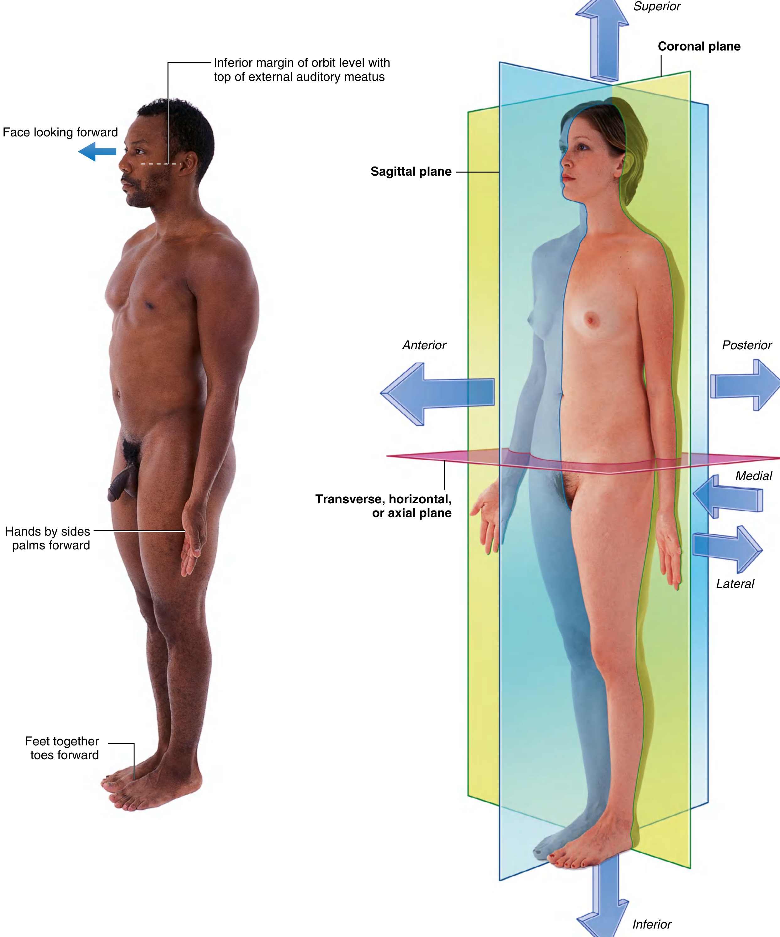
Fig. 1.1 - The Anatomical Position with body planes and directional terms. (Gray's Anatomy for Students)
2.2 Anatomical Planes
Three major planes divide the body:
| Plane | Orientation | Divides Body Into |
|---|---|---|
| Coronal (Frontal) | Vertical | Anterior (front) and posterior (back) parts |
| Sagittal | Vertical, at 90° to coronal | Right and left parts |
| Median Sagittal | Passes through exact midline | Equal right and left halves |
| Transverse (Horizontal / Axial) | Horizontal | Superior (upper) and inferior (lower) parts |
Memory tip: "Can She Trim?" = Coronal, Sagittal, Transverse
2.3 Terms of Direction and Location
Major Paired Terms
| Term | Meaning | Example |
|---|---|---|
| Anterior (Ventral) | Toward the front | Sternum is anterior to the heart |
| Posterior (Dorsal) | Toward the back | Vertebral column is posterior to the sternum |
| Superior (Cranial) | Toward the head/above | Head is superior to the shoulders |
| Inferior (Caudal) | Toward the feet/below | Knee is inferior to the hip |
| Medial | Toward the midline | Nose is medial to the eyes |
| Lateral | Away from the midline | Thumb is lateral to the little finger |
| Proximal | Closer to the origin/attachment | Shoulder is proximal to the elbow |
| Distal | Farther from origin/attachment | Hand is distal to the elbow |
| Superficial | Closer to body surface | Skin is superficial to muscle |
| Deep | Farther from body surface | Bone is deep to muscle |
Additional Terms
- Rostral - used in neuroscience; toward the nose/front of the brain
- Ipsilateral - on the same side
- Contralateral - on the opposite side
- Bilateral - on both sides
Terms Specific to the Hand and Foot
| Term | Used For | Meaning |
|---|---|---|
| Palmar (Volar) | Hand | Front surface of the hand |
| Dorsal | Hand / Foot | Back of the hand; top of the foot |
| Plantar | Foot | Sole of the foot |
2.4 Terms of Movement
| Movement | Definition |
|---|---|
| Flexion | Decreasing the angle between body parts |
| Extension | Increasing the angle between body parts |
| Abduction | Moving away from the midline |
| Adduction | Moving toward the midline |
| Medial (Internal) Rotation | Rotating toward the midline |
| Lateral (External) Rotation | Rotating away from the midline |
| Circumduction | Circular movement combining flexion, extension, abduction, adduction |
| Pronation | Rotating the forearm so the palm faces posteriorly/downward |
| Supination | Rotating the forearm so the palm faces anteriorly/upward |
| Inversion | Sole of foot turns medially (inward) |
| Eversion | Sole of foot turns laterally (outward) |
| Dorsiflexion | Foot pulled superiorly (toward shin) |
| Plantarflexion | Foot pushed inferiorly (pointing toes down) |
| Elevation | Moving a structure superiorly (e.g., shrugging the shoulder) |
| Depression | Moving a structure inferiorly |
| Protraction | Moving anteriorly (e.g., thrusting chin forward) |
| Retraction | Moving posteriorly (e.g., pulling chin back) |
CHAPTER 3: Osteology (Bones)
3.1 Functions of Bone
- Support - structural framework of the body
- Protection - skull protects brain; rib cage protects heart and lungs
- Movement - acts as levers for muscles
- Storage - calcium and phosphate reservoir
- Haematopoiesis - red blood cell production in red bone marrow
3.2 Classification of Bones by Shape
| Type | Shape | Example |
|---|---|---|
| Long bones | Longer than wide; shaft + two ends | Femur, humerus, radius, tibia |
| Short bones | Roughly cube-shaped | Carpal bones, tarsal bones |
| Flat bones | Thin, flattened; protect and provide large surface for muscle attachment | Skull, sternum, scapula |
| Irregular bones | Complex shapes not fitting other categories | Vertebrae, facial bones |
| Sesamoid bones | Develop within tendons | Patella (largest sesamoid) |
| Pneumatized bones | Contain air-filled sinuses | Maxilla, frontal, ethmoid bones |
3.3 Structure of a Long Bone
| Part | Description |
|---|---|
| Diaphysis | The shaft of the bone |
| Epiphysis | The expanded ends (proximal and distal) |
| Metaphysis | Region between diaphysis and epiphysis |
| Epiphyseal (Growth) Plate | Cartilaginous zone of active bone growth in children; becomes epiphyseal line in adults |
| Articular Cartilage | Hyaline cartilage covering the articular surfaces |
| Periosteum | Fibrous sheath covering the outer bone (except at articular surfaces); contains osteogenic cells; richly supplied with pain fibers |
| Endosteum | Lines the internal medullary cavity |
| Medullary (Marrow) Cavity | Hollow center of the shaft containing bone marrow |
3.4 Bone Marrow
| Type | Location | Contents |
|---|---|---|
| Red marrow (Myeloid tissue) | Flat bones, ends of long bones, vertebrae | Produces RBCs, WBCs, platelets (haematopoiesis) |
| Yellow marrow | Shaft of long bones in adults | Dominated by fat; few white cells |
- At birth, most marrow is red
- With age, red marrow converts to yellow marrow in the medullary cavities of long bones
- Red marrow persists in adults in: vertebrae, ribs, sternum, skull, ilium, proximal femur/humerus
3.5 Bone Blood Supply and Innervation
- Nutrient artery - enters through the nutrient foramen to supply the medullary cavity
- Periosteal vessels - supply the outer cortex and periosteum
- Periosteum - rich sensory nerve supply (explains why periosteal injury/fracture is very painful)
- Bone itself has very few sensory nerves
3.6 Ossification (Bone Formation)
| Type | Process |
|---|---|
| Intramembranous ossification | Mesenchymal cells directly form bone (no cartilage template). Examples: flat bones of skull, clavicle |
| Endochondral ossification | Cartilaginous template is replaced by bone. Most bones of the body form this way |
- Primary ossification centre - forms in the diaphysis
- Secondary ossification centres - form in the epiphyses after birth
- Skeletal maturity occurs between ages 20-25 years
- Bone age can be assessed by X-ray of the non-dominant (left) hand (comparing carpal bone ossification to standard charts)
3.7 Total Bone Count
- Adult human skeleton: 206 bones
- Divided into:
- Axial skeleton (80 bones): skull, vertebral column, ribs, sternum
- Appendicular skeleton (126 bones): limb bones + pectoral and pelvic girdles
CHAPTER 4: Arthrology (Joints)
4.1 Definition
A joint (articulation) is where two or more bones meet.
4.2 Classification of Joints
A. By Structure
| Type | Tissue Between Bones | Movement |
|---|---|---|
| Fibrous joints | Fibrous connective tissue | Little to none |
| Cartilaginous joints | Cartilage | Limited |
| Synovial joints | Synovial fluid in a joint cavity | Freely movable |
B. By Function (Amount of Movement)
| Term | Movement | Example |
|---|---|---|
| Synarthrosis | Immovable | Skull sutures |
| Amphiarthrosis | Slightly movable | Pubic symphysis, intervertebral discs |
| Diarthrosis | Freely movable | Knee, hip, shoulder |
4.3 Fibrous Joints (Synarthroses)
| Subtype | Description | Example |
|---|---|---|
| Sutures | Bones connected by thin fibrous tissue; interlocking edges | Skull bones |
| Syndesmoses | Bones connected by an interosseous membrane or ligament | Inferior tibiofibular joint |
| Gomphoses | Peg-and-socket joint | Tooth in jaw (dentoalveolar joint) |
4.4 Cartilaginous Joints
| Subtype | Tissue | Movement | Example |
|---|---|---|---|
| Primary (Synchondrosis) | Hyaline cartilage | Immovable; temporary (growth plates) | Epiphyseal plate, 1st sternocostal joint |
| Secondary (Symphysis) | Fibrocartilage | Slightly movable | Pubic symphysis, intervertebral discs |
4.5 Synovial Joints (Diarthroses) - Most Important
Components of a Synovial Joint:
| Component | Structure | Function |
|---|---|---|
| Articular cartilage | Hyaline cartilage | Reduces friction, absorbs shock |
| Joint cavity | Space containing synovial fluid | Allows movement |
| Synovial membrane | Inner lining of joint capsule | Secretes synovial fluid |
| Joint capsule | Fibrous outer layer + synovial inner layer | Encloses and stabilizes joint |
| Synovial fluid | Viscous fluid (filtrate of plasma + hyaluronic acid) | Lubrication, nutrition of cartilage |
| Ligaments | Fibrous bands (intrinsic or extrinsic) | Stabilize joint |
| Articular discs (menisci) | Fibrocartilage (in some joints) | Improve congruency, shock absorption |
| Bursae | Fluid-filled sacs near joint | Reduce friction between structures |
Types of Synovial Joints by Shape:
| Type | Movement | Example |
|---|---|---|
| Plane (gliding) | Gliding movements | Acromioclavicular, intercarpal joints |
| Hinge | Flexion/extension only (uniaxial) | Elbow, knee, interphalangeal joints |
| Pivot | Rotation only (uniaxial) | Atlantoaxial (C1-C2), proximal radioulnar |
| Condyloid (Ellipsoid) | Biaxial: flexion/extension + abduction/adduction | Radiocarpal (wrist), MCP joints |
| Saddle | Biaxial + rotation; concave-convex | 1st carpometacarpal (thumb) |
| Ball & Socket | Multiaxial (all movements) | Hip, shoulder (glenohumeral) |
Memory tip (most to least mobile): Ball & Socket > Saddle > Condyloid > Hinge/Pivot > Plane
CHAPTER 5: Myology (Muscles)
5.1 Types of Muscle Tissue
| Type | Location | Control | Striations |
|---|---|---|---|
| Skeletal (striated) | Attached to skeleton | Voluntary | Yes |
| Cardiac | Heart wall | Involuntary | Yes (intercalated discs) |
| Smooth | Walls of viscera, vessels | Involuntary | No |
5.2 Structure of Skeletal Muscle
- Epimysium - fibrous sheath around entire muscle
- Perimysium - surrounds bundles of muscle fibers (fascicles)
- Endomysium - surrounds individual muscle fibers
- Fascicle - bundle of muscle fibers surrounded by perimysium
- Muscle fiber (myocyte) - individual muscle cell; contains myofibrils
- Myofibril - contains the contractile proteins: actin (thin) and myosin (thick)
5.3 Muscle Attachments
| Term | Definition |
|---|---|
| Origin | The more fixed / proximal attachment of a muscle |
| Insertion | The more mobile / distal attachment of a muscle |
| Tendon | Cord of fibrous connective tissue attaching muscle to bone |
| Aponeurosis | Sheet-like fibrous expansion; broad tendon |
5.4 Classification of Muscles by Shape/Fiber Arrangement
| Type | Description | Example |
|---|---|---|
| Parallel (strap) | Fibers parallel to long axis | Sartorius |
| Fusiform | Spindle-shaped; tapered at ends | Biceps brachii |
| Pennate (unipennate) | Fibers attach obliquely on one side of tendon | Extensor digitorum longus |
| Bipennate | Fibers attach on both sides of a central tendon | Rectus femoris |
| Multipennate | Multiple tendinous insertions | Deltoid |
| Circular (sphincter) | Concentric fibers around an opening | Orbicularis oculi, pyloric sphincter |
Pennate muscles have more fibers per cross-section = greater strength but less range of motion
5.5 Roles of Muscles in Movement
| Role | Definition | Example |
|---|---|---|
| Prime mover (agonist) | Primary muscle producing a movement | Biceps brachii in elbow flexion |
| Antagonist | Opposes the prime mover | Triceps brachii during elbow flexion |
| Synergist | Assists prime mover | Brachialis during elbow flexion |
| Fixator (stabilizer) | Fixes the origin to allow insertion to move | Rotator cuff muscles stabilizing shoulder |
5.6 Nerve Supply to Muscles
- Each skeletal muscle is supplied by at least one nerve
- The nerve contains both motor fibers (to contract the muscle) and sensory fibers (from muscle spindles and Golgi tendon organs)
- Motor unit = one motor neuron + all the muscle fibers it innervates
- Neuromuscular junction = synapse between motor nerve terminal and muscle fiber
CHAPTER 6: Angiology (Blood Vessels)
6.1 Types of Blood Vessels
| Vessel | Function | Wall Structure |
|---|---|---|
| Arteries | Carry blood AWAY from the heart | Thick, muscular walls; withstand high pressure |
| Arterioles | Small arteries; regulate blood flow to capillaries | Smooth muscle-rich |
| Capillaries | Site of nutrient/gas exchange | Single endothelial cell layer |
| Venules | Drain capillaries into veins | Thin walls |
| Veins | Return blood TO the heart | Thin walls; contain valves (prevent backflow) |
6.2 Structure of Blood Vessel Walls
All vessels except capillaries have three layers (tunicae):
| Layer | Composition | Function |
|---|---|---|
| Tunica intima (interna) | Single layer of endothelium + basement membrane | Smooth lining; reduces friction |
| Tunica media | Smooth muscle + elastic fibers | Regulates vessel diameter (vasoconstriction/dilation) |
| Tunica adventitia (externa) | Fibrous connective tissue | Anchors vessel; contains nerves and vasa vasorum |
6.3 Arteries
- Elastic (conducting) arteries - large arteries (aorta, pulmonary); expand during systole, recoil during diastole (Windkessel effect)
- Muscular (distributing) arteries - medium arteries; rich in smooth muscle; distribute blood to organs
- Arterioles - major site of resistance; control blood pressure and flow distribution
- End arteries - arteries with no anastomoses; blockage leads to tissue death (e.g., central artery of retina)
- Anastomoses - connections between arterial branches; provide collateral circulation if one vessel is blocked
6.4 Veins
- Veins contain valves (especially in the limbs) to prevent backflow of blood against gravity
- Valves are bicuspid (two cusps)
- Varicose veins result from incompetent (leaky) valves causing venous pooling
- Deep veins are larger and accompany arteries; superficial veins lie in the subcutaneous tissue
6.5 Capillaries
- Diameter ~8-10 µm (just large enough for an RBC to pass)
- Site of gas exchange, nutrient delivery, and waste removal
- Types:
- Continuous capillaries - most common; tight junctions; CNS, muscle, lung
- Fenestrated capillaries - pores for rapid exchange; kidney, intestine, endocrine glands
- Sinusoidal capillaries - large gaps; liver, spleen, bone marrow
CHAPTER 7: Neurology (Nervous System)
7.1 Overview of the Nervous System
| Division | Components |
|---|---|
| Central Nervous System (CNS) | Brain + Spinal cord |
| Peripheral Nervous System (PNS) | All nerves outside CNS (cranial nerves, spinal nerves, ganglia) |
| Functional Division | Function |
|---|---|
| Somatic NS | Voluntary control of skeletal muscle; conscious sensation from skin/joints |
| Autonomic NS (ANS) | Involuntary control of smooth muscle, cardiac muscle, glands |
7.2 Neurons - The Basic Unit
| Part | Function |
|---|---|
| Cell body (soma) | Contains nucleus; metabolic center |
| Dendrites | Receive signals from other neurons |
| Axon | Transmits signals away from cell body |
| Axon terminals (synaptic boutons) | Release neurotransmitters at synapses |
| Myelin sheath | Insulates axon; speeds conduction; formed by Schwann cells (PNS) or oligodendrocytes (CNS) |
| Nodes of Ranvier | Gaps in myelin; site of saltatory conduction |
7.3 Nerve Fibers - Classification
| Type | Myelination | Diameter | Speed | Function |
|---|---|---|---|---|
| Aα (Ia/Ib) | Heavily myelinated | 13-20 µm | 80-120 m/s | Proprioception, motor to skeletal muscle |
| Aβ (II) | Myelinated | 6-12 µm | 35-75 m/s | Touch, pressure |
| Aδ (III) | Thinly myelinated | 1-5 µm | 5-30 m/s | Sharp (fast) pain, temperature |
| C (IV) | Unmyelinated | 0.2-1.5 µm | 0.5-2 m/s | Slow (burning) pain, temperature, postganglionic ANS |
7.4 Components of a Peripheral Nerve
| Component | Description |
|---|---|
| Endoneurium | Surrounds individual nerve fibers |
| Perineurium | Surrounds a fascicle (bundle of nerve fibers) |
| Epineurium | Surrounds the entire nerve trunk |
7.5 Spinal Nerves
-
31 pairs of spinal nerves:
- Cervical: 8 pairs (C1-C8)
- Thoracic: 12 pairs (T1-T12)
- Lumbar: 5 pairs (L1-L5)
- Sacral: 5 pairs (S1-S5)
- Coccygeal: 1 pair (Co1)
-
Each spinal nerve has two roots:
- Dorsal (posterior) root - sensory (afferent); carries signals TO the spinal cord; has a dorsal root ganglion (DRG) containing cell bodies of sensory neurons
- Ventral (anterior) root - motor (efferent); carries signals FROM the spinal cord to muscles/glands
Bell-Magendie Law: Dorsal roots are sensory; ventral roots are motor
7.6 Nerve Plexuses
| Plexus | Spinal Levels | Area Supplied |
|---|---|---|
| Cervical | C1-C4 | Neck, diaphragm (phrenic nerve C3,4,5) |
| Brachial | C5-T1 | Entire upper limb |
| Lumbar | L1-L4 | Anterior thigh, medial leg |
| Sacral | L4-S3 | Posterior thigh, leg, foot; perineum |
| Lumbosacral | L1-S3 | Lower limb combined |
7.7 Autonomic Nervous System (ANS)
| Feature | Sympathetic | Parasympathetic |
|---|---|---|
| Origin | Thoracolumbar (T1-L2) | Craniosacral (CN III, VII, IX, X; S2-S4) |
| Ganglia location | Close to spinal cord (paravertebral) | Close to or within target organ |
| Pre-ganglionic fiber | Short | Long |
| Post-ganglionic fiber | Long | Short |
| Neurotransmitter (pre) | Acetylcholine | Acetylcholine |
| Neurotransmitter (post) | Noradrenaline (NE) | Acetylcholine |
| General effect | "Fight or Flight" | "Rest and Digest" |
CHAPTER 8: The Lymphatic System
8.1 Components
- Lymphatic capillaries - blind-ended, very permeable vessels in tissues
- Lymphatic vessels - collect lymph from capillaries; have valves (similar to veins)
- Lymph nodes - small (0.1-2.5 cm), bean-shaped, encapsulated structures along lymphatic vessels
- Lymphatic trunks and ducts - large collecting vessels that drain into the venous system
- Lymphoid organs - thymus, spleen, tonsils, Peyer's patches
8.2 Functions of the Lymphatic System
- Returns fluid - recollects 2-4 L of fluid/day that leaks from capillaries back to the bloodstream
- Immune defense - lymph nodes filter pathogens; lymphocytes mount immune responses
- Lipid absorption - lacteals in small intestine absorb dietary fats (chylomicrons) as chyle
8.3 Key Lymph Node Groups
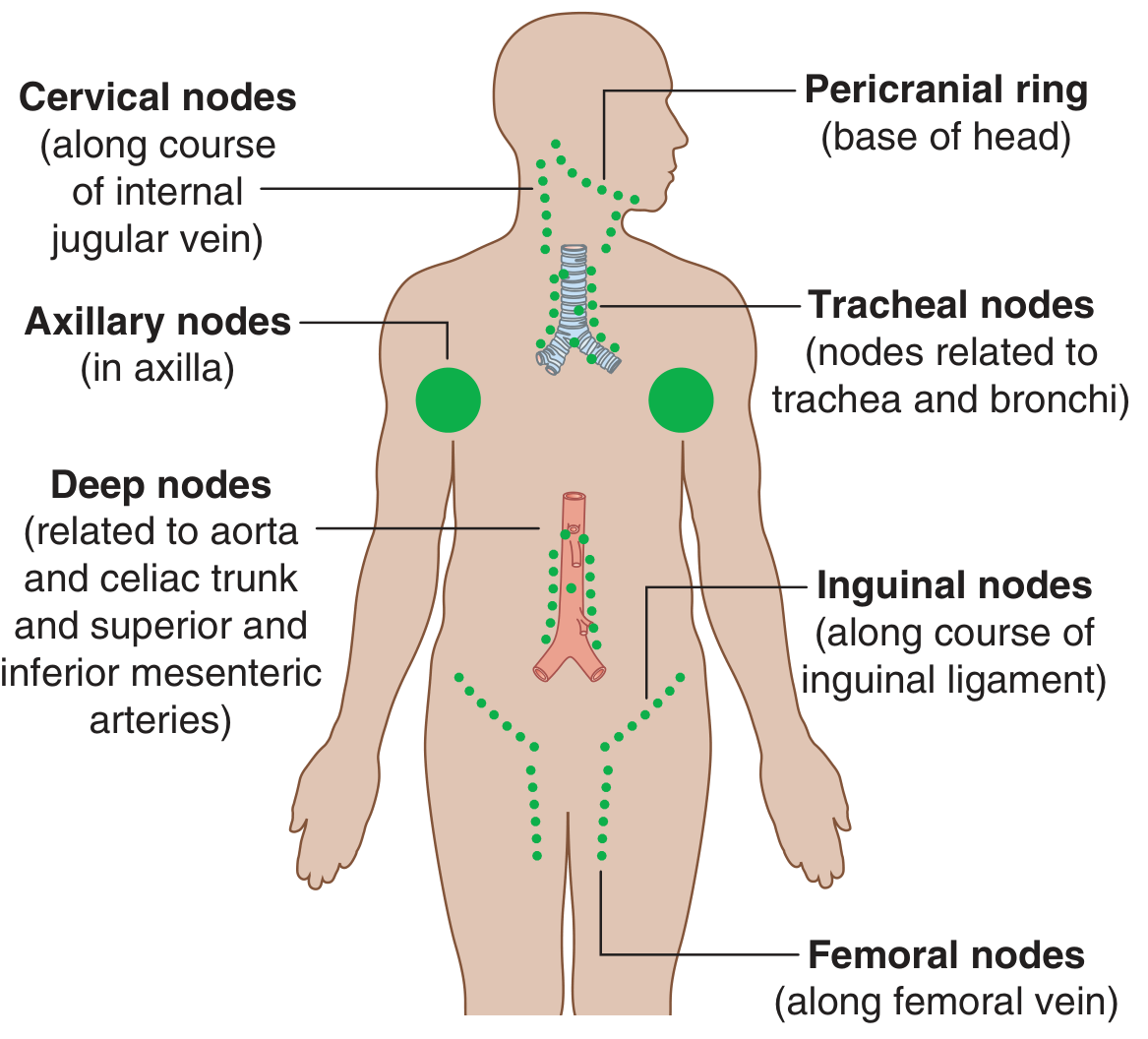
Fig. 1.31 - Major lymph node groups of the body (Gray's Anatomy for Students)
| Group | Location | Drains |
|---|---|---|
| Cervical nodes | Along internal jugular vein | Head and neck |
| Axillary nodes | In the axilla | Upper limb, breast, chest wall |
| Tracheal/Mediastinal | Around trachea and bronchi | Lungs, mediastinum |
| Para-aortic (deep) | Along aorta and its branches | Abdominal viscera |
| Inguinal nodes | Along inguinal ligament | Lower limb, perineum, lower abdomen |
| Femoral nodes | Along femoral vein | Lower limb |
8.4 Lymphatic Drainage - Major Ducts
| Duct | Drains |
|---|---|
| Thoracic duct (largest) | Everything EXCEPT right upper quadrant; drains into left brachiocephalic vein (junction of left internal jugular + left subclavian) |
| Right lymphatic duct | Right side of head/neck, right upper limb, right side of thorax; drains into right brachiocephalic vein |
Clinical point: Cancer cells that enter lymphatics often lodge in lymph nodes (first as sentinel nodes, then spread regionally). Enlarged (hard, non-tender) lymph nodes may indicate malignancy; tender, soft nodes suggest infection.
CHAPTER 9: Skin (Integumentary System)
9.1 Layers of the Skin
| Layer | Composition | Key Features |
|---|---|---|
| Epidermis | Stratified squamous epithelium (keratinized) | Avascular; 5 layers in thick skin; 4 in thin skin |
| Dermis | Dense irregular connective tissue | Contains hair follicles, sweat glands, blood vessels, nerves |
| Hypodermis (Subcutaneous tissue) | Loose connective tissue + fat | Insulation; shock absorption; not true skin |
9.2 Layers of the Epidermis (Deep to Superficial)
| Layer | Mnemonic |
|---|---|
| Stratum Basale (germinativum) | Boy |
| Stratum Spinosum | Sells |
| Stratum Granulosum | Green |
| Stratum Lucidum (thick skin only) | Leaves |
| Stratum Corneum | Clear |
Memory trick: "Baby Skin Grows Layers Continuously"
9.3 Dermatomes
A dermatome is an area of skin supplied by a single spinal nerve (its dorsal root).
Key dermatome landmarks:
| Level | Area |
|---|---|
| C3-C4 | Neck and clavicle region |
| C6 | Thumb |
| C7 | Middle finger |
| C8 | Little finger |
| T4 | Nipple level |
| T10 | Umbilicus |
| L1 | Inguinal region |
| L4 | Medial leg/great toe |
| S1 | Lateral foot/little toe |
| S3-S4 | Perineum (saddle area) |
Clinical significance: Dermatome mapping is used to localize the level of a spinal cord lesion or nerve root compression (e.g., disc herniation causing dermatomal pain).
CHAPTER 10: Body Cavities and Membranes
10.1 Major Body Cavities
| Cavity | Contents | Subdivisions |
|---|---|---|
| Cranial cavity | Brain | - |
| Vertebral (spinal) canal | Spinal cord | - |
| Thoracic cavity | Heart, lungs, great vessels | Right pleural, left pleural, mediastinum |
| Abdominal cavity | Digestive organs, liver, spleen, kidneys | - |
| Pelvic cavity | Bladder, rectum, reproductive organs | - |
The abdominal and pelvic cavities are often considered together as the abdominopelvic cavity
10.2 Serous Membranes (Double-Layered Membranes)
All serous membranes have:
- Visceral layer - closely covers the organ
- Parietal layer - lines the body wall
- Serous cavity - potential space between layers; contains a thin film of serous fluid for lubrication
| Membrane | Cavity | Organ |
|---|---|---|
| Pleura | Pleural cavity | Lungs |
| Pericardium | Pericardial cavity | Heart |
| Peritoneum | Peritoneal cavity | Abdominal organs |
CHAPTER 11: Imaging in Anatomy
11.1 Radiological Imaging Modalities
| Modality | Principle | Best For | Radiation |
|---|---|---|---|
| Plain X-ray (Radiography) | X-rays pass through tissues (dense structures = white/opaque) | Bones, chest, fractures | Yes (low dose) |
| Contrast Radiography | Radioopaque contrast used to outline hollow structures | GI tract, urinary system, blood vessels | Yes |
| Ultrasound (USG) | High-frequency sound waves; echoes create image | Soft tissues, abdominal organs, vessels, fetus | No (safe in pregnancy) |
| Computed Tomography (CT) | X-rays from multiple angles; computer reconstructs cross-sectional images | Detailed bone and organ anatomy, trauma, bleeding | Yes (higher dose) |
| Magnetic Resonance Imaging (MRI) | Magnetic fields + radio waves; images hydrogen atoms | Soft tissues, brain, spinal cord, joints | No |
| Nuclear Medicine (PET/SPECT) | Radioactive tracers; image metabolic activity | Cancer staging, cardiac function, brain activity | Yes (internal) |
| Doppler Ultrasound | Detects direction and speed of blood flow | Blood vessels, DVT, cardiac function | No |
11.2 X-Ray Densities (Radiographic Appearance)
| Structure | X-ray Appearance | Reason |
|---|---|---|
| Air / Gas | Black (radiolucent) | Low density; X-rays pass through easily |
| Fat | Dark gray | Low density |
| Soft tissues / Water | Gray | Intermediate density |
| Bone | White (radio-opaque) | Calcium blocks X-rays |
| Metal | Bright white | Highest density |
11.3 Reading a Chest X-ray (CXR) - Systematic Approach
P-A-R-T-S mnemonic:
- Patient details + quality (rotation, penetration, inspiration)
- Airways - trachea midline? Carina angle?
- Respiratory - lung fields, costophrenic angles, fissures
- Tissues - cardiac size (cardiothoracic ratio <0.5), mediastinum
- Skeleton - ribs, clavicles, vertebrae, shoulder joints
CHAPTER 12: Clinical Anatomy Pearls
| Concept | Clinical Relevance |
|---|---|
| Periosteum is pain-sensitive | Fractures are painful; bone tumors cause deep bone pain |
| Endochondral ossification | Growth plate injuries in children can affect limb length |
| Valves in veins | Varicose veins = incompetent valves; DVT diagnosis |
| End arteries | Central retinal artery occlusion = sudden blindness with no collateral recovery |
| Dermatomes | Localize disc herniation level (e.g., L4/L5 disc = L5 dermatome pain) |
| Lymph node enlargement | Hard, non-tender = malignancy; soft, tender = infection |
| Thoracic duct | Injured in left-sided chest surgery; leaks = chylothorax |
| Anatomical position | All anatomical descriptions refer to this position regardless of patient's actual position |
| Synovial fluid | Aspiration (arthrocentesis) is diagnostic for gout, infection, hemarthrosis |
| Bone marrow transplant | Used in leukemia; hematopoietic stem cells restore marrow function |
QUICK REFERENCE: Key Numbers to Memorize
| Fact | Number |
|---|---|
| Total bones in adult | 206 |
| Axial skeleton bones | 80 |
| Appendicular skeleton bones | 126 |
| Pairs of ribs | 12 |
| Cervical vertebrae | 7 |
| Thoracic vertebrae | 12 |
| Lumbar vertebrae | 5 |
| Sacral segments | 5 |
| Coccygeal segments | 4 (fused) |
| Total pairs of spinal nerves | 31 |
| Pairs of cranial nerves | 12 |
| Carpal bones | 8 |
| Tarsal bones | 7 |
| Skeletal maturity age | 20-25 years |
Sources:
- Gray's Anatomy for Students, 4th Ed. - Richard Drake et al.
- THIEME Atlas of Anatomy: General Anatomy and Musculoskeletal System
---These are your complete MBBS General Anatomy notes, covering all the core topics from basics to advanced clinical correlations. Here's a summary of what's covered:
What's Included
| Chapter | Topic |
|---|---|
| 1 | Introduction to Anatomy - definition, types, approaches |
| 2 | Anatomical Terminology - position, planes, directions, movements |
| 3 | Osteology - bone types, structure, ossification, marrow, blood supply |
| 4 | Arthrology - joint classification (fibrous, cartilaginous, synovial), synovial joint types |
| 5 | Myology - muscle types, structure, attachments, roles |
| 6 | Angiology - blood vessels, arterial/venous structure, capillary types |
| 7 | Neurology - CNS/PNS, neurons, nerve fibers, spinal nerves, dermatomes, ANS |
| 8 | Lymphatic System - components, drainage, major node groups with diagram |
| 9 | Skin - layers, epidermis layers, dermatomes |
| 10 | Body Cavities and Serous Membranes |
| 11 | Radiology - all imaging modalities, X-ray densities, CXR reading |
| 12 | Clinical Anatomy Pearls + Key Numbers to Memorize |
Tips for using these notes:
- Use the tables for quick revision before exams
- The memory mnemonics (e.g., bone types, epidermal layers) are exam-ready
- Clinical pearls in Chapter 12 directly connect anatomy to OSCEs and vivas
- Let me know which region (Upper Limb, Lower Limb, Head & Neck, Thorax, Abdomen) you want next for detailed regional anatomy notes!
This is a shared conversation. Sign in to Orris to start your own chat.