
Hashimoto thyroiditis
I'll start by reading the medical library skill, then search for comprehensive information on Hashimoto thyroiditis.
Reading File
Now let me search the medical library and PubMed in parallel for comprehensive coverage.
Finding Sources
Searching PubMed
"Hashimoto thyroiditis"[MeSH Terms]
Excellent results. Now let me read the most comprehensive sources in parallel - Robbins Pathology (Basic), Current Surgical Therapy, and Lee's Otolaryngology.
Reading File
Reading File
Reading File
Now let me get the treatment section from Current Surgical Therapy and also fetch the key pathogenesis diagram and the histology image.
Reading File
Loading Image
Finding Sources
Now let me fetch the histology image from Robbins and the ultrasound image, and also get the treatment section from Current Surgical Therapy.
Finding Sources
Loading Image
I now have all the information needed for a comprehensive answer. Let me also get the Robbins histology image.
Finding Sources
Reading File
I have enough comprehensive data to write a full, well-structured answer. Let me also fetch the Robbins histology image of Hashimoto for completeness.
Finding Sources
I have all the necessary information. Here is the comprehensive answer:
Hashimoto Thyroiditis
Hashimoto thyroiditis (chronic autoimmune thyroiditis / chronic lymphocytic thyroiditis) is the most common cause of hypothyroidism in iodine-sufficient regions, and the most prevalent autoimmune disease in the United States.
Epidemiology
- Most common cause of diffuse goiter and hypothyroidism in iodine-replete countries (responsible for ~85% of hypothyroid cases in those areas)
- Prevalence of hypothyroidism in iodine-replete populations: 1-2%
- Peak age: 45-65 years (also reported in 3rd-5th decades)
- Female predominance: female-to-male ratio of 10:1 to 20:1
- Clusters in families; co-occurs with Graves disease and other autoimmune diseases
Pathogenesis
The disease results from a breakdown of immune self-tolerance to thyroid antigens, with progressive autoimmune destruction of thyrocytes. Key mechanisms:
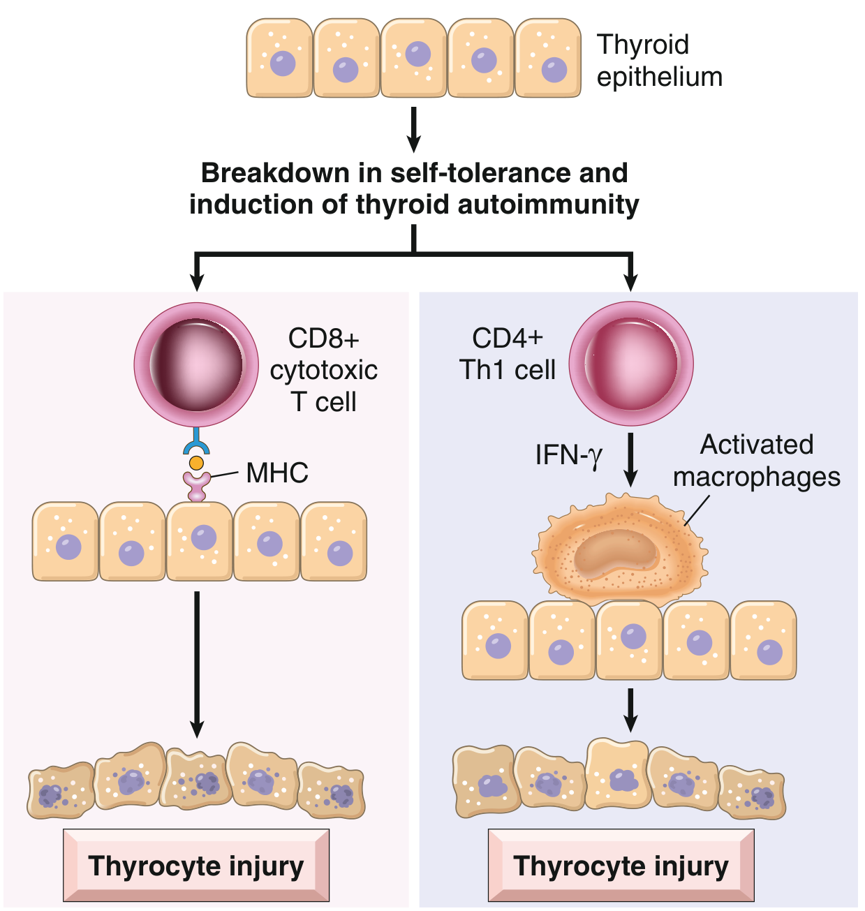
Fig. 18.4 - Pathogenesis of Hashimoto thyroiditis (Robbins & Kumar Basic Pathology)
- CD8+ cytotoxic T cells - kill thyroid epithelial cells directly via MHC-restricted mechanisms
- CD4+ Th1 cytokine-mediated damage - IFN-γ production recruits and activates macrophages, which destroy follicles
- Antithyroid antibodies - anti-thyroid peroxidase (anti-TPO, also called antimicrosomal antibody) and anti-thyroglobulin antibodies are present in nearly all patients. These may contribute via antibody-dependent cell-mediated cytotoxicity (ADCC) or complement-dependent mechanisms, though it remains unclear if they are the cause or consequence of injury.
Genetic factors:
- ~40% concordance in monozygotic twins
- ~50% of asymptomatic siblings carry antithyroid antibodies
- Associated with polymorphisms in CTLA4 (an inhibitor of T-cell responses) and other immune-regulatory genes
Morphology / Histology
- Thyroid is usually diffusely and symmetrically enlarged
- Microscopy (classic features):
- Widespread mononuclear inflammatory infiltrate: lymphocytes, plasma cells, macrophages
- Well-developed germinal centers (lymphoid follicle formation)
- Thyroid follicles are atrophic
- Hürthle (oxyphil) cell metaplasia - follicular epithelial cells with abundant eosinophilic, granular cytoplasm due to numerous mitochondria (metaplastic response to injury)
- Interstitial fibrosis (variable, may be abundant)
- Fibrosing variant - forms a massive, firm goiter; can be difficult to distinguish from Riedel thyroiditis
Clinical Features
| Feature | Detail |
|---|---|
| Presentation | Painless, firm, symmetric goiter in a middle-aged woman |
| Thyroid function | Usually euthyroid at presentation; hypothyroidism develops in ~20% and progresses over time |
| Hashitoxicosis | Transient thyrotoxicosis early in disease from follicular destruction releasing stored hormone; elevated free T4/T3, suppressed TSH, low radioiodine uptake |
| Compressive symptoms | Dyspnea, dysphagia, cough, hoarseness, choking (if goiter is large) |
| Persistent symptoms | Fatigue, muscle/joint pain, poor sleep, dry mouth/eyes - may not resolve with hormone replacement (thought to be autoimmune-mediated) |
On ultrasound: Diffuse heterogeneity of the gland is the hallmark finding.
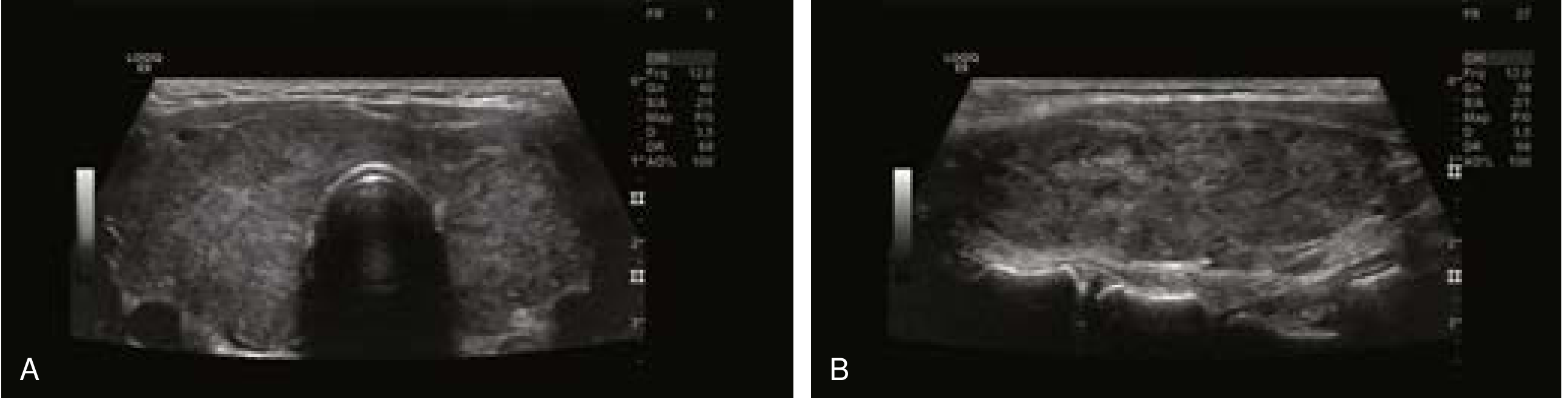
Fig. 3 - Diffuse heterogeneity characteristic of Hashimoto thyroiditis (Current Surgical Therapy, 14e)
Laboratory Findings
- Anti-TPO antibodies (antimicrosomal): elevated in 70-90% of patients - most sensitive marker
- Anti-thyroglobulin antibodies: also commonly elevated
- TSH: elevated in hypothyroid phase; suppressed in hashitoxicosis phase
- Free T4/T3: low in hypothyroid phase; elevated transiently in hashitoxicosis
- Inflammatory cytokines elevated
Thyroid Nodules and Cancer Risk
- Hürthle cell metaplasia leads to associated thyroid nodules (most are benign Hürthle cell nodules)
- FNAB findings: follicular epithelial cells, colloid, lymphocytes, Hürthle cells - classified as Bethesda II (benign)
- Predominance of Hürthle cells may be Bethesda III (AUS) or IV (Hürthle cell neoplasm)
- Can cause false-positive FNAB suspicious for papillary thyroid cancer (Bethesda V)
- Any discrete palpable nodule not part of diffuse goiter should be evaluated with FNA regardless of the Hashimoto diagnosis
- Increased risk of B-cell non-Hodgkin lymphoma (primary thyroid lymphoma) - a rapidly enlarging mass within a Hashimoto gland warrants urgent biopsy
- Possible increased predisposition to papillary thyroid carcinoma (controversial)
Treatment
1. Asymptomatic / Euthyroid
- Observation; no treatment required
2. Hypothyroidism
- Levothyroxine (T4) replacement - resolves hypothyroid symptoms and typically reduces goiter size
- Dose titrated to normalize TSH
3. Persistent Symptoms Despite Adequate Hormone Replacement
- Many symptoms (fatigue, myalgia, dry eyes) may be autoimmune-mediated and do not resolve with T4 alone
- Total thyroidectomy is an option in this setting: removes the antigenic target, reduces the systemic inflammatory response, and lowers anti-TPO titers and pro-inflammatory cytokines
- The Norwegian Trial on Surgery for Hashimoto Disease (prospective RCT) demonstrated improved health-related quality of life, reduced fatigue, and normalization of anti-TPO titers in surgically treated patients vs. controls
4. Compressive Goiter / Surgical Indications
- Surgery for large, symptomatic, or refractory goiter
- Thyroid lobectomy + isthmusectomy for unilateral disease; total thyroidectomy for bilateral or extensive disease
- Intraoperative nerve monitoring recommended (inflamed, firm gland increases risk of recurrent laryngeal nerve traction injury)
- Enlarged reactive central compartment lymph nodes are common; frozen section can exclude metastatic thyroid cancer
5. Radioiodine (I-131)
- Typically contributes little useful information to the workup (I-123 scanning also not usually helpful)
Associated Conditions and Variants
| Condition | Notes |
|---|---|
| Painless (silent) thyroiditis | Variant of Hashimoto; autoimmune follicular destruction with spontaneously resolving hyperthyroidism |
| Postpartum thyroiditis | Specific type of painless thyroiditis; occurs within 1 year of delivery or abortion; up to 10% of pregnancies; may recur with subsequent pregnancies |
| Fibrosing variant | Massive firm goiter; mimics Riedel thyroiditis |
| Graves disease | Can coexist (both share autoimmune thyroid pathogenesis) |
| Other autoimmune diseases | Type 1 DM, Addison disease, pernicious anemia, vitiligo, etc. |
Sources: Robbins & Kumar Basic Pathology, p. 731 | Current Surgical Therapy, 14e, pp. 885-886 | K.J. Lee's Essential Otolaryngology, p. 727
This is a shared conversation. Sign in to Orris to start your own chat.