Show me branches and blood supply of brachial artery around the elbow.
Reading File
Finding Sources
Finding Sources
Reading File
Reading File
Reading File
Reading File
Loading Image
Loading Image
Reading File
Reading File
Loading Image
Loading Image
Loading Image
I now have comprehensive information and excellent anatomical images. Here is a complete, well-illustrated answer.
Brachial Artery: Branches and Blood Supply Around the Elbow
Overview and Course
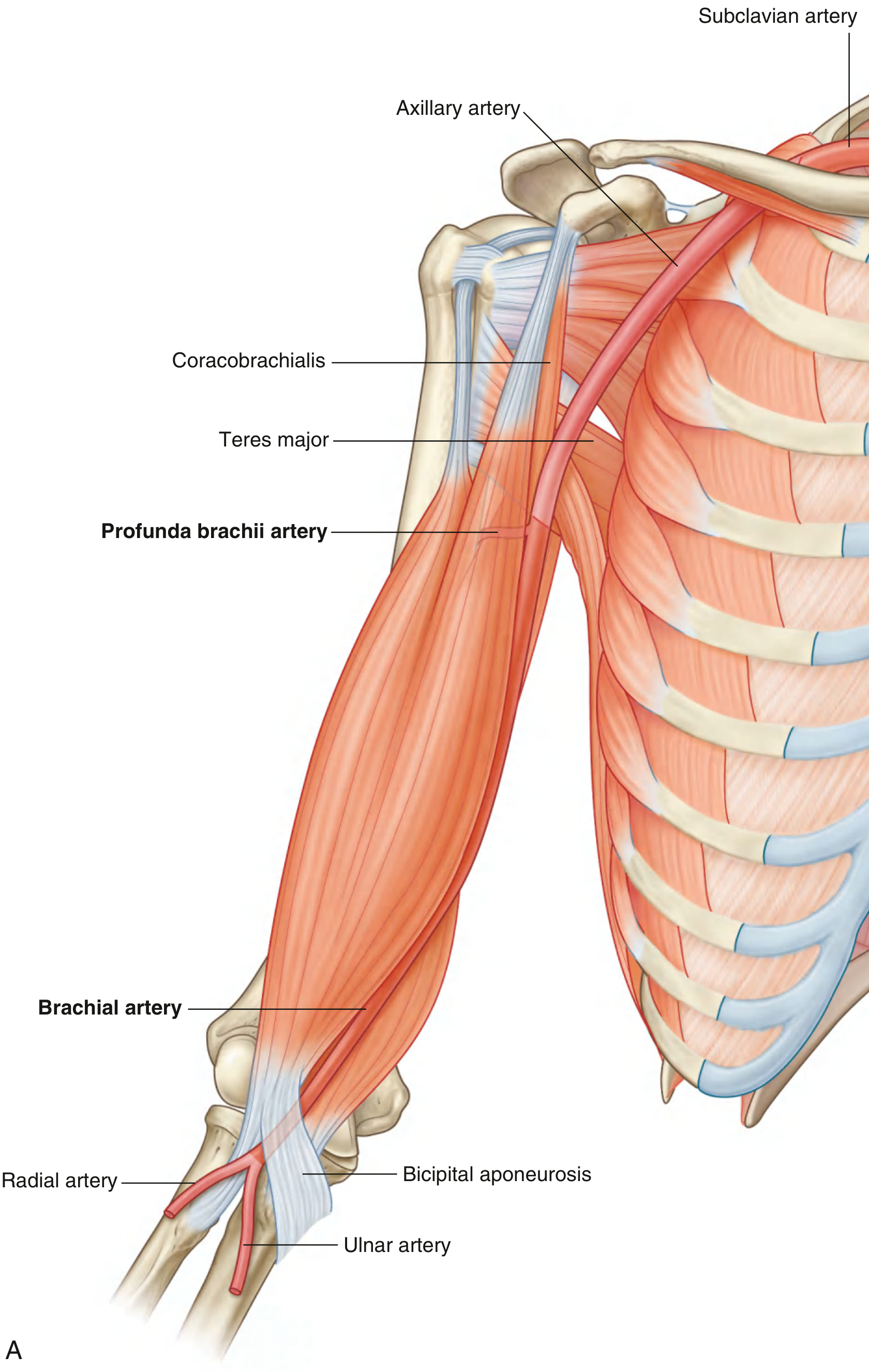
The brachial artery is the continuation of the axillary artery, starting at the lower border of the teres major muscle. It descends in the medial bicipital groove, crossing anteriorly to the elbow joint where it lies immediately medial to the biceps brachii tendon, roughly midway between the medial and lateral epicondyles. It terminates in the cubital fossa at the level of the neck of the radius by dividing into the radial and ulnar arteries.
Branches of the Brachial Artery
| Branch | Origin | Course and Supply |
|---|---|---|
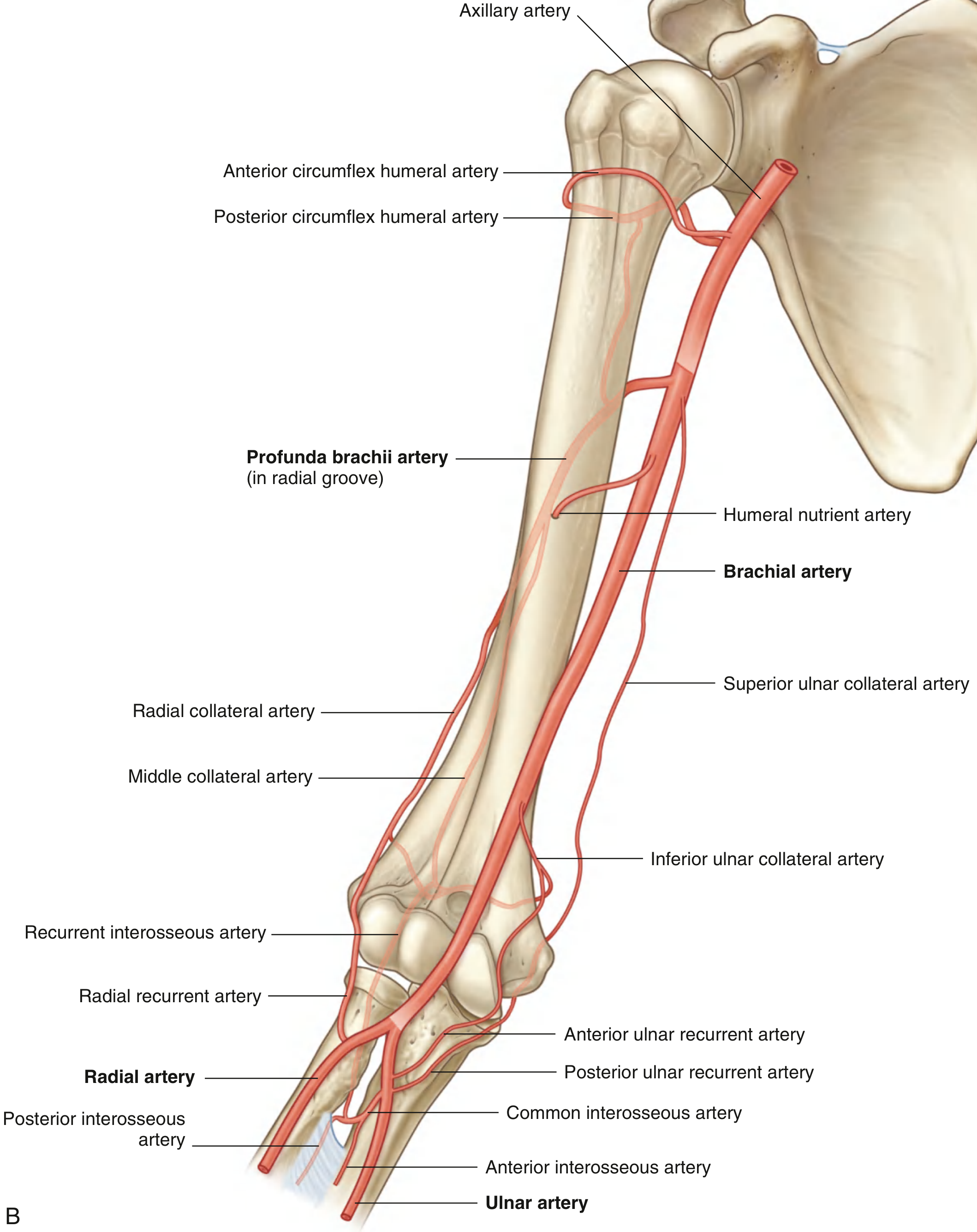
| Profunda brachii (deep brachial) artery | Largest branch; arises proximally | Enters posterior compartment with the radial nerve through the triangular interval; runs in the radial groove deep to the lateral head of triceps; supplies posterior arm muscles and anastomoses with the posterior circumflex humeral artery |
| Middle collateral artery | Terminal branch of profunda brachii | Descends to lateral/posterior elbow; anastomoses with the interosseous recurrent artery |
| Radial collateral artery | Terminal branch of profunda brachii | Accompanies radial nerve anteriorly to the lateral epicondyle; anastomoses with the radial recurrent artery |
| Superior ulnar collateral artery | Brachial artery, mid-arm | Follows ulnar nerve around medial epicondyle to posterior elbow; anastomoses with posterior ulnar recurrent artery |
| Inferior ulnar collateral artery | Brachial artery, distal arm | Passes anterior to medial epicondyle; anastomoses with anterior ulnar recurrent artery |
| Muscular branches | Along the course | Supply the biceps brachii, brachialis, and coracobrachialis |
| Humeral nutrient artery | Variable | Enters nutrient foramen on anteromedial surface of humeral shaft |
The Arterial Network (Anastomosis) Around the Elbow
This periarticular anastomotic network is formed by descending branches from above meeting ascending recurrent branches from below:

Contributions to the network:
From the brachial artery / profunda brachii (above):
- Radial collateral artery (lateral side)
- Middle collateral artery (posterior side)
- Superior ulnar collateral artery (medial/posterior)
- Inferior ulnar collateral artery (medial/anterior)
From the radial and ulnar arteries (below):
- Radial recurrent artery (from radial a.) - anastomoses with radial collateral
- Interosseous recurrent artery (from posterior interosseous a.) - anastomoses with middle collateral
- Anterior ulnar recurrent artery (from ulnar a.) - anastomoses with inferior ulnar collateral
- Posterior ulnar recurrent artery (from ulnar a.) - anastomoses with superior ulnar collateral
Cubital Fossa Relationships
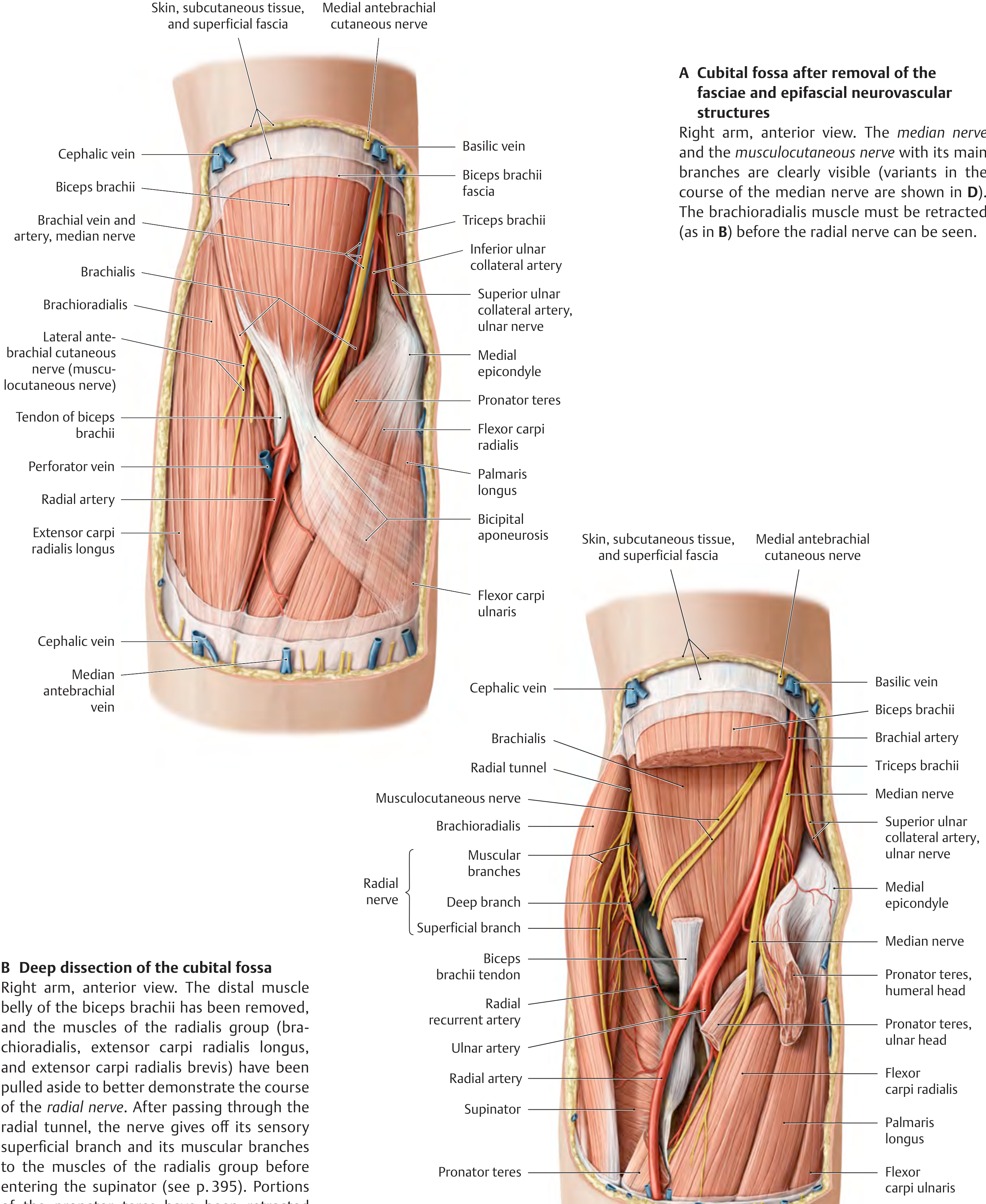
In the cubital fossa, from lateral to medial: biceps tendon → brachial artery → median nerve (mnemonic: TAN - Tendon, Artery, Nerve). The bicipital aponeurosis covers all these structures superficially.
Clinical Significance
-
Ligation safety: The brachial artery can be safely ligated distal to the origin of the profunda brachii because the periarticular anastomotic network provides adequate collateral circulation to the forearm. - THIEME Atlas of Anatomy, p. 394
-
Blood pressure measurement: The brachial artery is compressed against the humerus in the mid-arm when using a sphygmomanometer; the pulse is auscultated in the cubital fossa. - Gray's Anatomy for Students, p. 876
-
Variant anatomy: In ~20% of individuals, the brachial artery gives off a superficial brachial artery that courses superficial to the median nerve. This can present as a high-origin radial artery (10%), high-origin ulnar artery (3%), or early bifurcation (7%). - Fischer's Mastery of Surgery, p. 6991
-
Supracondylar process: A rare anomaly (0.7%) - a bony outgrowth above the medial epicondyle connected by Struthers' ligament can create a fibro-osseous canal that traps both the brachial artery and median nerve.
-
Elbow dislocation/fracture: Despite complete brachial artery disruption, the hand may remain well perfused due to the rich collateral network. - Campbell's Operative Orthopaedics, 15th Ed 2026
Sources: Gray's Anatomy for Students (9780323934237); THIEME Atlas of Anatomy - General Anatomy & Musculoskeletal System (9781626237186); Fischer's Mastery of Surgery 8th Ed (9781975176433); Campbell's Operative Orthopaedics 15th Ed (9780443117633)
Just draw me a table for management of pink pulseless supracondylar humerus fracture.
Finding Sources
Finding Sources
Reading File
Reading File
Finding Sources
Reading File
Reading File
I have enough from the textbooks. Here is the table:
Management of Pink Pulseless Hand in Supracondylar Humerus Fracture
Definition: The hand is pink and warm (adequate perfusion via collaterals) but the radial pulse is absent. This distinguishes it from the white/pale pulseless hand, which is a true vascular emergency with absent perfusion.
| Step | Action | Detail |
|---|---|---|
| 1. Initial assessment | Confirm hand is pink and warm | Check capillary refill, skin colour, temperature - perfusion present despite absent pulse |
| 2. First-line treatment | Urgent CRPP (Closed Reduction + Percutaneous Pinning) | Reduction often relieves arterial kinking or spasm; pulse returns in the majority of cases after reduction |
| 3. Post-reduction: pulse returns | Inpatient observation + splinting | Monitor hourly neurovascular checks; no further vascular intervention needed |
| 4. Post-reduction: pulse still absent but hand remains pink | Inpatient observation + splinting | Continue close monitoring; collateral flow is maintaining perfusion |
| 5. Hand becomes poorly perfused at any point | Vascular surgery consultation | Proceed to surgical exploration of brachial artery (arteriotomy / repair) |
| 6. Arteriography | Generally not warranted | Location of injury (brachial artery at fracture site) is almost always known |
Key Points
| Feature | Pink Pulseless | White/Pale Pulseless |
|---|---|---|
| Hand perfusion | Maintained (collaterals intact) | Absent |
| Urgency | Urgent (not emergent) | Emergent |
| First step | CRPP | Emergent CRPP |
| Vascular exploration | Only if hand becomes poorly perfused | If perfusion not restored after CRPP |
| Prognosis | Pulse returns in most after reduction | Higher risk of ischaemia / arterial repair needed |
Sources: Miller's Review of Orthopaedics 9th Ed, p. 950; Campbell's Operative Orthopaedics 15th Ed 2026
Create a quick-reference PDF on supracondylar fracture complications
Finding Sources
Reading File
Reading File
Finding Sources
Reading File
Reading File
Finding Sources
Finding Sources
Finding Sources
Reading File
Reading File
Reading File
Reading File
Reading File
Reading File
Reading File
Reading File
Running Command
Your PDF is ready:
Supracondylar Fracture Complications
PDF Document · PDF
What's inside (single-page A4):
| Section | Content |
|---|---|
| 1. Overview | Epidemiology, mechanism, Gartland classification (I–IV) |
| 2. Complications at a Glance | 14-row colour-coded table - vascular injury, compartment syndrome, Volkmann contracture, all 4 nerve injuries, cubitus varus/valgus, recurvatum, pin complications, stiffness, AVN, growth disturbance |
| 3. Vascular Injury Algorithm | Pink pulseless vs white pulseless - urgency, first step, post-reduction management |
| 4. Nerve Injury Summary | AIN, radial, median, ulnar - clinical test, prognosis (AIN/radial excellent → ulnar only 25%) |
| 5. Angular Deformity | Cubitus varus, valgus, recurvatum - cause, features, treatment |
| 6. Clinical Pearls | 8 high-yield points including the 3 As of compartment syndrome, pin configuration, arteriography rule, and cubitus varus ≠ growth arrest |
Sources: Campbell's Operative Orthopaedics 15th Ed 2026, Miller's Review of Orthopaedics 9th Ed, Bailey & Love's 28th Ed, Rosen's Emergency Medicine.
Detailed anatomy of posterolateral corner of knee joint.
Finding Sources
Finding Sources
Finding Sources
Finding Sources
Finding Sources
Finding Sources
Reading File
Reading File
Reading File
Reading File
Reading File
Reading File
Reading File
Loading Image
Loading Image
Loading Image
Loading Image
Loading Image
Reading File
Loading Image
Reading File
I now have thorough content and all the key images. Here is the detailed anatomy of the posterolateral corner:
Posterolateral Corner (PLC) of the Knee - Detailed Anatomy
The posterolateral corner (PLC) is a complex region bounded by the lateral femoral condyle, the fibular head, and the posterior aspect of the lateral tibial plateau. It is the primary stabiliser against external tibial rotation, and also resists varus stress and posterior tibial translation, especially in the mid-ranges of flexion.
Anatomical Overview
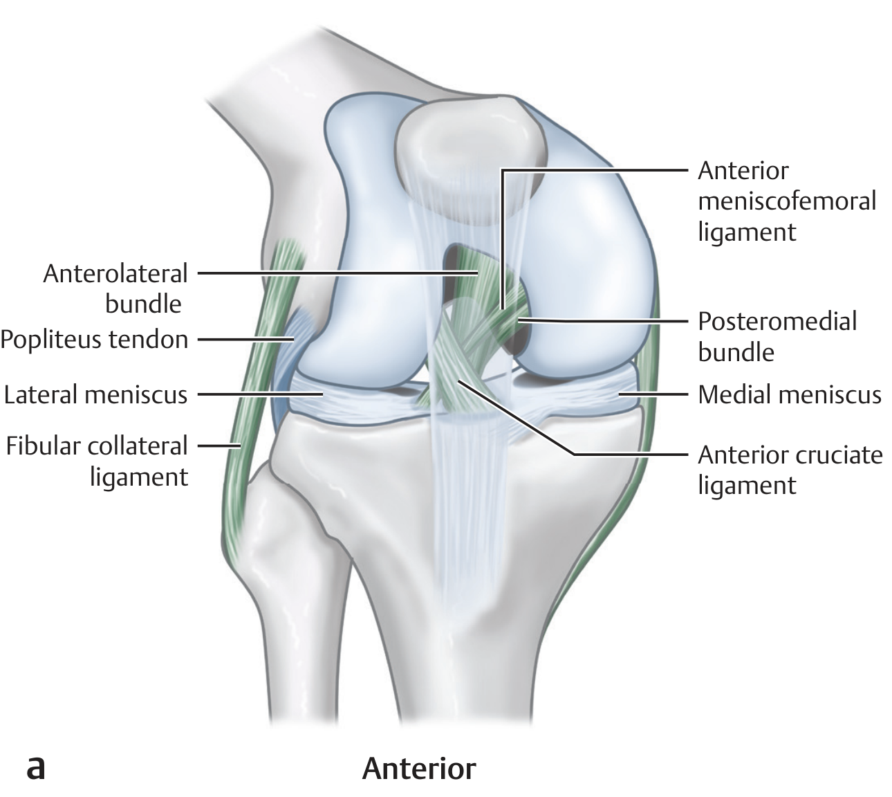
Anterior view - fibular collateral ligament, popliteus tendon, and their relationship to the lateral meniscus:
Posterior view - popliteus muscle, popliteofibular ligament, and fibular collateral ligament:
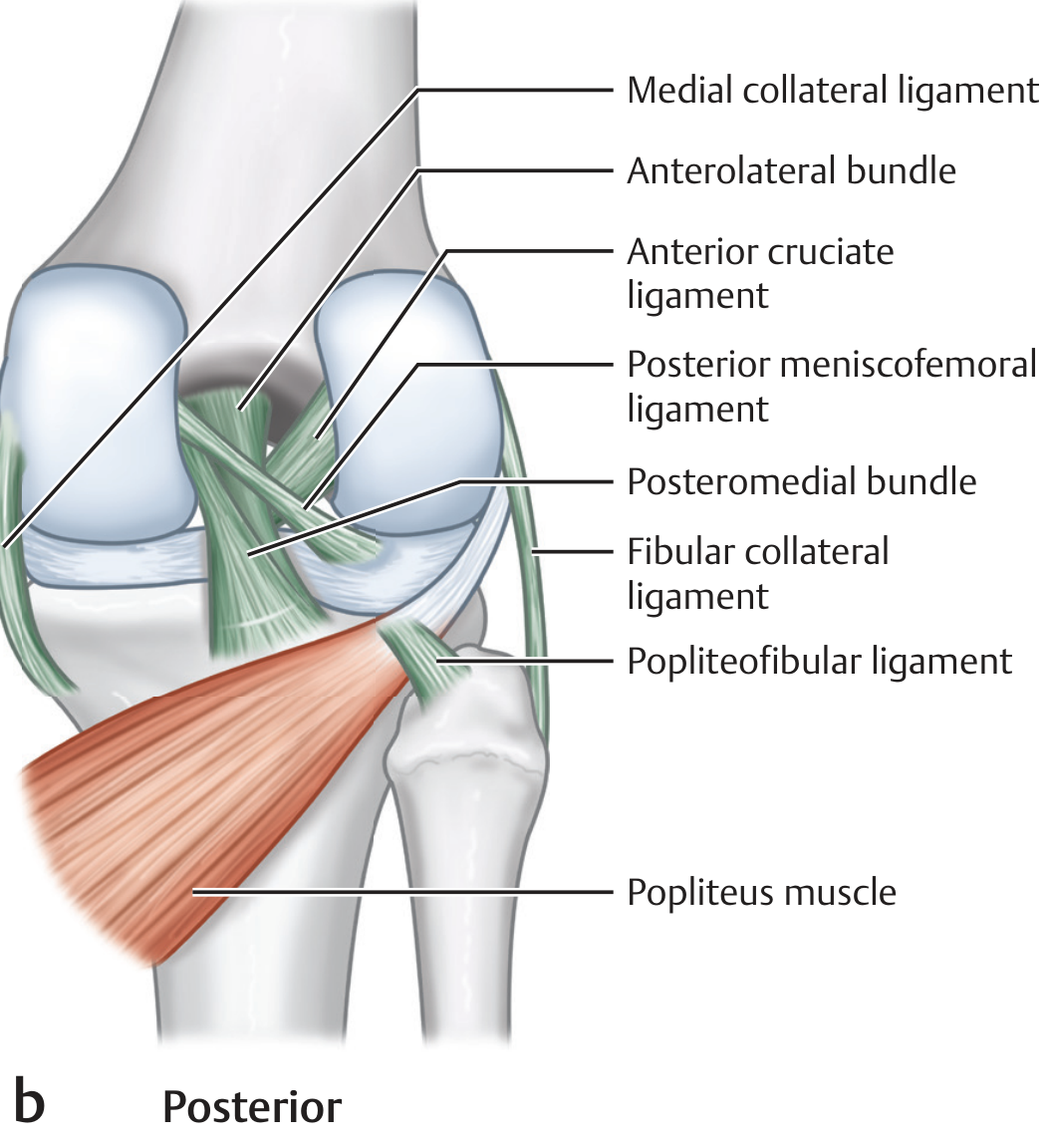
Primary Structures of the PLC
There are three structures universally recognised as the primary stabilisers:
1. Fibular (Lateral) Collateral Ligament (FCL / LCL)
- Femoral origin: Proximal and posterior to the lateral femoral epicondyle; posterior, superficial, and proximal to the insertion of the popliteus tendon
- Distal insertion: Anterior to the midpoint of the lateral fibular head (8 mm posterior to the anterior margin of the fibular head, 28 mm distal to the tip of the fibular styloid) - it is the most anterior structure inserting on the proximal fibula
- Length: 63-71 mm
- Mechanics: Tight in extension, lax in flexion (lies posterior to the axis of knee rotation). Primary restraint to varus stress in all degrees of knee flexion
- Relation to biceps: The FCL cleaves the distal biceps femoris tendon immediately proximal to the fibular head - the direct and anterior arms of biceps long head become lateral to it, while the short head arms sit medial to it
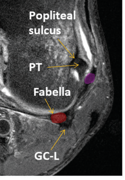
2. Popliteus Tendon
- Femoral attachment: Anterior aspect of the femoral popliteal sulcus, on the lateral femoral condyle - distal, anterior, and deep to the LCL (mnemonic: "DAD")
- Course: The tendon is intracapsular but extra-articular and extrasynovial. It passes from the popliteal sulcus inferolaterally, then curves inferomedially toward the posterior proximal tibia
- Hiatus: Traverses the coronary ligament via the popliteal hiatus (bounded by popliteomeniscal fascicles) to become intraarticular
- Muscle belly: Originates on the posterior tibia above the soleal line; the myotendinous junction lies at the lateral third of the popliteal fossa
- Function: Internally rotates the tibia; acts as a retractor of the lateral meniscus preventing impingement
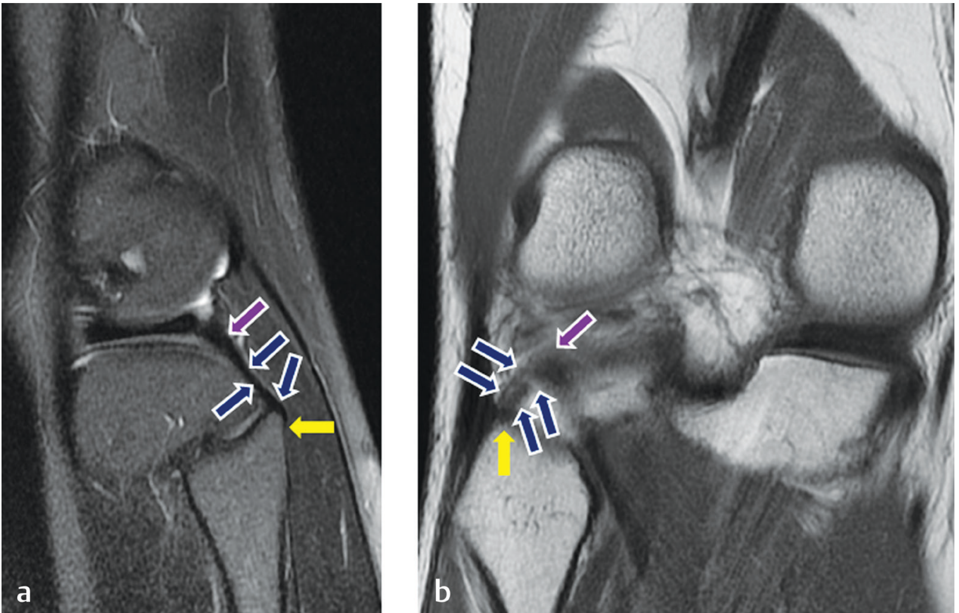
3. Popliteofibular Ligament (PFL)
- Origin: Inferolateral aspect of the popliteus myotendinous junction
- Insertion: Apex of the fibular styloid process, and slightly medial to this apex - posteromedial to both the FCL and biceps femoris fibular attachments
- Fibular attachment order (anterior to posterior): LCL → PFL → Biceps femoris
- Structure: Two divisions (anterior and posterior) described; the posterior is more substantive
- Significance: A fairly constant structure; biomechanical studies show the FCL, popliteus tendon, and PFL together are the most important stabilisers of the PLC
MRI - popliteofibular ligament (dark blue arrows) anchored at fibular styloid apex (yellow arrows) extending to popliteus myotendinous junction (purple arrows):
Secondary / Accessory Structures
| Structure | Description | Status |
|---|---|---|
| Iliotibial band (ITB) | Runs from Gerdy's tubercle to the lateral femoral epicondyle; contributes to anterolateral stability | Constant |
| Biceps femoris tendon | Long and short heads converge and insert on the fibular head; reinforces the PLC laterally | Constant |
| Anterolateral ligament (ALL) | Origin: immediately anterior to popliteus tendon insertion on femur. Insertion: midway between Gerdy's tubercle and fibular head with firm attachments to lateral meniscus. Stabilises against internal tibial rotation | Constant |
| Fabellofibular ligament | Runs from the fabella (or lateral gastrocnemius tendon if fabella absent) to the fibular styloid; reinforces the posterolateral capsule | Variable (~20-35%) |
| Arcuate ligament | Y-shaped thickening of the posterolateral capsule; lateral limb to posterolateral capsule and oblique popliteal ligament, base to fibular styloid apex. Some recommend retiring this term as it is inconsistent | Variable; often absent |
| Lateral gastrocnemius tendon | Arises from the fabella/posterolateral femur; contributes to posterolateral capsule reinforcement | Constant |
| Mid-third lateral capsular ligament | Thickening of the mid-lateral capsule | Variable |
| Coronary ligament of lateral meniscus | Meniscotibial attachment; contributes to lateral meniscal stabilisation | Constant |
| Popliteomeniscal fascicles | Three fascicles (posteroinferior, posterosuperior, anteroinferior) from popliteus to posterior horn of lateral meniscus; form the popliteal hiatus | Posterosuperior + anteroinferior constant on MRI |
| Oblique popliteal ligament (Winslow's) | Extension of semimembranosus tendon; crosses the posterior capsule obliquely; reinforces the posterocentral capsule | Constant |
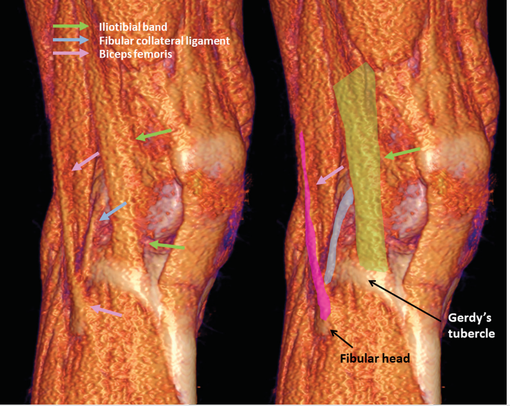
3D CT showing ITB (green), FCL (blue), and biceps femoris (pink) at the lateral knee - with Gerdy's tubercle and fibular head:
Layers of the Lateral Knee (Seebacher Classification)
The lateral knee is organised into three layers (deep to superficial):
| Layer | Contents |
|---|---|
| Layer I (superficial) | Iliotibial band anteriorly; biceps femoris and its expansion posteriorly |
| Layer II (middle) | Quadriceps retinaculum anteriorly; two patellofemoral ligaments |
| Layer III (deep / capsular) | Joint capsule; FCL (superficial lamina); popliteus tendon; fabellofibular + arcuate ligaments (deep lamina) |
Biomechanics and Functional Anatomy
| Motion Resisted | Primary Restraint | Secondary Restraint |
|---|---|---|
| External tibial rotation | Popliteus tendon + PFL | FCL, biceps femoris, ALL |
| Varus stress | FCL | Popliteus, IT band |
| Posterior tibial translation | PCL (primary) | PLC structures (secondary) |
| Internal tibial rotation | ACL (primary) | ALL, IT band |
| Genu recurvatum | PLC + PCL | Posterior capsule |
- The PLC provides its greatest contribution to posterolateral stability between 30° and 60° of knee flexion
- The FCL is tight in extension and lax in flexion, while the popliteus tendon shows increasing tension with internal tibial rotation at all degrees of flexion
- Isolated PLC injury rarely occurs; it is most commonly associated with PCL tears and/or ACL tears
- Brachial artery can be safely ligated: the common peroneal (fibular) nerve is the critical neural structure at risk in the PLC region, passing posterior to the biceps femoris tendon and around the neck of the fibula
Vascular Relations
The inferior lateral genicular artery (branch of the popliteal artery) runs along the lateral meniscus, superficial to the popliteus tendon and PFL, and deep to the FCL. It is relevant surgically when performing posterolateral corner reconstruction or lateral meniscus surgery.
Nerve at Risk
The common peroneal nerve runs along the posterior border of the biceps femoris tendon, passes around the fibular neck just medial to the biceps insertion, and is at risk in:
- PLC injuries (direct trauma / traction)
- Surgical approaches to the posterolateral knee
- Varus corrective osteotomies
Clinical Assessment Points
- Varus stress test: 0° and 30° flexion - isolates FCL (LCL) at 30°
- Dial test (external rotation): Asymmetric external rotation >10-15° at 30° only = isolated PLC; at 30° and 90° = combined PLC + PCL injury
- Posterolateral drawer test: External tibial rotation at 80-90° flexion
- Reverse pivot shift: Reduction of externally rotated/posterolaterally subluxed tibia during knee extension
Sources: Miller's Review of Orthopaedics 9th Ed (9780443112140, p. 338-339); Imaging Anatomy Vol 3 - Bones, Joints, Vessels & Nerves (9781626239845, pp. 288-299, 804-806); Campbell's Operative Orthopaedics 15th Ed 2026 (9780443117633); Grainger & Allison's Diagnostic Radiology (9780323760751, p. 996)
This is a shared conversation. Sign in to Orris to start your own chat.