
Type 1 dm investigations
I'll read the medical library skill first, then search for Type 1 DM investigations.
Reading File
Now searching the library for Type 1 DM investigations in parallel:
Finding Sources
Finding Sources
Finding Sources
Good results. Now reading the key sections in parallel:
Reading File
Reading File
Reading File
Excellent content. Let me get more detail from the Tietz and Henry's blocks on monitoring and HLA:
Reading File
Reading File
Finding Sources
I now have comprehensive data from multiple authoritative textbooks. Let me also get the Tietz data on lab table and Goldman-Cecil for clinical overview:
Reading File
Reading File
I now have comprehensive data to compile a detailed, well-referenced answer. Let me also read a bit more from Goldman-Cecil on clinical investigations and get the Tietz table of lab variables:
Reading File
Finding Sources
I also noticed a clinical diagram image in Goldman-Cecil. Let me fetch it for the response, and also get the beta-hydroxybutyrate data from Tietz:
Loading Image
Reading File
I now have all the information needed for a comprehensive, well-cited answer. Here is the full response:
Type 1 Diabetes Mellitus - Investigations
Type 1 DM (T1DM) accounts for 5-10% of all diabetes cases. Investigations are divided into those used for diagnosis, differentiating T1DM from other types, monitoring glycemic control, and assessing complications.
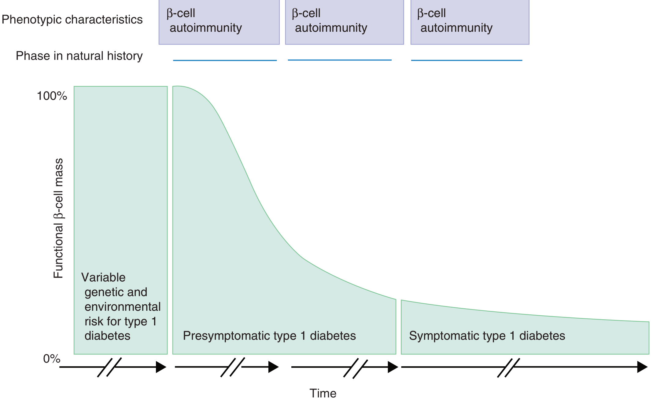
Figure: Progressive loss of functional β-cell mass from the presymptomatic to symptomatic phase of T1DM. Throughout all phases, β-cell autoimmunity is present. (Goldman-Cecil Medicine)
1. Investigations for Diagnosis of Diabetes
These establish whether diabetes is present (applicable to T1DM and all types). Two abnormal results are required (same day or two different days):
| Test | Normal | Pre-diabetes | Diabetes |
|---|---|---|---|
| Fasting plasma glucose (FPG) (fasting ≥8h) | <100 mg/dL | 100-125 mg/dL | ≥126 mg/dL (≥7.0 mmol/L) |
| 2-hour OGTT (75 g glucose load) | <140 mg/dL | 140-199 mg/dL | ≥200 mg/dL (≥11.1 mmol/L) |
| HbA1c (NGSP-certified, DCCT-standardized) | <5.7% | 5.7-6.4% | ≥6.5% (≥48 mmol/mol) |
| Random plasma glucose + symptoms | - | - | ≥200 mg/dL (≥11.1 mmol/L) |
Important: Point-of-care capillary glucose and CGM readings are useful for monitoring but should NOT be used to diagnose diabetes - laboratory plasma glucose confirmation is required due to greater accuracy. - Henry's Clinical Diagnosis and Management by Laboratory Methods
2. Tests Specific to Type 1 DM (to confirm autoimmune etiology and differentiate from T2DM)
A. Pancreatic Autoantibodies
These are the hallmark investigations for T1DM. They may be present before clinical onset and help differentiate T1DM from T2DM:
| Autoantibody | Notes |
|---|---|
| Anti-GAD65 (Glutamic Acid Decarboxylase) | Highest sensitivity as a single marker (91%); more common in adults with T1DM |
| Insulin Autoantibodies (IAA) | More common in young children who develop T1DM |
| IA-2 / ICA512 (Insulinoma-Associated Protein 2, a tyrosine phosphatase) | Located on surface membrane of β-cell secretory granules |
| ZnT8A (Zinc Transporter 8 Antibodies) | Also on secretory granule membranes; composite assay improves prediction |
| Islet Cell Antibodies (ICA) | Older assay; detected by immunofluorescence |
- The presence of ≥2 autoantibodies is strongly associated with progression to T1DM.
- GAD65 has the highest single-marker sensitivity (91%) for detecting multi-antibody-positive individuals.
- The absence of antibodies does not exclude T1DM - about 10% of patients (idiopathic T1DM) are antibody-negative. - Henry's Clinical Diagnosis and Management by Laboratory Methods, Goldman-Cecil Medicine
Clinical use: When a patient presents in DKA but has features of T2DM (obese, over 40, strong family history), antibody testing may be the only way to confirm T1DM. - Goldman-Cecil Medicine
B. C-Peptide
C-peptide is the connecting peptide cleaved from proinsulin and reflects endogenous insulin secretion:
- Low or undetectable C-peptide is characteristic of the absolute insulin deficiency in T1DM.
- Measured after an overnight fast, then after stimulation by a mixed meal tolerance test or glucagon stimulation test.
- The mixed meal tolerance test C-peptide is used as an endpoint in immunomodulatory clinical trials for T1DM prevention.
- Urine C-peptide:creatinine ratio can also be used to assess residual β-cell function.
- Unlike insulin, C-peptide is primarily degraded in the kidneys - levels are elevated in renal failure. - Henry's Clinical Diagnosis and Management by Laboratory Methods, Tietz Textbook of Laboratory Medicine
T1DM vs T2DM: Low/absent C-peptide → T1DM. Preserved/elevated C-peptide → T2DM.
C. HLA Typing (Genetic/Research Setting)
- About 60 T1DM genes are known; HLA genes contribute ~50% of genetic susceptibility.
- ~95% of white patients with T1DM express HLA-DR3 or HLA-DR4 (but note: up to 40% of the nondiabetic population also carry these alleles).
- The DR4-DQ8 and DR3-DQ2 haplotypes are present in ~90% of children with T1DM.
- DR15-DQ6 (DQB1*0602) is highly protective - found in only 1% of children with T1DM vs 20% of the general population.
- Routine HLA typing is not recommended for clinical diagnosis or management; used primarily in research and family screening for potential pancreatic transplant donors. - Tietz Textbook of Laboratory Medicine, Goldman-Cecil Medicine
3. Monitoring Investigations
A. HbA1c (Glycated Hemoglobin)
- Reflects average blood glucose over the preceding 2-3 months (lifespan of RBCs).
- Should be measured every 3-6 months in patients with T1DM.
- Target: <7% for most T1DM patients (from DCCT trial data).
- False results occur with: hemoglobinopathies, hemolytic anemia, blood transfusions, uremia (shortened RBC lifespan). - Henry's Clinical Diagnosis and Management, Tietz Textbook
B. Blood Glucose Monitoring
- Self-monitoring by point-of-care capillary glucose.
- Continuous Glucose Monitoring (CGM) - measures interstitial fluid glucose; useful for detecting hypo/hyperglycemic trends, guiding insulin dosing.
- Neither replaces plasma glucose for diagnosis, but both are essential for management.
C. Ketone Testing
- T1DM patients are prone to DKA due to absolute insulin deficiency.
- When to test: during acute illness, blood glucose consistently >300 mg/dL (>16.7 mmol/L), during pregnancy, or when ketoacidosis symptoms are present.
- Blood β-hydroxybutyrate is preferred over urine ketone strips - it is more accurate, reduces hospital admissions, and shortens recovery from DKA.
- Urine nitroprusside strips (Ketostix) detect acetoacetate/acetone but do NOT detect β-hydroxybutyrate (the predominant ketone in DKA) - a negative result does not rule out ketoacidosis. - Tietz Textbook of Laboratory Medicine
D. Lipid Profile
- Dyslipidemia is common; baseline and periodic monitoring are standard.
E. Renal Function / Urine Albumin
- Annual urine albumin-to-creatinine ratio (UACR) and serum creatinine/eGFR to screen for diabetic nephropathy.
F. Thyroid Function (TSH)
- T1DM has a high association with autoimmune thyroid disease (Hashimoto's, Graves') - screening TSH is recommended at diagnosis and periodically.
4. Investigations at Presentation / When DKA is Suspected
| Test | Purpose |
|---|---|
| Serum glucose | Hyperglycemia (>200 mg/dL in DKA) |
| Blood/urine ketones | Confirm ketosis |
| Arterial blood gas (ABG) | pH <7.3 confirms metabolic acidosis in DKA |
| Serum bicarbonate | <15 mEq/L in DKA |
| Anion gap | Elevated in DKA; used to monitor recovery |
| Serum electrolytes (Na, K, Cl) | K depletion despite high serum K initially |
| Serum creatinine/BUN | Assess renal function and hydration |
| CBC | Leukocytosis may indicate infection as precipitant |
| Urine analysis | Glycosuria, ketonuria |
Summary
| Investigation | Purpose |
|---|---|
| FPG / OGTT / HbA1c / random glucose | Diagnose diabetes |
| Anti-GAD65, IA-2, ZnT8A, IAA, ICA | Confirm autoimmune T1DM, differentiate from T2DM |
| C-peptide (fasting + stimulated) | Assess residual β-cell function |
| HLA typing | Research/family screening only |
| HbA1c (every 3-6 months) | Monitor glycemic control |
| Blood β-hydroxybutyrate / urine ketones | Detect and monitor DKA |
| ABG, anion gap, electrolytes | Manage acute DKA |
| UACR, eGFR, lipids, TSH | Screen for complications and comorbidities |
Sources: Henry's Clinical Diagnosis and Management by Laboratory Methods; Tietz Textbook of Laboratory Medicine, 7th ed.; Goldman-Cecil Medicine; Quick Compendium of Clinical Pathology, 5th ed.
This is a shared conversation. Sign in to Orris to start your own chat.