
Generate a short answer on this question for MBBS biochemistry exam based on Vasudevan with simple cycle(easily understandable) with simple diagram Regulation of Gene Expression Principle of Electrophoresis Northern Blotting
Simple educational biochemistry diagram showing Regulation of Gene Expression: Two panels side by side. Left panel labeled "PROKARYOTE - Lac Operon" showing: DNA strand with labeled boxes: lacI gene, Promoter (P), Operator (O), lacZ, lacY, lacA genes. Two states shown with arrows: State 1 (No lactose): Repressor protein binds Operator → RNA Polymerase BLOCKED → No transcription. State 2 (Lactose present): Allolactose binds Repressor → Repressor released from Operator → RNA Polymerase proceeds → mRNA produced → Proteins β-galactosidase, Permease, Transacetylase. Right panel labeled "EUKARYOTE" showing: Chromatin remodeling (histone acetylation) → DNA accessible → Transcription factors bind enhancer/promoter → RNA Polymerase II recruited → mRNA → Post-transcriptional regulation → Translation. Clean white background, blue and green color scheme, clearly labeled arrows, exam-friendly style
Simple educational biochemistry diagram showing two panels. Left panel: "PRINCIPLE OF ELECTROPHORESIS" - shows a horizontal gel tank with buffer, negative electrode (cathode) on left, positive electrode (anode) on right, electric current arrow pointing right. DNA/protein sample loaded in wells at top. Molecules separated by size: large molecules stay near top (wells), small molecules migrate further toward positive end. Labels: "Migration depends on: 1. Size (smaller = faster) 2. Charge (negative = moves to +ve) 3. Shape". Right panel: "NORTHERN BLOTTING STEPS" - numbered flowchart: Step 1: Extract mRNA from tissue, Step 2: Run mRNA on agarose gel (electrophoresis), Step 3: Transfer (blot) mRNA to nitrocellulose/nylon membrane, Step 4: Add labeled DNA probe (hybridization), Step 5: Wash off unbound probe, Step 6: Autoradiography - bands visible showing mRNA size and amount. Clean white background, simple arrows connecting each step, exam-friendly style, labeled clearly
1. Regulation of Gene Expression
Prokaryotic Regulation - The LAC Operon Model (Jacob & Monod, 1961)
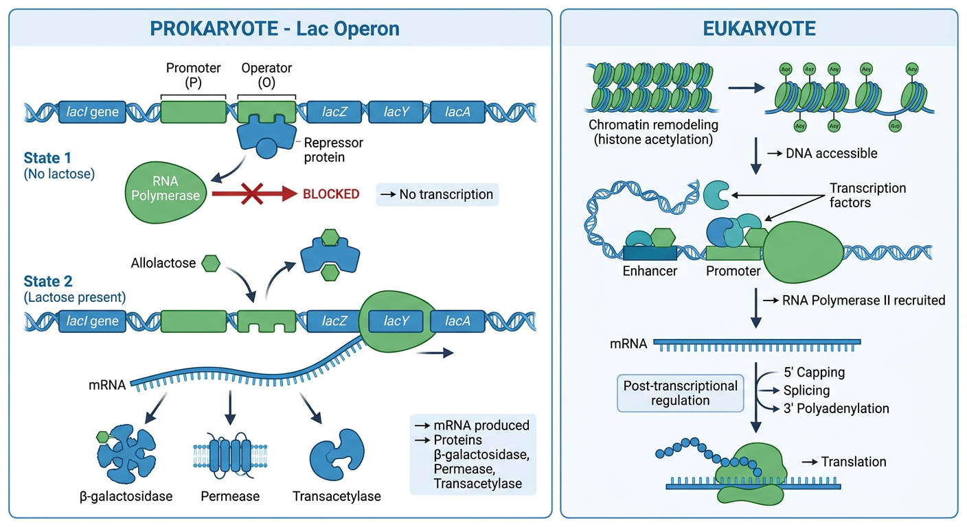
Lac Operon Components:
| Component | Function |
|---|---|
| lacI gene | Codes for repressor protein |
| Promoter (P) | Site where RNA polymerase binds |
| Operator (O) | Regulatory region; repressor binding site |
| lacZ, lacY, lacA | Structural genes (β-galactosidase, Permease, Transacetylase) |
Two States:
Repressor → binds Operator → blocks RNA Polymerase → NO transcription
Allolactose (inducer) → binds Repressor → Repressor leaves Operator → RNA Polymerase proceeds → Polycistronic mRNA produced → 3 enzymes synthesized
Types of Control:
- Negative control: Repressor blocks transcription (as above)
- Positive control: CAP-cAMP complex activates transcription (when glucose is absent)
- Constitutive expression: Housekeeping genes - always expressed at constant rate
Eukaryotic Regulation
| Level | Mechanism |
|---|---|
| DNA level | Chromatin remodeling - histone acetylation opens chromatin; DNA methylation silences genes |
| Transcription | Specific transcription factors (transactivators) bind enhancers/promoters to recruit RNA Polymerase II |
| Post-transcriptional | RNA splicing, capping, polyadenylation |
| Translation | miRNA, translational repressors |
Key difference: Eukaryotes do NOT have operons. Each gene has its own promoter.
2. Principle of Electrophoresis
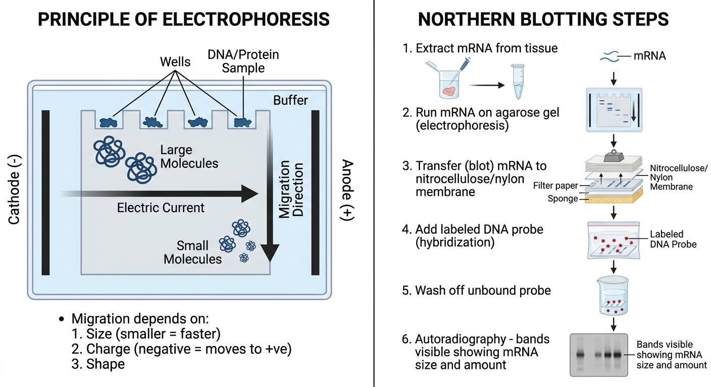
Basic Principle:
Charged molecules placed in an electric field migrate toward the opposite pole.
(−) Cathode  ←—[gel/medium]—→  Anode (+)
              ↑
         Sample loaded here
         (DNA/RNA are -ve charged,
          migrate TOWARD +ve pole)
Factors Governing Migration:
| Factor | Effect |
|---|---|
| Size | Smaller molecules migrate FASTER (less resistance) |
| Charge | More negative charge = faster movement toward anode |
| Shape | Linear > branched > circular (same size) |
| Gel concentration | Higher % = slower migration, better resolution of small molecules |
Types of Electrophoresis:
- Agarose gel electrophoresis - separates nucleic acids (DNA, RNA)
- Polyacrylamide gel electrophoresis (PAGE) - separates proteins
- SDS-PAGE - sodium dodecyl sulphate denatures proteins; separation purely by size
- Capillary electrophoresis (CE) - separates proteins in a capillary tube
- Immunofixation electrophoresis - detects monoclonal proteins (M-band in myeloma)
Visualization:
- DNA/RNA - Ethidium bromide stain (fluoresces under UV)
- Proteins - Coomassie blue, Amido black, Ponceau S stain
3. Northern Blotting
Named "Northern" as a directional analogy to "Southern" blotting (which uses DNA).
Steps (Mnemonic: ETHER-H)
Simple Flow Diagram:
Tissue Sample
     ↓
Extract mRNA
     ↓
Run on Agarose-Formaldehyde Gel
     ↓
Transfer mRNA to Nitrocellulose Membrane
     ↓
Hybridize with Labeled DNA Probe
     ↓
Wash (remove unbound probe)
     ↓
Autoradiography / Fluorescence Detection
     ↓
BAND = Size and Amount of specific mRNA
What Northern Blot Tells Us:
| Information | How obtained |
|---|---|
| Size of mRNA | Position of band on gel |
| Amount of mRNA | Intensity of band |
| Gene expression pattern | Which tissue expresses the gene |
Comparison of Blotting Techniques:
| Blot | Target | Probe |
|---|---|---|
| Southern | DNA | DNA probe |
| Northern | RNA (mRNA) | DNA probe |
| Western | Protein | Antibody |
Clinical Significance:
- Detects abnormal mRNA in genetic diseases
- Studies gene expression in cancer vs normal tissue
- Confirms results of PCR or microarray experiments
- Now largely replaced by RT-PCR and RNA sequencing (but still the only method giving mRNA size information)
Sources: Harper's Illustrated Biochemistry 32nd Ed; Basic Medical Biochemistry - A Clinical Approach 6e; Emery's Medical Genetics; Schwartz's Principles of Surgery 11th Ed
Chromatography — Principle & Types Products derived from Glycine Salient Features of Genetic Code Quality Control
Simple educational biochemistry diagram "CHROMATOGRAPHY - Principle and Types". Top section shows principle: horizontal tube/column with "Stationary Phase" inside, arrow showing "Mobile Phase flows through", molecules shown as different colored circles - some stick to stationary phase longer (slow, retained) vs others move faster (weak interaction). Labels: "Sample injected" at start, "Detector" at end, showing a chromatogram peak output. Below: table-style boxes showing 4 types: 1) Ion Exchange Chromatography - ions separated by charge, cation vs anion exchange resin shown; 2) Gel Filtration (Size Exclusion) - large molecules exit first, small molecules get trapped in pores exit last, diagram of column with pores; 3) Affinity Chromatography - specific ligand on column, only target molecule binds, eluted by salt, diagram showing lock-key binding; 4) Adsorption/Partition Chromatography - paper/TLC shown with Rf value formula Rf = distance moved by solute / distance moved by solvent. Clean white background, blue color scheme, exam-style labeled arrows
Simple educational biochemistry diagram with two panels. Left panel: "PRODUCTS DERIVED FROM GLYCINE" - central oval labeled GLYCINE with arrows pointing outward to: 1) HEME (porphyrin ring - shown as diamond shape) labeled "with Succinyl CoA", 2) CREATINE/CREATININE (muscle energy) labeled "with Arginine + SAM", 3) GLUTATHIONE (antioxidant tripeptide) labeled "with Glu + Cys", 4) BILE SALTS labeled "conjugated with bile acids", 5) PURINE BASES (for DNA/RNA) labeled "C4, C5, N7 of purine ring", 6) HIPPURIC ACID labeled "detoxification in liver", 7) SERINE labeled "via serine hydroxymethyltransferase". Right panel: "SALIENT FEATURES OF GENETIC CODE" - numbered list: 1) Triplet code - 3 bases = 1 codon, 2) Degenerate/Redundant - 64 codons for 20 amino acids, 3) Non-overlapping - read continuously, 4) Comma-less - no punctuation, 5) Universal - same in all organisms (except mitochondria), 6) Non-ambiguous/Specific - one codon = one amino acid, 7) Start codon: AUG (Met), 8) Stop codons: UAA, UAG, UGA. Clean white background, colorful arrows, exam-friendly style
Simple educational biochemistry diagram "QUALITY CONTROL in Biochemistry Laboratory". Central flowchart showing: Step 1: PREANALYTICAL - Patient preparation, sample collection, labeling, transport (icons of blood tube, patient). Step 2: ANALYTICAL - Instrument calibration, reagent quality, control samples run alongside patient samples, showing LEVEY-JENNINGS CHART with mean line and +1SD, +2SD, +3SD warning lines plotted with dots, WESTGARD RULES shown: 1-2s warning, 1-3s rejection, 2-2s rule, R-4s rule. Step 3: POSTANALYTICAL - Result interpretation, reference ranges, critical value reporting, delta check. Bottom: two columns - Internal QC (run control sera each day, in-house monitoring) vs External QC/EQA (interlaboratory comparison, proficiency testing). Colors: blue for preanalytical, green for analytical, orange for postanalytical. Clean white background, exam-friendly
1. Chromatography - Principle & Types
Principle
Chromatography is a separation technique based on the differential distribution of compounds between a mobile phase and a stationary phase as they travel through a support medium.
- Compounds that interact more strongly with the stationary phase are retained longer (elute later)
- Compounds that interact more with the mobile phase move faster (elute earlier)
- Each compound has a characteristic retention time (tR) - used to identify it
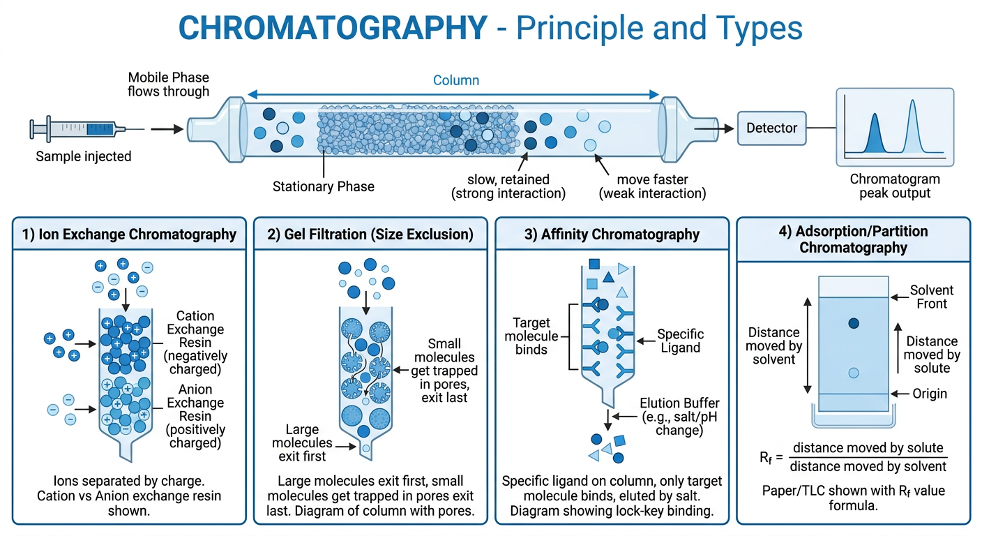
Key Terms:
| Term | Meaning |
|---|---|
| Retention time (tR) | Time for a compound to elute from the column |
| Theoretical plates (N) | Measure of column efficiency; N = 16(tR/Wb)² |
| Selectivity factor (α) | Ratio of capacity factors of two compounds |
| Rf value | Distance moved by solute / Distance moved by solvent (paper/TLC) |
Types of Chromatography
1. Ion Exchange Chromatography
- Separates molecules based on charge
- Cation exchange resin (negatively charged) - binds positively charged molecules
- Anion exchange resin (positively charged) - binds negatively charged molecules
- Elution done by changing salt concentration or pH
- Use: Purification of proteins, amino acids, nucleotides
2. Gel Filtration / Size Exclusion Chromatography
- Separates molecules based on size (molecular weight)
- Column packed with porous beads
- Large molecules - cannot enter pores → elute FIRST
- Small molecules - enter pores → elute LAST
- Use: Determining molecular weight, protein purification
Large molecule → excluded from pores → elutes FIRST
Small molecule → enters pores → elutes LAST
3. Affinity Chromatography
- Most specific type of chromatography
- Column has a specific ligand (e.g., antibody, enzyme substrate) covalently attached
- Only the target molecule binds; others pass through
- Target eluted by salt gradient or specific substrate
- Use: Purification of antibodies, enzymes, receptors, hormones
4. Adsorption / Partition Chromatography
- Paper Chromatography: Stationary = water in paper; Mobile = organic solvent
- TLC (Thin Layer): Stationary = silica on plate; Mobile = solvent
- Separation by polarity - polar compounds stay near origin; nonpolar move far
- Use: Amino acid analysis (ninhydrin spray), lipid separation
5. Gas Chromatography (GC)
- Mobile phase = inert gas (He, N₂)
- Stationary phase = liquid on column packing
- Requires volatile compounds; temperature-controlled oven
- Use: Drug detection, toxicology, amino acid analysis, fatty acid profiling
6. High-Performance Liquid Chromatography (HPLC)
- Mobile phase = liquid pumped under high pressure
- Very high resolution, sensitivity, speed
- Use: Drug levels (immunosuppressants, antibiotics), hormone assays, HbA1c
2. Products Derived from Glycine
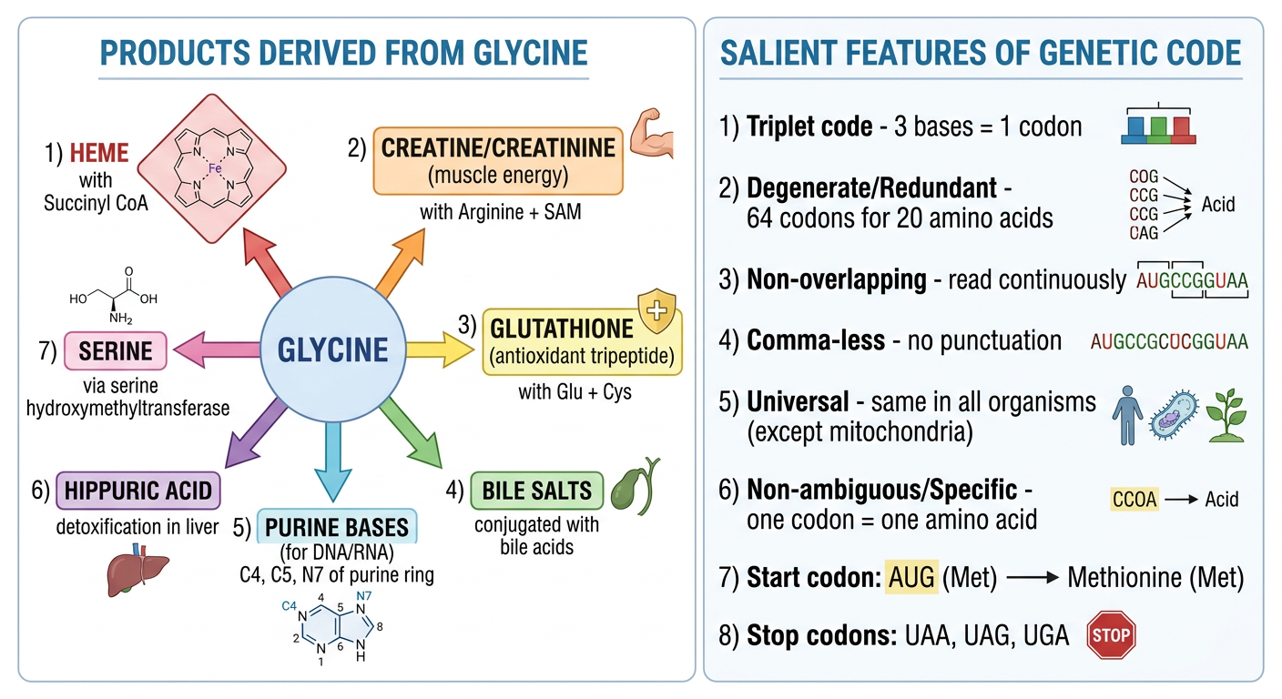
Summary Table:
| Product | Pathway / Partner | Significance |
|---|---|---|
| Heme / Porphyrins | Glycine + Succinyl-CoA → δ-ALA (rate-limiting: ALA synthase, requires B6) | Oxygen transport (hemoglobin), cytochromes |
| Creatine / Creatinine | Glycine + Arginine → Guanidinoacetate → + SAM → Creatine → Creatinine | Muscle energy reserve (phosphocreatine); creatinine = kidney function marker |
| Glutathione (GSH) | Glu + Cys + Glycine → GSH (γ-glutamyl-cysteinyl-glycine) | Major intracellular antioxidant; protects RBCs |
| Purines (A, G) | Glycine contributes C4, C5, N7 of purine ring (de novo synthesis) | Building blocks of DNA, RNA, ATP |
| Bile salts | Bile acids (cholate, chenodeoxycholate) conjugated with Glycine or Taurine | Fat emulsification and absorption (glycocholate, glycochenodeoxycholate) |
| Hippuric acid | Glycine + Benzoic acid (in liver) | Detoxification of benzoate; used in liver function test (Quick's test) |
| Serine | Glycine + CH₂-THF ⇌ Serine (via serine hydroxymethyltransferase, B6) | Non-essential amino acid; reversible reaction |
| Glyoxylate / Oxalate | Glycine → Glyoxylate → Oxalate | Excess → hyperoxaluria → renal stones |
| One-carbon units | Glycine → methylene-THF | For methylation reactions |
Memory Aid - "HCGBPSO":
3. Salient Features of the Genetic Code
- 4 bases → 3-base codons → 4³ = 64 possible codons for 20 amino acids
- First cracked by Marshall Nirenberg (1961) - poly(U) → polyphenylalanine (UUU = Phe)
The 8 Salient Features (Mnemonic: "T-DUNDCN"):
| # | Feature | Explanation |
|---|---|---|
| 1 | Triplet | Each codon = 3 nucleotides; minimum needed to code 20 amino acids |
| 2 | Degenerate (Redundant) | 61 codons code for only 20 amino acids; most amino acids have >1 codon. Only Met & Trp have single codons |
| 3 | Universal | Same genetic code in all organisms (bacteria to humans). Exception: Mitochondria (UGA = Trp, not stop) |
| 4 | Non-ambiguous (Specific) | One codon always codes for the same amino acid (never two different amino acids) |
| 5 | Non-overlapping | Each base belongs to only one codon; read in sequence without sharing |
| 6 | Comma-less | No punctuation between codons; read continuously from start |
| 7 | Collinear | Sequence of codons in mRNA corresponds to sequence of amino acids in protein |
| 8 | Has Start & Stop codons | Start: AUG (methionine) · Stop: UAA, UAG, UGA (nonsense codons - no amino acid) |
Codon Chart Summary:
64 total codons:
  ├── 61 → amino acids (sense codons)
  │     ├── AUG → Methionine (START, only codon for Met)
  │     └── UGG → Tryptophan (only codon for Trp)
  └──  3 → STOP codons (UAA, UAG, UGA)
        (Mnemonic: "U Are Away", "U Are Gone", "U Go Away")
Mutations and Consequences:
- Silent mutation - codon changed but same amino acid (e.g., UCA → UCU = still Ser)
- Missense mutation - different amino acid coded (e.g., Sickle cell: GAG → GUG = Glu → Val)
- Nonsense mutation - premature stop codon → truncated protein
- Frameshift - insertion/deletion shifts reading frame → completely different protein downstream
4. Quality Control (QC) in Biochemistry Laboratory
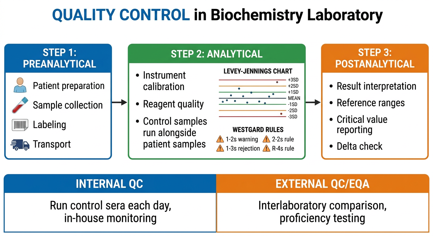
Three Phases of Quality Control:
A. Pre-analytical QC
- Patient preparation (fasting, medication effects)
- Correct sample collection (right tube, anticoagulant)
- Proper labeling
- Transport and storage conditions (temperature, time)
- Hemolysis, lipemia, icterus - pre-analytical interferences
B. Analytical QC (most important for exam)
- Normal control - values in normal range
- Abnormal control - values in high/low range
+3 SD  ----warning----
+2 SD  ----warning----
+1 SD
Mean   ═══════════════
-1 SD
-2 SD  ----warning----
-3 SD  ----warning----
| Rule | Meaning | Action |
|---|---|---|
| 1₂ₛ | 1 control exceeds mean ± 2SD | Warning only |
| 1₃ₛ | 1 control exceeds mean ± 3SD | Reject run |
| 2₂ₛ | 2 consecutive controls exceed same ±2SD | Reject (systematic error) |
| R₄ₛ | Range between 2 controls >4SD | Reject (random error) |
- Random error - unpredictable, affects precision (e.g., air bubble in pipette)
- Systematic error - consistent bias, affects accuracy (e.g., wrong calibrator, reagent deterioration)
C. Post-analytical QC
- Result interpretation with proper reference ranges
- Reporting critical (panic) values immediately
- Delta check - compare with patient's previous result to detect specimen mix-up
- Proper transcription and reporting
Internal vs External QC:
| Internal QC | External QC (EQA/PT) | |
|---|---|---|
| What | Run control sera daily in your lab | Send unknown samples to central body |
| Purpose | Day-to-day precision monitoring | Interlaboratory comparison |
| Frequency | Daily | Monthly/quarterly |
| Examples | Westgard rules, L-J chart | EQAS, RIQAS, CAP proficiency testing |
Key Exam Fact: Accuracy is assessed by bias from true value; Precision is assessed by CV% (coefficient of variation) = (SD/mean) × 100. A good lab has CV% < 5% for most assays.
Sources: Henry's Clinical Diagnosis & Management by Laboratory Methods; Tietz Textbook of Laboratory Medicine 7th Ed; Lippincott Illustrated Reviews Biochemistry 8th Ed; Basic Medical Biochemistry - A Clinical Approach 6e
25 Tumour Markers Gene Therapy Quaternary Structure of Protein Free Radicals
Simple educational MBBS biochemistry diagram "TUMOUR MARKERS" showing a central table/chart layout. Title at top: "25 Important Tumour Markers". Three columns: MARKER | CANCER | NORMAL VALUE. Rows listing: AFP (Alpha-fetoprotein) - Liver (HCC), Testicular germ cell - less than 8.4 ng/mL; CEA (Carcinoembryonic antigen) - Colorectal, Pancreatic, Lung - less than 3 ng/mL; CA-125 - Ovarian cancer - less than 46 U/mL; CA 19-9 - Pancreatic cancer, Cholangiocarcinoma - less than 35 U/mL; PSA (Prostate-specific antigen) - Prostate cancer; beta-HCG - Choriocarcinoma, Testicular; LDH - Lymphoma, Testicular; CA 15-3 - Breast cancer; CA 72-4 - Gastric cancer; Calcitonin - Medullary thyroid cancer. Bottom section: Two boxes labeled "Uses of Tumour Markers": 1) Screening/Diagnosis, 2) Monitoring treatment response, 3) Detecting recurrence. Note: "No tumour marker is 100% specific". Clean white background, color-coded rows (blue header), exam-friendly style
Simple educational MBBS biochemistry diagram "GENE THERAPY" showing a clear flowchart. Top: Definition box "Introduction of functional therapeutic gene into patient cells to treat disease". Two main columns below: LEFT COLUMN "Types of Gene Therapy": 1) Somatic gene therapy - modifies non-reproductive body cells, changes not inherited, APPROVED FOR USE; 2) Germline gene therapy - modifies germ cells/embryo, changes are inherited, NOT APPROVED (ethical issues). CENTER: "Methods of Gene Delivery" with two branches: A) VIRAL VECTORS (most efficient): Retrovirus - integrates into genome - used for SCID; Adenovirus - does not integrate - used for cystic fibrosis; Adeno-Associated Virus (AAV) - episomal, long-lasting - used for hemophilia, SMA, retinal dystrophy; Lentivirus - infects non-dividing cells. B) NON-VIRAL VECTORS: Liposomes, Naked DNA, Electroporation. RIGHT: "Examples" box: ADA-SCID - adenosine deaminase deficiency; SMA - Zolgensma (FDA approved); Hemophilia B - Factor IX; Leber congenital amaurosis. Bottom warning box: "Limitations: Insertional mutagenesis, immune reaction, transient expression". Clean white background, blue-green color scheme, exam-style
Simple educational MBBS biochemistry diagram two panels. LEFT panel "QUATERNARY STRUCTURE OF PROTEIN": Top shows ladder/hierarchy of protein structure: Primary (amino acid sequence) → Secondary (alpha helix + beta sheet) → Tertiary (3D folding of single chain) → QUATERNARY (assembly of multiple subunits). Main diagram shows hemoglobin as classic example: four circles representing subunits - two alpha chains (blue) and two beta chains (red) arranged together, labeled "Tetramer = 4 subunits (2α + 2β)". Each subunit has a heme group shown as small diamond. Bonds holding subunits: hydrogen bonds, ionic bonds, hydrophobic interactions, Van der Waals forces (NO covalent bonds except disulfide). Box: "Cooperativity: binding of O2 to one subunit increases affinity of other subunits". Other examples listed: Collagen (trimer), Insulin (dimer), Immunoglobulins. RIGHT panel "FREE RADICALS": Central definition: "Atoms/molecules with unpaired electron in outer orbit - highly reactive". Types listed with symbols: Superoxide (O2-), Hydrogen peroxide (H2O2), Hydroxyl radical (OH-) most reactive, Peroxynitrite (ONOO-). Sources: mitochondria, radiation, inflammation, iron (Fenton reaction). Removal enzymes: SOD, Catalase, Glutathione peroxidase. Damage: lipid peroxidation, DNA damage, protein oxidation. Clean exam-style white background
1. Tumour Markers (25 Important Markers)
Key Point: No tumour marker is 100% specific or 100% sensitive. They are NEVER used alone for diagnosis.
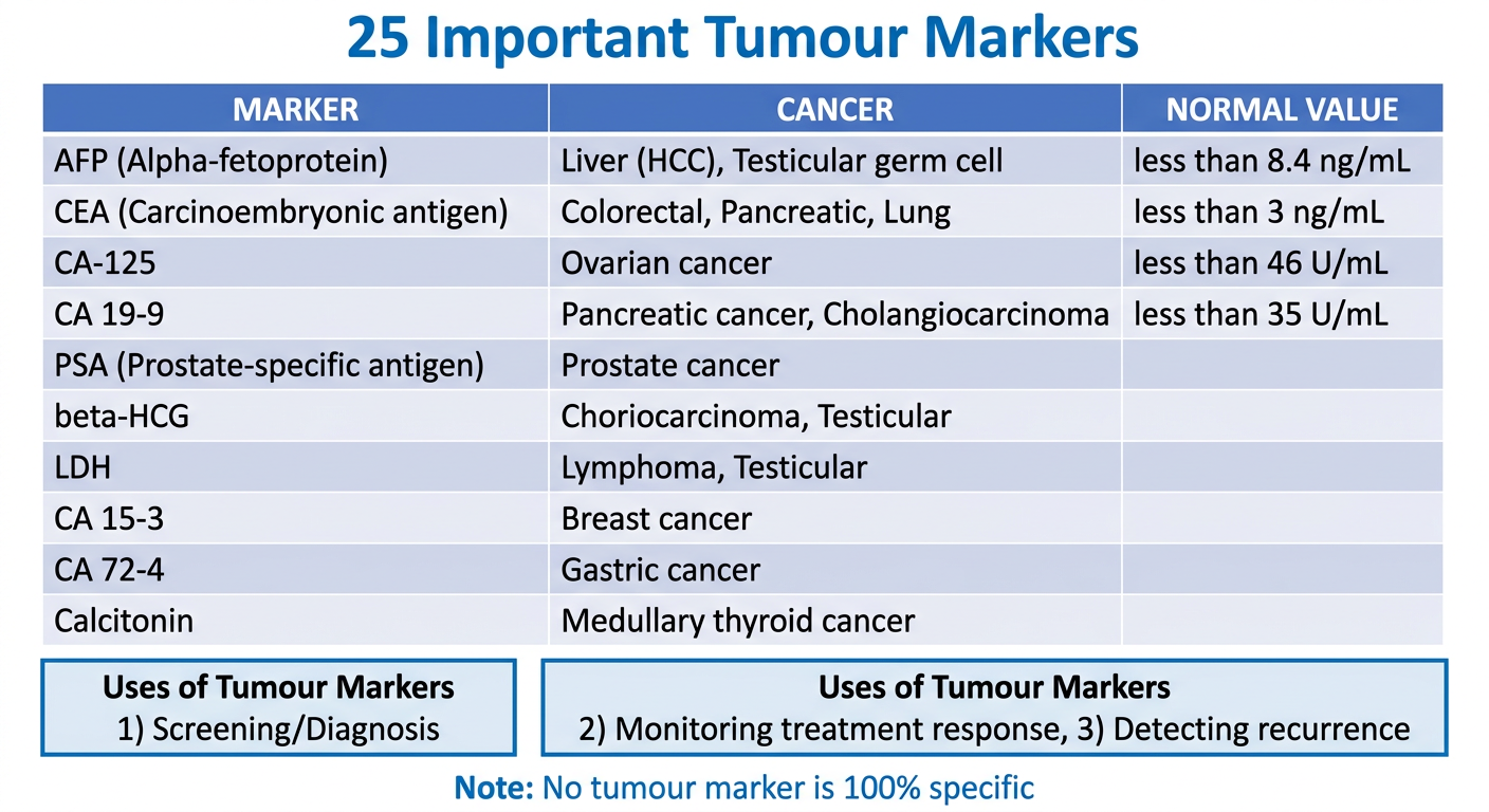
Complete Table of 25 Important Tumour Markers:
| # | Marker | Full Name | Associated Cancer | Normal Value |
|---|---|---|---|---|
| 1 | AFP | Alpha-fetoprotein | Hepatocellular carcinoma (HCC), Testicular (yolk sac, embryonal), Ovarian | <8.4 ng/mL |
| 2 | CEA | Carcinoembryonic antigen | Colorectal, Pancreatic, Lung, Breast | <3 ng/mL (smokers <5) |
| 3 | PSA | Prostate-specific antigen | Prostate cancer | <4 ng/mL |
| 4 | CA-125 | Cancer antigen 125 | Ovarian cancer (epithelial) | <46 U/mL |
| 5 | CA 19-9 | Carbohydrate antigen 19-9 | Pancreatic adenocarcinoma, Cholangiocarcinoma | <35 U/mL |
| 6 | CA 15-3 | Cancer antigen 15-3 | Breast cancer | <30 U/mL |
| 7 | CA 72-4 | Cancer antigen 72-4 | Gastric (stomach) cancer | <6.9 U/mL |
| 8 | β-HCG | Beta-human chorionic gonadotropin | Choriocarcinoma, Testicular (non-seminoma), Hydatidiform mole | <5 mIU/mL |
| 9 | LDH | Lactate dehydrogenase | Lymphoma, Testicular cancer, general tumour bulk | 140-280 U/L |
| 10 | Calcitonin | Calcitonin | Medullary thyroid carcinoma (MTC) | <10 pg/mL |
| 11 | Thyroglobulin | Thyroglobulin | Differentiated thyroid cancer (follicular, papillary) | Varies |
| 12 | PAP | Prostatic acid phosphatase | Prostate cancer | Replaced by PSA |
| 13 | 5-HIAA | 5-Hydroxyindoleacetic acid (urine) | Carcinoid tumour | <9 mg/24h |
| 14 | VMA | Vanillylmandelic acid (urine) | Pheochromocytoma, Neuroblastoma | <7 mg/24h |
| 15 | Catecholamines | Epinephrine/Norepinephrine | Pheochromocytoma | Varies |
| 16 | Chromogranin A | Chromogranin A | Neuroendocrine tumours, Carcinoid | <93 ng/mL |
| 17 | S-100 | S-100 protein | Melanoma, Brain tumours | Negative |
| 18 | NSE | Neuron-specific enolase | Neuroblastoma, Small cell lung cancer | <16.3 ng/mL |
| 19 | BRCA1/2 | Breast cancer gene 1/2 | Breast, Ovarian cancer (hereditary) | No mutation |
| 20 | ER/PR | Estrogen/Progesterone receptors | Breast cancer (hormone receptor status) | Negative |
| 21 | HER-2/neu | Human epidermal growth factor receptor 2 | Breast cancer, Gastric cancer | Negative |
| 22 | Bence Jones Protein | Immunoglobulin light chains (urine) | Multiple myeloma, Waldenstrom | Absent |
| 23 | M-protein/Paraprotein | Monoclonal immunoglobulin | Multiple myeloma | Absent |
| 24 | Ferritin | Ferritin | Hepatoma, Lymphoma (elevated, nonspecific) | 12-300 ng/mL |
| 25 | PD-L1 | Programmed death ligand 1 | Melanoma, NSCLC (immunotherapy target) | Varies by tumour |
Uses of Tumour Markers:
- Screening - population at high risk (e.g., AFP in cirrhosis for HCC)
- Diagnosis - support, never sole basis
- Staging - gauge extent of disease
- Monitoring treatment - falling levels = good response
- Detecting recurrence - rising levels post-treatment = relapse
- Prognosis - e.g., LDH in lymphoma staging
Oncofoetal Antigens (important exam category):
- AFP - produced by fetal liver / yolk sac; re-expressed in HCC
- CEA - produced during fetal development; re-expressed in GI cancers
2. Gene Therapy
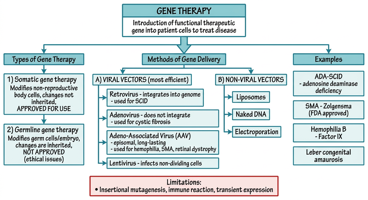
Types:
| Type | Target Cells | Inheritance | Status |
|---|---|---|---|
| Somatic gene therapy | Body cells (liver, muscle, stem cells) | Changes NOT inherited | APPROVED |
| Germline gene therapy | Egg/sperm/embryo | Changes ARE inherited | NOT APPROVED (ethical) |
Requirements Before Gene Therapy Can Be Applied (Box 14.1):
- Identity of the molecular defect must be known
- A functional cDNA/gene clone must be available
- An appropriate vector must be available
- A target cell accessible for gene delivery must exist
Methods of Gene Delivery (Vectors):
A. Viral Vectors (most efficient):
RETROVIRUS
  → Integrates into host genome
  → Dividing cells only
  → Used for: SCID (X-linked), ADA-SCID
  → Risk: insertional mutagenesis

ADENOVIRUS
  → Does NOT integrate (episomal)
  → Infects non-dividing cells too
  → Used for: Cystic fibrosis, cancer (transient effect)
  → Risk: immune reaction

ADENO-ASSOCIATED VIRUS (AAV)
  → Episomal (stable without integration)
  → Long-term expression in non-dividing cells
  → Used for: Hemophilia B, SMA (Zolgensma), Leber amaurosis
  → FDA APPROVED products available

LENTIVIRUS
  → Integrates into genome
  → Infects non-dividing cells
  → Used for: X-linked adrenoleukodystrophy, SCID (SIN vectors)
B. Non-viral Vectors (less efficient, safer):
- Liposomes - lipid vesicles carry DNA into cell
- Naked DNA injection - direct injection into muscle
- Electroporation - electric pulses open cell membrane pores
- Gene gun - biolistic particle delivery
Examples of Approved/Successful Gene Therapy:
| Disease | Defective Gene | Vector | Outcome |
|---|---|---|---|
| X-linked SCID | IL2RG | Retroviral → stem cells | 27/32 improved |
| ADA-SCID | ADA | Retroviral → stem cells | 29/40 off enzyme therapy |
| SMA (Spinal muscular atrophy) | SMN1 | AAV (IV injection) | Marked improvement; >1800 patients |
| Hemophilia B | Factor IX | AAV | Stable Factor IX expression; >20 off prophylaxis |
| Leber congenital amaurosis | RPE65 | AAV → retinal cells | FDA approved; vision improved |
| X-linked adrenoleukodystrophy | ABCD1 | Lentiviral → stem cells | Arrested demyelination in 17/19 |
Limitations:
- Insertional mutagenesis - viral integration can activate oncogenes (5 SCID patients got leukemia-like disorder)
- Immune response to viral vector
- Transient expression (for non-integrating vectors)
- Large genes cannot fit in current vectors
- High cost (Zolgensma = ~$2 million per dose)
3. Quaternary Structure of Protein
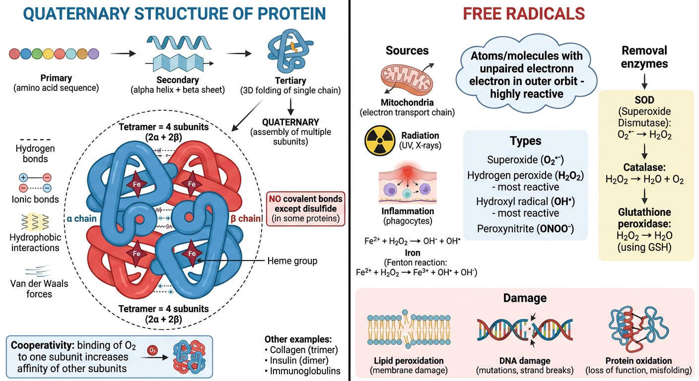
Key Points:
- Occurs when a protein has more than one polypeptide chain
- Each polypeptide chain = a subunit (monomer)
- Subunits may be identical (homo-multimer) or different (hetero-multimer)
- Subunits held together by non-covalent bonds primarily
Bonds Holding Subunits Together:
| Bond Type | Nature |
|---|---|
| Hydrogen bonds | Non-covalent |
| Ionic bonds | Non-covalent |
| Hydrophobic interactions | Non-covalent |
| Van der Waals forces | Non-covalent |
| Disulfide bonds | Covalent (rare; e.g., immunoglobulins) |
Classic Example - Hemoglobin:
Hemoglobin = TETRAMER
    ┌──────────────────────────────┐
    │   α₁ subunit + heme group   │
    │   α₂ subunit + heme group   │
    │   β₁ subunit + heme group   │
    │   β₂ subunit + heme group   │
    └──────────────────────────────┘
    Total = 4 subunits (2α + 2β)
Other Examples:
| Protein | Subunits | Type |
|---|---|---|
| Hemoglobin | 2α + 2β (tetramer) | Hetero-tetramer |
| Collagen | 3 chains (trimer) | Homo/Hetero-trimer |
| Insulin | A + B chains (dimer in storage) | Hetero-dimer |
| Immunoglobulin IgG | 2H + 2L chains | Hetero-tetramer |
| LDH (isoenzymes) | M and H chains (tetramer) | Hetero-tetramer |
| Creatine kinase (CK) | M and B chains (dimer) | Hetero-dimer |
Isoenzymes:
- Proteins with same function but different primary structure
- Arise from different genes OR tissue-specific processing
- Have different subunit compositions
- Example: LDH₁ (HHHH - heart) vs LDH₅ (MMMM - liver/muscle)
Significance:
- Quaternary structure allows cooperative interactions
- Enables allosteric regulation
- Provides structural stability
- Isoenzyme patterns used as diagnostic markers (CK-MB = heart; LDH₁ > LDH₂ = MI)
4. Free Radicals
Types of Reactive Oxygen Species (ROS):
| Free Radical | Symbol | Properties | Inactivated By |
|---|---|---|---|
| Superoxide anion | O₂•⁻ | Moderately reactive; from mitochondria, phagocytes | SOD |
| Hydrogen peroxide | H₂O₂ | Not a free radical but reactive; can diffuse away from site | Catalase, GSH peroxidase |
| Hydroxyl radical | •OH | MOST REACTIVE; principal ROS causing lipid/protein/DNA damage | None enzymatic |
| Peroxynitrite | ONOO⁻ | From O₂•⁻ + NO; damages lipids, proteins, DNA | Peroxiredoxins |
| Nitric oxide | NO• | From endothelium, macrophages, neurons; mediator and free radical | - |
Generation of Free Radicals:
Sources of Free Radicals:

1. Normal metabolism
   O₂ → (4 electrons) → H₂O (normal respiration)
   Partial reduction → O₂•⁻, H₂O₂, •OH (byproducts)

2. Fenton Reaction (Iron catalyzed):
   H₂O₂ + Fe²⁺ → Fe³⁺ + •OH + OH⁻

3. Radiation / UV light:
   H₂O → •OH + H•

4. Leukocyte activation (NADPH oxidase):
   Burst → O₂•⁻ (kills microbes)

5. Drugs/Chemicals:
   CCl₄ → •CCl₃ (liver toxin)
Removal (Defense Mechanisms):
FREE RADICAL REMOVAL SYSTEMS:

1. SOD (Superoxide dismutase)
   2O₂•⁻ + 2H⁺ → H₂O₂ + O₂
   (Mn-SOD = mitochondria; Cu/Zn-SOD = cytoplasm)

2. Catalase (in peroxisomes)
   2H₂O₂ → O₂ + 2H₂O

3. Glutathione Peroxidase
   H₂O₂ + 2GSH → GSSG + 2H₂O

4. Antioxidants (non-enzymatic):
   → Vitamin E (fat-soluble, membrane protector)
   → Vitamin C (ascorbic acid, water-soluble)
   → Vitamin A / Beta-carotene
   → Glutathione (GSH) in cytosol

5. Metal binding proteins:
   → Transferrin, Ferritin (iron) - prevent Fenton reaction
   → Ceruloplasmin (copper)
Pathological Effects (Three Main Reactions):
| Effect | Mechanism | Consequence |
|---|---|---|
| Lipid peroxidation | •OH attacks double bonds of unsaturated fatty acids in membranes | Cell membrane destruction, autocatalytic chain reaction |
| DNA damage | •OH attacks DNA bases and sugar-phosphate backbone | Strand breaks, mutations, carcinogenesis |
| Protein oxidation | Cross-linking, fragmentation of polypeptides | Enzyme inactivation, receptor damage |
Clinical Significance:
| Disease | Role of Free Radicals |
|---|---|
| Ischaemia-reperfusion injury | Burst of ROS when blood flow restored |
| Atherosclerosis | LDL oxidation by ROS → foam cells |
| Aging | Cumulative oxidative damage |
| Cancer | DNA mutations by ROS |
| Alzheimer's disease | Aβ plaques associated with oxidative stress |
| Diabetes mellitus | Chronic hyperglycemia → excess ROS |
| Chronic granulomatous disease | NADPH oxidase defect → defective ROS in phagocytes → recurrent infections |
| Paracetamol/CCl₄ toxicity | Drug-generated free radicals → hepatocyte death |
Memory Aid for ROS scavengers: "SOD-CAT-GLUT" → SOD → Catalase → Glutathione peroxidase
Sources: Robbins & Kumar Basic Pathology (Robbins Pathology); Robbins, Cotran & Kumar Pathologic Basis of Disease; Lippincott Illustrated Reviews Biochemistry 8th Ed; Thompson & Thompson Genetics & Genomics in Medicine 9th Ed; Bailey & Love's Short Practice of Surgery 28th Ed
Toxins — Causes & Deficiency miRNA & ncRNA Branched Chain Amino Acids Hyperuricaemia Blotting Techniques
Simple educational MBBS biochemistry diagram "miRNA AND ncRNA". Left panel: miRNA BIOGENESIS PATHWAY - step by step flowchart: Step 1: Gene transcribed → pri-miRNA (primary miRNA, long transcript) in NUCLEUS. Step 2: Microprocessor complex (Drosha + DGCR8) processes → pre-miRNA (70 nucleotide stem-loop hairpin). Step 3: Exportin-5 exports pre-miRNA to CYTOPLASM. Step 4: DICER enzyme cleaves pre-miRNA → mature miRNA duplex (21-23 nucleotides). Step 5: One strand loaded into RISC (RNA-Induced Silencing Complex). Step 6: miRNA-RISC binds to complementary 3'UTR of target mRNA → either mRNA DEGRADATION or TRANSLATIONAL REPRESSION → Gene silenced. Right panel: TYPES OF ncRNA (Non-Coding RNA) - table showing: miRNA (microRNA) - 21-23 nt - gene silencing; siRNA (small interfering RNA) - 20-25 nt, double-stranded, synthetic, therapeutic use; lncRNA (long non-coding RNA) - >200 nt, chromatin remodeling; rRNA (ribosomal RNA) - translation machinery; tRNA (transfer RNA) - amino acid carrier; snRNA - splicing. Clinical significance: dysregulated in cancer, heart disease. Clean white/blue background, exam-friendly arrows and labels
Simple educational MBBS biochemistry diagram "BRANCHED CHAIN AMINO ACIDS (BCAAs)". Top: Three BCAAs in colored ovals: VALINE (glucogenic, blue), LEUCINE (purely ketogenic, red), ISOLEUCINE (both glucogenic + ketogenic, green). Below: Common first two steps box: Step 1: TRANSAMINATION (BCAA transaminase, requires B6) → BCKA (branched-chain alpha-keto acids) + Glutamate. Step 2: OXIDATIVE DECARBOXYLATION by BCKDH complex (branched-chain keto acid dehydrogenase) → acyl-CoA derivatives. Then diverging paths: Valine → Succinyl-CoA (enters TCA) → GLUCOGENIC; Leucine → Acetyl-CoA + Acetoacetate → KETOGENIC; Isoleucine → Succinyl-CoA + Acetyl-CoA → BOTH. Special features box: Metabolized primarily in MUSCLE (not liver); major muscle fuel during fasting/exercise; nitrogen transport as ALANINE (glucose-alanine cycle) and GLUTAMINE. Disease box: MSUD (Maple Syrup Urine Disease) - BCKDH deficiency - sweet-smelling urine - treat with BCAA-restricted diet. Clean white background, color-coded, exam-style
Simple educational MBBS biochemistry diagram with two panels. LEFT panel "HYPERURICEMIA": Definition box: Serum uric acid greater than 7 mg/dL in men, greater than 6 mg/dL in women. Central flowchart showing two causes with arrows pointing down: CAUSE 1 - UNDEREXCRETION (more than 90% of cases): Thiazide diuretics, Renal failure, Lead toxicity (saturnine gout), Lactic acidosis, Cyclosporine. CAUSE 2 - OVERPRODUCTION (less than 10%): PRPP synthetase overactivity, Lesch-Nyhan syndrome (HGPRT deficiency), Myeloproliferative disorders, Chemotherapy (tumor lysis), Von Gierke disease. Below: Consequences: Acute gouty arthritis → monosodium urate crystals in joints, needle-shaped, negatively birefringent; Tophi (MSU deposits in soft tissue); Uric acid nephrolithiasis (kidney stones). Treatment: Allopurinol (xanthine oxidase inhibitor), Febuxostat, Probenecid. RIGHT panel "BLOTTING TECHNIQUES" comparison table: Row headers: Southern, Northern, Western, Southwestern, Far-Western. Columns: Target molecule, Probe used, Purpose. Southern: DNA, labeled DNA probe, detect specific DNA sequences; Northern: mRNA/RNA, labeled DNA probe, detect gene expression/mRNA size; Western: Protein, specific antibody (immunoblot), detect proteins; Southwestern: DNA-binding proteins, labeled DNA, study transcription factors; Far-Western: Protein-protein interactions, labeled protein, study protein interactions. Clean white background, color rows, exam-friendly
1. Toxins - Causes & Deficiency
Note: In Vasudevan-based biochemistry exams, "Toxins" as a topic is typically asked in the context of vitamin/nutrient deficiency caused by toxins, metabolic toxins (ammonia, bilirubin, homocysteine), and drug/chemical toxins and their biochemical effects.
A. Vitamin Deficiency Caused by Toxins / Drugs
| Toxin/Drug | Vitamin/Nutrient Affected | Mechanism | Deficiency Disease |
|---|---|---|---|
| Alcohol (ethanol) | Thiamine (B1), Folate, B6, B12 | Impaired absorption + increased urinary excretion | Wernicke-Korsakoff (B1); Megaloblastic anemia (Folate) |
| Isoniazid (INH) | Pyridoxine (B6) | INH binds pyridoxal phosphate, inactivates it | Peripheral neuropathy |
| Methotrexate | Folate | Inhibits DHFR (dihydrofolate reductase) | Megaloblastic anemia |
| Metformin | Vitamin B12 | Impairs ileal absorption of B12-IF complex | Megaloblastic anemia, neuropathy |
| Cholestyramine | Fat-soluble vitamins (A, D, E, K) | Bile acid sequestrant reduces fat absorption | Night blindness (A), rickets (D), coagulopathy (K) |
| Anticonvulsants (phenytoin, phenobarbitone) | Folate, Vitamin D | Enhanced hepatic catabolism | Megaloblastic anemia; osteomalacia |
| Penicillamine | Pyridoxine (B6) | Forms pyridoxal complex, inactivates it | B6 deficiency |
| Sulfasalazine | Folate | Impairs folate absorption | Megaloblastic anemia |
| CCl₄ (carbon tetrachloride) | - | Generates free radicals (•CCl₃) → hepatocyte necrosis | Liver damage, lipid peroxidation |
| Lead | Heme synthesis | Inhibits ALA dehydratase and ferrochelatase | Microcytic anemia (iron in ring sideroblasts) |
| Cyanide | - | Inhibits Complex IV (cytochrome oxidase) → blocks oxidative phosphorylation | Histotoxic hypoxia, death |
| CO (Carbon monoxide) | Hemoglobin | Binds Hb with 250x affinity > O₂ → carboxyhemoglobin | Tissue hypoxia |
B. Metabolic Toxins (Endogenous):
| Toxin | Produced From | Toxic Effect |
|---|---|---|
| Ammonia (NH₃) | Amino acid catabolism, gut bacteria | Inhibits α-ketoglutarate → TCA cycle failure; cerebral edema → hepatic encephalopathy |
| Homocysteine | Methionine metabolism | Endothelial damage → atherosclerosis, thrombosis; elevated in B6/B12/folate deficiency |
| Bilirubin (unconjugated) | Heme catabolism | Lipid soluble → crosses BBB → kernicterus (in neonates) |
| Oxalate | Glycine/Glyoxylate | Renal stones (calcium oxalate), hyperoxaluria |
| Uric acid | Purine catabolism | Gout, nephrolithiasis |
| Lactic acid | Anaerobic glycolysis | Metabolic acidosis (lactic acidosis in shock) |
C. Classic Enzyme Inhibitions by Toxins:
TOXIN              →  ENZYME INHIBITED      →  EFFECT
─────────────────────────────────────────────────────────
Cyanide (CN⁻)     →  Cytochrome c oxidase  →  No ATP (histotoxic hypoxia)
Fluoride (F⁻)     →  Enolase               →  No glycolysis
Lead (Pb²⁺)       →  ALA dehydratase       →  No heme → anemia
Malonate           →  Succinate dehydrogenase →  TCA blocked
Arsenic           →  Pyruvate dehydrogenase →  No acetyl CoA
Mercury           →  SH-group enzymes       →  Widespread enzyme inhibition
2. miRNA & ncRNA
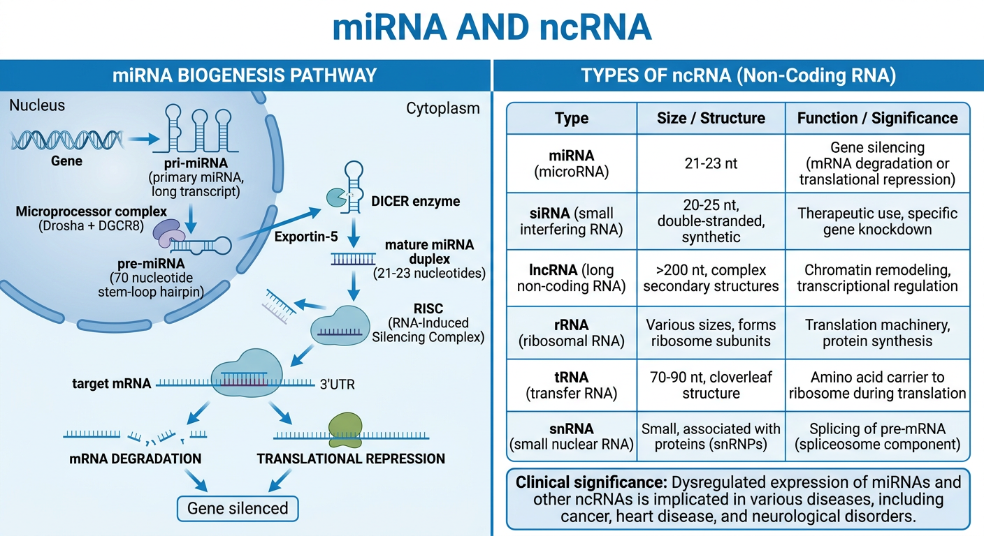
miRNA (microRNA) - Biogenesis Pathway:
NUCLEUS
  │
  ▼
Gene → transcribed → pri-miRNA (primary miRNA, long hairpin transcript)
  │
  ▼ Microprocessor complex (Drosha + DGCR8)
  │
pre-miRNA (precursor, ~70 nt stem-loop structure)
  │
  ▼ Exportin-5 exports to CYTOPLASM
  │
  ▼ DICER enzyme cleaves pre-miRNA
  │
Mature miRNA duplex (~21-23 nucleotides, single-stranded)
  │
  ▼ Loaded into RISC (RNA-Induced Silencing Complex)
  │
miRNA-RISC binds complementary 3'UTR of target mRNA
  │
  ├── Perfect complementarity → mRNA CLEAVAGE/DEGRADATION
  └── Imperfect complementarity → TRANSLATIONAL REPRESSION
  │
  ▼
GENE SILENCED (protein not produced)
Key Features of miRNA:
- Size: 21-23 nucleotides (single-stranded)
- Location: Encoded in genome; found in nucleus and cytoplasm
- Function: Post-transcriptional gene silencing - one miRNA can regulate hundreds of target mRNAs
- Targets: 3'UTR (untranslated region) of mRNA
- Clinical role: Dysregulated in cancer, cardiac disease; potential biomarkers and therapeutic targets
Types of Non-Coding RNA (ncRNA):
| Type | Size | Function | Clinical Relevance |
|---|---|---|---|
| miRNA | 21-23 nt | Post-transcriptional gene silencing via RISC | Dysregulated in cancer, heart failure |
| siRNA (small interfering RNA) | 20-25 nt, double-stranded | Sequence-specific mRNA degradation; synthetic/therapeutic | RNAi therapy (Patisiran for TTR amyloidosis - FDA approved) |
| lncRNA (long ncRNA) | >200 nt | Chromatin remodeling, gene regulation, splicing | HOTAIR, XIST (X-inactivation) |
| rRNA | Variable | Structural & catalytic component of ribosome | Essential for translation |
| tRNA | 73-93 nt | Amino acid carrier; anticodon-codon recognition | Essential for translation |
| snRNA | ~150 nt | Pre-mRNA splicing (spliceosome component) | Mutations → spinal muscular atrophy |
| snoRNA | ~60-300 nt | rRNA processing and modification | - |
| piRNA | 26-31 nt | Silences transposons in germline | - |
siRNA vs miRNA:
| Feature | miRNA | siRNA |
|---|---|---|
| Origin | Endogenous (own genome) | Exogenous / Synthetic |
| Structure | Single-stranded | Double-stranded |
| Complementarity | Imperfect | Perfect |
| Effect | Translational repression OR degradation | mRNA cleavage |
| Nuclear processing | Yes (Drosha) | Bypasses Drosha |
| Targets | Multiple mRNAs | Single specific mRNA |
3. Branched Chain Amino Acids (BCAAs)
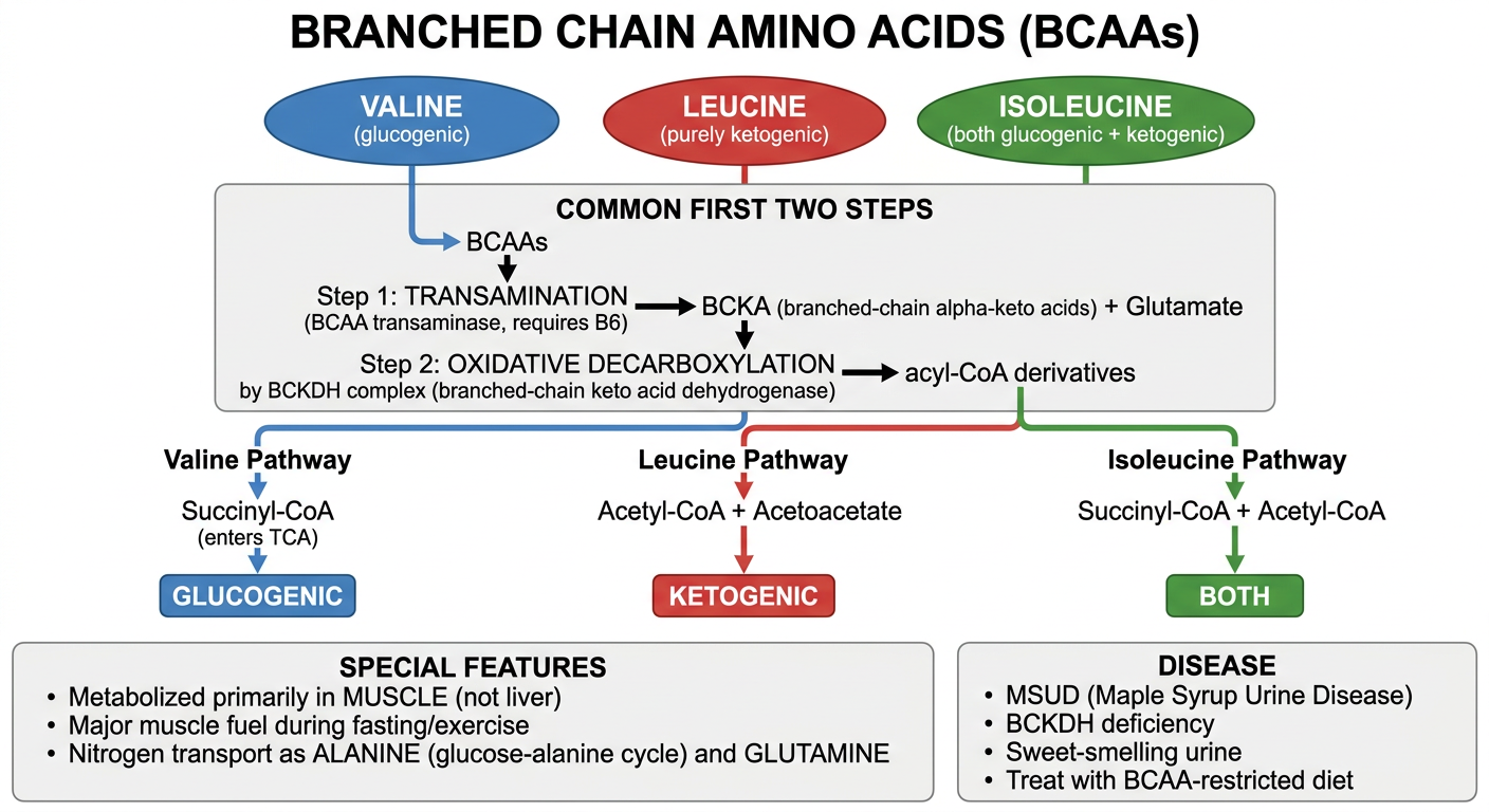
Special Features:
- The only amino acids metabolized primarily in skeletal muscle (not liver)
- Liver has very low BCAA transaminase activity
- Major fuel for muscle during fasting, exercise, and trauma
- Provide nitrogen for glutamine and alanine synthesis
Common Catabolic Pathway (First Two Steps Shared):
Step 1: TRANSAMINATION
BCAAs + α-Ketoglutarate
        ↓ BCAA Transaminase (requires Pyridoxal phosphate, B6)
α-Keto acids (BCKAs) + Glutamate

Step 2: OXIDATIVE DECARBOXYLATION (rate-limiting)
BCKAs + CoA + NAD⁺
        ↓ BCKDH Complex (Branched-Chain Keto Acid Dehydrogenase)
           (requires: TPP, Lipoate, FAD, NAD⁺, CoA - same as PDH)
Acyl-CoA derivatives + CO₂ + NADH
Individual Fate (After Common Steps):
| BCAA | Final Products | Metabolic Category |
|---|---|---|
| Valine | Succinyl-CoA (via propionyl-CoA) | Purely Glucogenic |
| Leucine | Acetyl-CoA + Acetoacetate | Purely Ketogenic (only BCAA that is) |
| Isoleucine | Succinyl-CoA + Acetyl-CoA | Both Glucogenic + Ketogenic |
Role in Inter-organ Nitrogen Transport:
Muscle: Glucose → Pyruvate
        BCAAs donate amino group to pyruvate → ALANINE
        Alanine released into blood → travels to Liver
Liver: Alanine → Pyruvate + NH₃ (→ Urea)
       Pyruvate → Glucose (gluconeogenesis) → back to muscle
BCAAs → transamination → Glutamate → Glutamine (via glutamine synthetase)
Glutamine carried to liver, kidney, gut for nitrogen disposal
BCAA Deficiency Disease:
MSUD - Maple Syrup Urine Disease:
- Defect: BCKDH complex (branched-chain keto acid dehydrogenase) - deficient
- Inheritance: Autosomal recessive
- Accumulation: Leucine, Isoleucine, Valine + their keto acids
- Features: Sweet maple syrup odor in urine and cerumen; encephalopathy, seizures, mental retardation
- Treatment: BCAA-restricted diet (especially Leucine); thiamine supplementation; liver transplant
- Toxic metabolite: Leucine most neurotoxic
4. Hyperuricaemia
Causes of Hyperuricaemia:
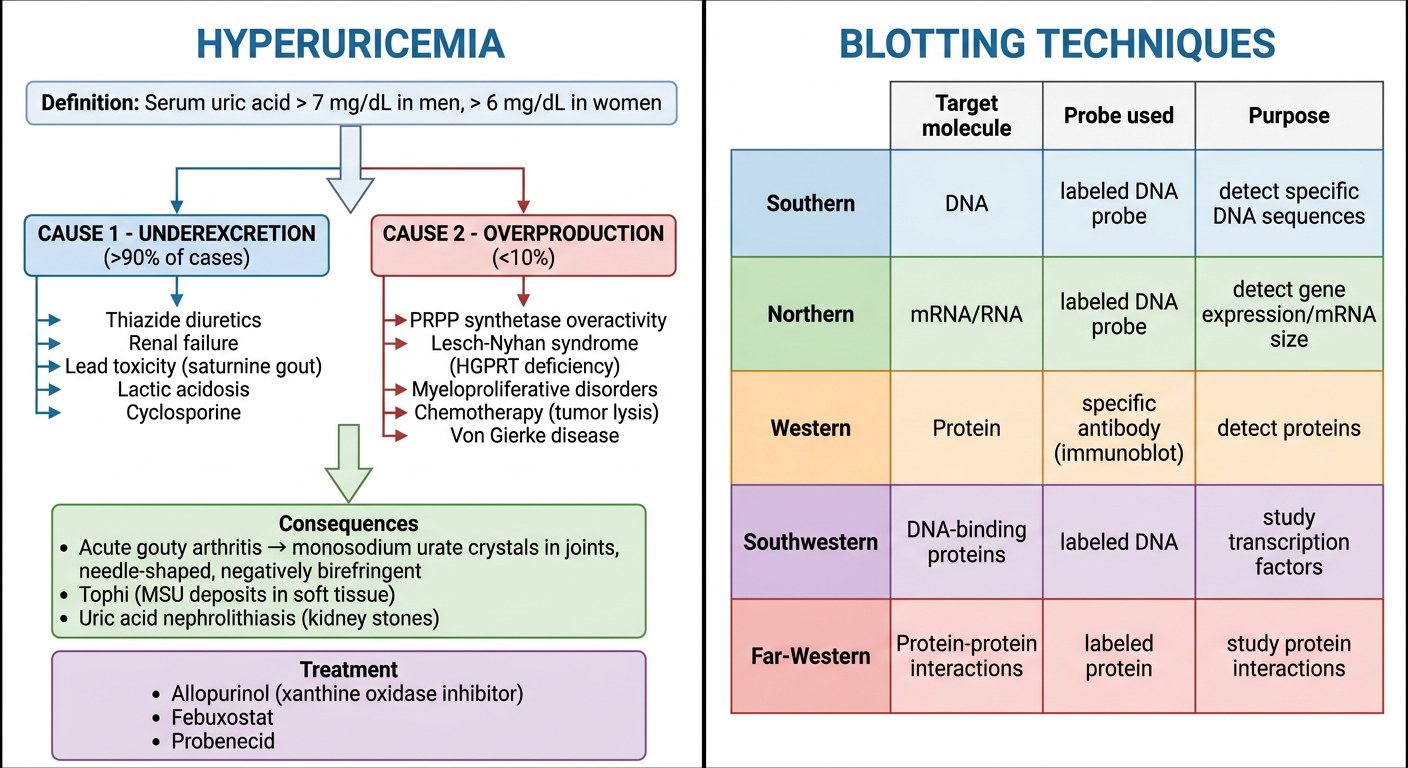
1. Underexcretion (>90% of cases):
| Type | Causes |
|---|---|
| Primary | Idiopathic excretory defects |
| Secondary | Renal failure (decreased GFR), Thiazide/loop diuretics, Aspirin (low dose), Lactic acidosis (lactate competes with urate for tubular secretion), Lead poisoning (Saturnine gout), Cyclosporine, Ethambutol |
2. Overproduction (<10% of cases):
| Type | Causes |
|---|---|
| Primary (genetic) | PRPP synthetase overactivity (X-linked) - increased PRPP → increased purine synthesis; Lesch-Nyhan syndrome (HGPRT deficiency - X-linked) - purines not salvaged → increased degradation to uric acid |
| Secondary | Myeloproliferative/lymphoproliferative disorders (increased cell turnover), Chemotherapy/radiation (tumour lysis syndrome), Psoriasis (increased skin cell turnover), Von Gierke disease (G6Pase deficiency - increased PRPP), Hereditary fructose intolerance |
Consequences (Clinical Features of Gout):
Hyperuricaemia
      ↓
Monosodium Urate (MSU) crystal deposition
      ├── JOINTS → ACUTE GOUTY ARTHRITIS
      │    (podagra = 1st MTP joint; needle-shaped, negatively birefringent crystals)
      │
      ├── SOFT TISSUES → TOPHI
      │    (ear pinna, tendons, olecranon bursa)
      │
      └── KIDNEY → URIC ACID NEPHROLITHIASIS
           (radiolucent stones on X-ray)
Key exam fact: Diagnosis confirmed by synovial fluid aspiration showing needle-shaped MSU crystals that are negatively birefringent under polarized light.
Treatment:
| Drug | Mechanism | Use |
|---|---|---|
| Allopurinol | Xanthine oxidase inhibitor → reduces uric acid production | Chronic prevention |
| Febuxostat | Xanthine oxidase inhibitor (non-purine analog) | Chronic prevention |
| Probenecid | Blocks tubular reabsorption of urate → increases excretion | Underexcretors |
| Colchicine | Inhibits tubulin polymerization → impairs neutrophil chemotaxis | Acute attack |
| NSAIDs/Steroids | Anti-inflammatory | Acute attack |
| Rasburicase | Recombinant uricase → converts uric acid to allantoin | Tumour lysis syndrome |
5. Blotting Techniques
Complete Comparison Table:
| Blot | Target Molecule | Separation By | Probe Used | Purpose |
|---|---|---|---|---|
| Southern | DNA | Agarose gel; size | Labeled DNA probe | Detect specific DNA sequences, mutations, RFLP |
| Northern | RNA (mRNA) | Agarose-formaldehyde gel; size | Labeled DNA probe | Detect gene expression; determine mRNA size and quantity |
| Western (Immunoblot) | Protein | SDS-PAGE; size | Specific antibody | Detect and quantify specific proteins |
| Southwestern | DNA-binding proteins | SDS-PAGE | Labeled DNA | Study transcription factors |
| Far-Western | Protein-protein interactions | SDS-PAGE | Labeled protein | Study protein-protein binding |
Southern Blotting - Step by Step:
1. Extract DNA from cells (e.g., WBCs)
2. Cleave with Restriction Endonuclease → millions of fragments
3. Separate by Agarose Gel Electrophoresis (size-based)
4. Denature DNA (NaOH) → single stranded
5. Transfer (blot) to Nitrocellulose/Nylon Membrane
6. Hybridize with Radiolabeled (³²P) or fluorescent DNA Probe
7. Wash off unbound probe
8. Autoradiography → Bands revealed
   ↓
   Band pattern = specific DNA sequence identified
- Detection of gene mutations (large deletions, insertions)
- Trinucleotide repeat expansion (e.g., Fragile X, Huntington)
- RFLP analysis for genetic mapping
- Diagnosing infections (e.g., HIV proviral DNA)
Northern Blotting:
- Same as Southern but uses mRNA as target (in formaldehyde gel to denature RNA)
- Uses RNase inhibitors during RNA extraction
- Gives mRNA size AND quantity - unique advantage over RT-PCR
- Now largely replaced by RT-PCR and RNA sequencing
Western Blotting (Immunoblot):
- Proteins separated by SDS-PAGE (by molecular weight)
- Transferred to membrane → incubated with primary antibody → secondary antibody (enzyme/fluorescent labeled) → detection
- Confirmatory test for HIV (detects anti-HIV antibodies against specific viral proteins)
- Used for: protein identification, post-translational modifications, protein levels in disease
Memory Aid:
"Some Nights Will Shower Frequently"
Southern → DNA
Northern → RNA
Western → Protein
Southwestern → DNA-binding protein
Far-Western → Protein-protein interaction
Sources: Guyton & Hall Medical Physiology; Lippincott Illustrated Reviews Biochemistry 8th Ed; Basic Medical Biochemistry - A Clinical Approach 6e; Harper's Illustrated Biochemistry 32nd Ed; Goldman-Cecil Medicine; Robbins & Cotran Pathologic Basis of Disease
Summary for all these questions
🧬 MBBS Biochemistry - Master Summary Sheet
SESSION 1 TOPICS
1. Regulation of Gene Expression
| Level | Key Point |
|---|---|
| Prokaryote | Lac operon - repressor binds operator → blocks RNA pol; allolactose removes repressor → transcription ON |
| Controls | Negative (repressor) + Positive (CAP-cAMP) |
| Eukaryote | Chromatin remodeling (histone acetylation) → transcription factors → RNA Pol II |
| Other levels | mRNA processing, transport, translation, miRNA |
2. Principle of Electrophoresis
| Factor | Effect |
|---|---|
| Size | Smaller = faster migration |
| Charge | More negative = moves faster to anode (+) |
| Shape | Linear > branched > circular |
3. Northern Blotting
SESSION 2 TOPICS
4. Chromatography - Principle & Types
| Type | Basis | Use |
|---|---|---|
| Ion exchange | Charge | Protein/amino acid purification |
| Gel filtration | Size | MW determination (large elutes FIRST) |
| Affinity | Specific binding (lock-key) | Most specific; antibody/enzyme purification |
| Paper/TLC | Polarity | Rf = solute distance / solvent distance |
| HPLC | High pressure liquid | Drug levels, HbA1c |
5. Products Derived from Glycine
| Product | Partner/Path |
|---|---|
| Heme | + Succinyl-CoA (ALA synthase, B6) |
| Creatine | + Arginine + SAM |
| Glutathione | + Glu + Cys |
| Bile salts | Conjugated with bile acids |
| Purines | C4, C5, N7 of purine ring |
| Serine | ⇌ via serine hydroxymethyltransferase (reversible) |
| Oxalate | Via glyoxylate → renal stones |
6. Salient Features of Genetic Code
| Feature | Meaning |
|---|---|
| Triplet | 3 bases = 1 codon |
| Degenerate | 64 codons for 20 AAs; multiple codons per AA |
| Universal | Same in all organisms (except mitochondria) |
| Non-ambiguous | 1 codon = 1 amino acid only |
| Direction | Read 5' → 3' |
| Commaless | No punctuation between codons |
| Non-overlapping | Each base belongs to one codon |
| Start/Stop | AUG (Met); UAA, UAG, UGA (stop) |
7. Quality Control
- Levey-Jennings chart - daily QC plotted around mean ± SD
- Westgard rules - 1₂ₛ = warning; 1₃ₛ = reject; 2₂ₛ = systematic error; R₄ₛ = random error
- Internal QC = daily control sera (precision)
- External QC (EQA) = interlaboratory comparison (accuracy)
- CV% = (SD/Mean) × 100; good lab < 5%
SESSION 3 TOPICS
8. 25 Tumour Markers
| Marker | Cancer |
|---|---|
| AFP | HCC, Testicular (yolk sac) |
| CEA | Colorectal, Pancreatic |
| CA-125 | Ovarian |
| CA 19-9 | Pancreatic, Cholangiocarcinoma |
| PSA | Prostate |
| β-HCG | Choriocarcinoma, Testicular |
| LDH | Lymphoma, Testicular |
| Calcitonin | Medullary thyroid |
| 5-HIAA | Carcinoid |
| VMA | Pheochromocytoma, Neuroblastoma |
| NSE | Neuroblastoma, SCLC |
| Bence Jones protein | Multiple myeloma |
9. Gene Therapy
| Type | Target | Inherited? | Status |
|---|---|---|---|
| Somatic | Body cells | No | Approved |
| Germline | Egg/sperm | Yes | Not approved |
10. Quaternary Structure of Protein
| Protein | Subunits | Type |
|---|---|---|
| Hemoglobin | 2α + 2β | Hetero-tetramer |
| Collagen | 3 chains | Trimer |
| IgG | 2H + 2L | Hetero-tetramer |
| LDH | 4 (M/H mix) | Isoenzymes |
11. Free Radicals
| ROS | Source | Removed By |
|---|---|---|
| O₂•⁻ Superoxide | Mitochondria, NADPH oxidase | SOD |
| H₂O₂ | SOD reaction, peroxisomes | Catalase, GSH peroxidase |
| •OH Hydroxyl | Fenton reaction (Fe²⁺+H₂O₂), radiation | None enzymatic (most dangerous) |
| ONOO⁻ Peroxynitrite | O₂•⁻ + NO | Peroxiredoxins |
SESSION 4 TOPICS
12. Toxins - Causes & Deficiency
| Toxin | Deficiency | Disease |
|---|---|---|
| Alcohol | B1 (Thiamine), Folate | Wernicke-Korsakoff, Megaloblastic anemia |
| INH | B6 (Pyridoxine) | Peripheral neuropathy |
| Methotrexate | Folate (DHFR inhibition) | Megaloblastic anemia |
| Metformin | B12 | Neuropathy |
| Lead | Heme synthesis (ALA dehydratase↓) | Microcytic anemia (sideroblastic) |
| Cyanide | Blocks Complex IV | Histotoxic hypoxia |
13. miRNA & ncRNA
Gene → pri-miRNA → (Drosha/DGCR8) → pre-miRNA → Exportin-5 →
Cytoplasm → DICER → mature miRNA → RISC → mRNA silenced
14. Branched Chain Amino Acids
BCAA → (BCAA transaminase, B6) → BCKA → (BCKDH complex) → Acyl-CoA
15. Hyperuricaemia
| Cause | Examples |
|---|---|
| Underexcretion (>90%) | Thiazides, Renal failure, Lead, Lactic acidosis, Cyclosporine |
| Overproduction (<10%) | PRPP synthetase overactivity, Lesch-Nyhan (HGPRT↓), Myeloproliferative disorders, Chemotherapy (tumour lysis), Von Gierke disease |
16. Blotting Techniques
| Blot | Target | Probe | Key Use |
|---|---|---|---|
| Southern | DNA | Labeled DNA | Mutation detection, RFLP |
| Northern | mRNA | Labeled DNA | Gene expression + mRNA size |
| Western | Protein | Antibody | Protein detection (HIV confirmatory) |
| Southwestern | DNA-binding proteins | Labeled DNA | Transcription factor study |
| Far-Western | Protein-protein | Labeled protein | Protein interactions |
QUICK REVISION TABLE - All 16 Topics
| # | Topic | Key One-liner |
|---|---|---|
| 1 | Gene Regulation | Lac operon (prokaryote); histone acetylation (eukaryote) |
| 2 | Electrophoresis | Size + charge + shape determine migration |
| 3 | Northern Blot | mRNA detection; size + quantity |
| 4 | Chromatography | Affinity=most specific; gel filtration=size; HPLC=clinical |
| 5 | Glycine products | HCGBPSO - Heme, Creatine, GSH, Bile, Purines, Serine, Oxalate |
| 6 | Genetic Code | 64 codons; degenerate; universal; AUG start; UAA/UAG/UGA stop |
| 7 | Quality Control | Westgard rules; L-J chart; Internal + External QC |
| 8 | Tumour Markers | AFP=liver/testis; CA-125=ovary; PSA=prostate; CEA=colon |
| 9 | Gene Therapy | AAV=best vector; somatic only approved; SMA/Hemophilia B treated |
| 10 | Quaternary Structure | 2+ subunits; non-covalent bonds; Hb=cooperativity |
| 11 | Free Radicals | •OH most dangerous; SOD→Catalase→GSH peroxidase defense |
| 12 | Toxins/Deficiency | INH→B6; MTX→Folate; Lead→heme; Cyanide→Complex IV |
| 13 | miRNA & ncRNA | pri→pre→mature miRNA→RISC→gene silenced |
| 14 | BCAAs | Val/Leu/Ile; muscle metabolism; BCKDH defect=MSUD |
| 15 | Hyperuricaemia | >90% underexcretion; Lesch-Nyhan; Allopurinol inhibits XO |
| 16 | Blotting Techniques | S=DNA, N=RNA, W=Protein; all need gel + membrane + probe |
Exam Tip: For 5-mark short answers in Vasudevan-based exams: Write definition → mechanism/pathway → table/diagram → clinical significance in every answer. This format reliably scores full marks.