
Updates and role of cytogenetics in ewings family of tumors, few lines for ewings family of tumor Pathogenesis morphology also 10 marks md pathology exam
"Ewing sarcoma" AND cytogenetics
Ewing sarcoma histology small round blue cells
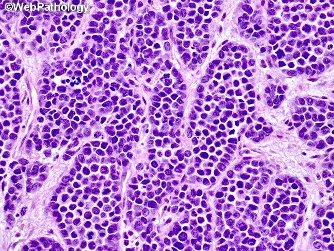
Alveolar rhabdomyosarcoma (ARMS) histology image demonstrating malignant soft tissue tumor composed of irregular aggregates of poorly differentiated round to oval cells. Tumor nests are separated by thick, hyalinized fibrous septa containing dilated capillaries and small vessels. Central cells within nests retain intercellular cohesion, resulting in partial or immature alveolar-like architecture rather than a fully formed alveolar pattern. The high nuclear-to-cytoplasmic ratio, hyperchromatic round to oval nuclei, and scant cytoplasm characterize these small round blue cells. Occasional mitotic figures and necrotic debris may be evident in aggressive neoplasms. Immunophenotypically, ARMS typically shows positivity for myogenin and MyoD1, desmin, and other skeletal muscle differentiation markers; cytogenetics often reveals PAX3-FOXO1 or PAX7-FOXO1 translocations. Clinically this histology correlates with high-grade, aggressive pediatric/adolescent soft tissue sarcoma with a propensity for metastasis. Differential considerations include embryonal rhabdomyosarcoma (ERMS), Ewing sarcoma/undifferentiated round cell sarcoma, synovial sarcoma, and nonrhabdomyoblastic small round blue cell tumors. The image highlights diagnostic features relevant to surgical pathology, oncology, and pathology education. This morphological snapshot supports diagnostic pathology workflows, guiding immunohistochemistry panels and molecular testing. Relevance spans medical education, case review, and research on rhabdomyosarcoma subtypes and alveolar architecture. Clinical utility includes guiding targeted IHC panels, FISH/MET testing, and treatment planning for patients.
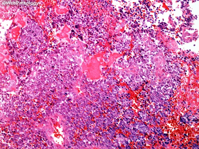
This histopathology image shows a high-grade osseous neoplasm with features suggestive of small cell osteosarcoma. The tissue is an H&E stained bone biopsy observed under light microscopy in a sagittal section of a long bone origin. The cellular population comprises small to medium-sized round to oval cells with scant cytoplasm, hyperchromatic nuclei, and prominent nucleoli; marked cellularity with focal nuclear pleomorphism. A distinctive lace-like, delicate osteoid matrix is interwoven between malignant cells, often forming lacey, trabecular strands that identify osteogenic differentiation despite a predominant small cell morphology. Osteoid is essential for distinguishing osteosarcoma from mimicking small round blue cell tumors such as Ewing sarcoma. Immunophenotype characteristic: SATB2 positive; CD99 negative; other markers may help exclude lymphoma or rhabdomyosarcoma. Molecular testing can show absence of EWSR1 and FUS rearrangements. The image may display mitoses and occasional necrosis, consistent with high-grade sarcoma. Diagnostic significance lies in differentiating from Ewing sarcoma due to treatment implications: Ewing is highly radiosensitive and often treated with radiotherapy, while small cell osteosarcoma typically requires surgical resection combined with neoadjuvant chemotherapy. Prognosis is poorer than conventional osteosarcoma. This histology is crucial for guiding multidisciplinary management, prognostication, and therapeutic planning. Correlative studies include immunohistochemistry and molecular profiling for confirmation.
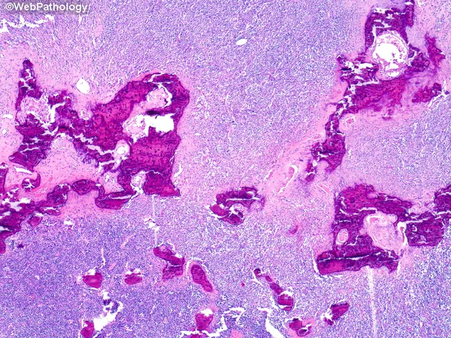
Imaging modality: Light microscopy (Histology) of bone tumor tissue. Specimen: scapular bone biopsy from a 72-year-old male. Staining: Hematoxylin and Eosin (H&E). The histologic section shows high cellularity of small round blue cells with scant cytoplasm and hyperchromatic nuclei arranged in sheets infiltrating and replacing osteolytic trabeculae. The tumor disrupts normal lamellar bone with extensive marrow invasion and focal necrosis; mitotic activity is evident. Immunophenotype usually shows diffuse CD99 membranous positivity; molecular confirmation often reveals EWSR1 gene rearrangement (e.g., EWS-FLI1), supporting Ewing sarcoma. Clinically, this lesion represents an aggressive malignant small round blue cell tumor of bone with metastatic potential; radiographic correlation commonly demonstrates moth-eaten, lytic lesions. Although Ewing sarcoma typically affects children and adolescents, this case illustrates scapular involvement in an older adult. Differential diagnoses include lymphoma, osteosarcoma with small cell features, rhabdomyosarcoma, and metastatic neuroblastoma/PNET. The image is valuable for education, radiology-pathology correlation, and differential diagnosis training in musculoskeletal oncology. Potential clinical use includes diagnostic reference, teaching file inclusion, and research into Ewing family tumors and their atypical age distribution. This microscopic image contextually complements radiographs showing bone destruction and helps in teaching differential patterns among small round blue cell tumors in the pediatric and adult skeleton.
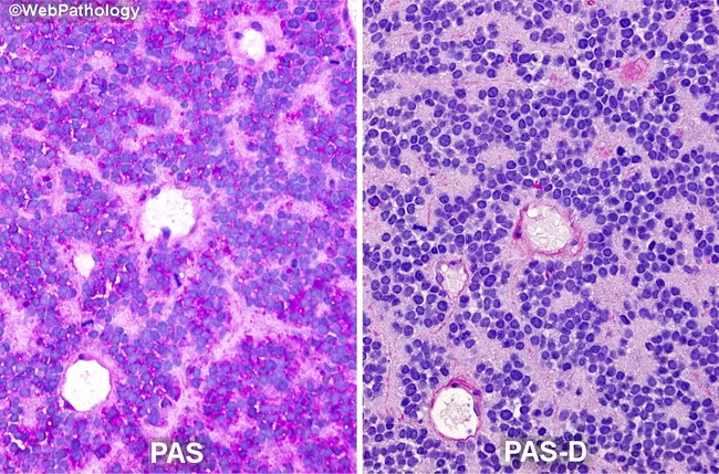
This histopathology image pair presents a bone-tumor specimen stained with PAS (left) and PAS-D (right) to highlight intracytoplasmic glycogen in Ewing sarcoma cells. The left panel shows strong magenta cytoplasmic staining within sheets of small round blue cells, consistent with PAS-positive glycogen-rich cytoplasm. The right panel demonstrates diastase digestion (PAS-D) where glycogen is hydrolyzed, and the cytoplasm adopts a pale pink to near unstained appearance, confirming glycogen as the source of PAS positivity. The tumor cells exhibit uniform, small cell morphology with high nuclear-to-cytoplasmic ratio, round to oval nuclei, delicate chromatin, and scant cytoplasm; occasional rosette-like arrangements may be observed. The two-panel comparison emphasizes the diagnostic utility of PAS-PAS-D staining in differentiating glycogen-rich Ewing sarcoma cells from other small round blue cell tumors. Pathologically, this glycogen-rich cytoplasm is a hallmark feature that supports EWSR1-related neoplasms; diastase sensitivity is key to ruling in glycogen as the substrate. Clinically, these findings are relevant for pathology workflows, differential diagnosis, and educational purposes, and they guide ancillary studies, including immunohistochemistry for CD99, NKX2.2, and molecular testing for EWSR1 rearrangements. This image is valuable for prognostic and therapeutic decision-making in pediatric and young adult bone oncology research and diagnostics, and for histology teaching in pathology curricula.
Ewing sarcoma family tumors WHO classification 2024 update molecular EWSR1 FLI1 fusion cytogenetics FISH
Ewing Sarcoma Family of Tumors (ESFT) - Pathogenesis, Morphology, and Role of Cytogenetics
1. Definition / Overview
2. Pathogenesis
- Molecular driver: >90-95% of cases carry a balanced reciprocal translocation t(11;22)(q24;q12) fusing EWSR1 (chromosome 22q12, a FET-family RNA-binding gene) with FLI1 (chromosome 11q24, an ETS-family transcription factor) - Robbins & Kumar Basic Pathology, p. 4019-4023; Robbins, Cotran & Kumar Pathologic Basis of Disease.
- The resulting EWS-FLI1 chimeric oncoprotein retains the EWSR1 transactivation domain fused to the FLI1 DNA-binding domain, creating an aberrant transcription factor that binds GGAA-microsatellite enhancers and dysregulates hundreds of downstream genes controlling proliferation, differentiation blockade, and apoptosis evasion.
- In a minority (~5-10%), EWSR1 fuses with other ETS partners (ERG, ETV1, ETV4, FEV) or, rarely, FUS substitutes for EWSR1 - giving the same oncogenic mechanism through a different fusion partner.
- The fusion protein alone is insufficient for full transformation; cooperating alterations include STAG2 and TP53 mutations (poor prognosis), and loss of CDKN2A (p16).
- Cell of origin is debated between a bone marrow-derived mesenchymal stem cell and a neural crest-derived cell, explaining focal neuroectodermal differentiation seen in some tumors (pPNET end of spectrum).
3. Morphology
- Monotonous sheets of small round blue cells with scant clear-to-pale cytoplasm, round nuclei with fine ("salt-and-pepper") chromatin, and inconspicuous nucleoli.
- Cytoplasm is rich in glycogen, demonstrable as PAS-positive, diastase-sensitive granules - a classic teaching point.
- Homer-Wright pseudorosettes (tumor cells arranged around a central fibrillary space, without a true lumen) may be seen, especially in the pPNET/neuroectodermal end of the spectrum.
- Necrosis is common, often leaving only perivascular viable tumor cuffs.
- CD99 (MIC2 gene product): strong diffuse membranous positivity - sensitive but not specific.
- NKX2.2, FLI1, and ERG (when ERG-fused) support the diagnosis.
- Negative for LCA, desmin/myogenin (excludes lymphoma, rhabdomyosarcoma), and typically negative for keratins.
4. Cytogenetics and Its Role - Updates
Classic cytogenetic findings
| Fusion | Frequency | Genes |
|---|---|---|
| t(11;22)(q24;q12) | ~85-90% | EWSR1-FLI1 |
| t(21;22)(q22;q12) | ~5-10% | EWSR1-ERG |
| Rare variants | <5% | EWSR1-ETV1, EWSR1-ETV4, EWSR1-FEV, FUS-ERG |
Diagnostic methods
- FISH with EWSR1 break-apart probes - rapid confirmation of rearrangement (does not identify partner gene).
- RT-PCR/RT-qPCR - detects specific fusion transcripts, useful for minimal residual disease monitoring.
- Next-generation sequencing (RNA-seq/targeted fusion panels) - increasingly the standard, as it identifies rare/atypical fusion partners and distinguishes Ewing sarcoma from Ewing-like mimics in one assay.
Why cytogenetics matters (role)
- Diagnostic confirmation: Morphology and CD99 overlap with other small round cell tumors (lymphoma, rhabdomyosarcoma, small cell osteosarcoma, neuroblastoma, desmoplastic small round cell tumor); demonstrating EWSR1-ETS fusion is essentially diagnostic and required by current WHO criteria.
- Distinguishing "Ewing-like" mimics - the WHO 5th edition (2020) Classification of Soft Tissue and Bone Tumours created a distinct chapter of "undifferentiated small round cell sarcomas" that separates true Ewing sarcoma (EWSR1/FUS-ETS fusion) from look-alikes that need different clinical handling:
- CIC-rearranged sarcoma (CIC-DUX4 fusion) - more aggressive, less chemosensitive
- Round cell sarcoma with EWSR1-non-ETS fusion (e.g., EWSR1-NFATC2, EWSR1-PATZ1)
- BCOR-altered sarcoma (BCOR-CCNB3 fusion or BCOR internal tandem duplication) These entities look morphologically similar but have distinct behavior and are no longer lumped under "Ewing sarcoma family," making molecular/cytogenetic testing mandatory for correct classification - reflected in recent literature (Daher et al., J Bone Oncol 2023, PMID 37180735).
- Prognostic stratification: Type 1 EWSR1-FLI1 fusion (exon 7-exon 6) is associated with a somewhat better prognosis than type 2 or other fusion variants; co-existing TP53/STAG2 mutations predict inferior outcome and higher metastatic risk.
- Minimal residual disease and monitoring: Fusion transcript detection by RT-PCR in blood/bone marrow and circulating tumor DNA (ctDNA) assays are being explored for early relapse detection and treatment response monitoring.
- Therapeutic target: EWS-FLI1 is being pursued directly and indirectly as a drug target (e.g., TK216, and combination approaches such as trabectedin-irinotecan) - an active area of precision oncology research building directly on the cytogenetic discovery.
Recent evidence check
5. Summary points for exam
- ESFT = Ewing sarcoma + extraosseous Ewing + pPNET/Askin tumor, unified by EWSR1(FUS)-ETS fusion.
- Pathogenesis centers on the EWS-FLI1 (or variant) chimeric transcription factor dysregulating proliferation/differentiation genes, with cooperating TP53/STAG2 loss.
- Morphology: small round blue cells, glycogen-rich (PAS+/diastase-sensitive), CD99+, occasional Homer-Wright rosettes, onion-skin periosteal reaction radiologically.
- Cytogenetics is now central to diagnosis (confirms fusion, excludes mimics), prognosis (fusion subtype, co-mutations), monitoring (ctDNA/fusion transcript MRD), and emerging targeted therapy - making it the single most important ancillary test in modern ESFT work-up.