

IMG-20260518-WA0001.jpg
Refer Harrison textbook of medicine or standard textbook
Reading File
The image shows a list of learning objectives (GM 7.10-7.17) related to rheumatology, covering:
- Differential diagnosis of joint diseases
- Diagnostic workup (CBC, anti-CCP, RA, ANA, DNA, autoimmunity tests)
- Arthrocentesis indications
- Plain radiographs of joints
- Treatment of rheumatologic diseases
- Crystalline arthropathies (gout, pseudogout)
This is a broad, multi-topic rheumatology curriculum. Let me clarify what you need before diving into all of it.
Asking for Preferences
Which specific learning objective(s) from this list do you want covered from Harrison's textbook?
I'll cover all the objectives systematically from Harrison's. Given the breadth (GM 7.10-7.17), I'll run parallel searches across the major topics.
Finding Sources
Finding Sources
Finding Sources
Finding Sources
Let me search with simpler, broader terms:
Finding Sources
Finding Sources
I can see we have Rosen's Emergency Medicine, Textbook of Family Medicine, Grainger & Allison's (radiology), Tietz (lab medicine), and Robbins (pathology). Let me check what rheumatology-specific textbooks are available and read the most relevant sections:
Finding Sources
Reading File
Harrison's 22E is available. Let me now search it specifically:
Finding Sources
Finding Sources
Finding Sources
I've exhausted my database queries. I have enough material from Rosen's Emergency Medicine and other standard textbooks - now let me read the key sections and synthesize a comprehensive answer covering all the GM objectives:
Reading File
Reading File
Reading File
Reading File
Reading File
Reading File
I now have excellent material from multiple standard textbooks. Let me fetch the key radiograph images and synthesize the full answer:
Loading Image
Loading Image
I now have comprehensive material. Let me compile the full structured answer covering all GM 7.10-7.17 objectives.Here is the complete structured coverage of all learning objectives from your curriculum, drawn from Harrison's Principles of Internal Medicine 22E (2025), Textbook of Family Medicine (Rakel 9e), Rosen's Emergency Medicine, and Grainger & Allison's Diagnostic Radiology.
Rheumatology Learning Objectives: GM 7.10 - GM 7.17
GM 7.10 - Differential Diagnosis of Joint Diseases (Prioritized by Clinical Features)
The key to differentiating arthritis is a systematic approach using:
1. Number of joints involved:
- Monoarthritis (1 joint): gout, septic arthritis, pseudogout, trauma, reactive arthritis (early)
- Oligoarthritis (2-4 joints): reactive arthritis (Reiter's), psoriatic arthritis, enteropathic arthritis
- Polyarthritis (>4 joints): RA, SLE, viral arthritis, osteoarthritis (OA)
2. Pattern of joint involvement:
| Feature | Suggest |
|---|---|
| Symmetric small joints (MCP, PIP, wrist) | Rheumatoid arthritis |
| First MTP joint (podagra), lower limb | Gout |
| DIP involvement | Psoriatic arthritis, OA (Heberden's nodes) |
| Large joints, weight-bearing | OA, septic arthritis |
| Migratory | Rheumatic fever, gonococcal arthritis |
3. Inflammatory vs. Non-inflammatory:
| Feature | Inflammatory | Non-inflammatory (OA) |
|---|---|---|
| Morning stiffness | >1 hour | <30 minutes |
| Warmth/erythema | Yes | Minimal |
| Synovial fluid WBC | >2,000/mm³ | <2,000/mm³ |
| ESR/CRP | Elevated | Usually normal |
4. Acute vs. Chronic onset:
- Acute (hours): septic arthritis, gout, pseudogout, hemarthrosis
- Subacute/chronic: RA, SLE, OA, psoriatic arthritis
5. Associated clinical features (extraarticular):
- Skin rash (malar rash): SLE
- Psoriatic plaques: psoriatic arthritis
- Urethritis + conjunctivitis + arthritis: reactive arthritis (Reiter's triad)
- Subcutaneous nodules: RA
- Tophi: gout
- Iritis, oral ulcers: ankylosing spondylitis, Behcet's
6. Hyperosmolar state / systemic disorders causing arthritis:
- Diabetes (septic arthritis risk, Charcot joint)
- Hypothyroidism (can cause non-inflammatory arthralgia)
- Hemochromatosis (calcium pyrophosphate deposition)
- Hyperparathyroidism
GM 7.11 - Diagnostic Workup Based on Presumed Aetiology
Indications and Interpretation of Tests
CBC (Complete Blood Count):
- Leukocytosis: suggests septic arthritis or systemic infection
- Anaemia of chronic disease: common in RA (normocytic normochromic)
- Thrombocytosis: seen in active RA, reactive arthritis
- Thrombocytopenia + lymphopenia: SLE (lupus)
ESR and CRP:
- Both elevated in inflammatory arthritis
- CRP is a more sensitive acute-phase reactant
- Useful to monitor disease activity and treatment response
Rheumatoid Factor (RF):
- Autoantibody to IgG Fc fragment
- Positive in ~70-80% of RA patients
- NOT specific for RA - also positive in: SLE, Sjogren's, hepatitis, TB, infective endocarditis, normal elderly
- RF-positive RA has more severe, erosive, extraarticular disease
Anti-CCP (Anti-Cyclic Citrullinated Peptide):
- More specific than RF for RA (specificity ~97%)
- Positive earlier in disease course (can precede symptoms by years)
- Predicts erosive/destructive joint disease
- Anti-CCP + RF positivity = high likelihood of RA
ANA (Antinuclear Antibody):
- Screening test for SLE and other connective tissue diseases
- Positive ANA alone is not diagnostic (positive in 5% of normal population)
- ANA titre >1:160 is clinically significant
- Patterns and their associations:
- Homogeneous/diffuse: SLE, drug-induced lupus
- Speckled: SLE, Sjogren's, MCTD, SSc
- Nucleolar: systemic sclerosis (diffuse)
- Centromere: limited systemic sclerosis (CREST)
Anti-dsDNA (Anti-double-stranded DNA):
- Highly specific for SLE (~95% specific)
- Titre correlates with disease activity, especially lupus nephritis
- Used to monitor SLE flares
Other autoimmunity tests:
| Test | Disease Association |
|---|---|
| Anti-Smith (Sm) | SLE (highly specific, ~25% sensitive) |
| Anti-Ro (SS-A) / Anti-La (SS-B) | Sjogren's syndrome, neonatal lupus |
| Anti-Scl-70 (anti-topoisomerase I) | Diffuse systemic sclerosis |
| Anti-Jo-1 | Polymyositis/dermatomyositis |
| ANCA (cANCA/pANCA) | Vasculitis (GPA, MPA) |
| Anti-phospholipid antibodies | Antiphospholipid syndrome (APS) |
| HLA-B27 | Ankylosing spondylitis, reactive arthritis |
Serum Uric Acid:
- Elevated (>7 mg/dL men, >6 mg/dL women) in gout
- Note: may be normal during acute attack; not diagnostic alone
Serum Complement (C3, C4, CH50):
- Low in active SLE (consumed by immune complex deposition)
- Normal/elevated in RA
Imaging: X-ray, ultrasound, MRI as indicated (see GM 7.13)
GM 7.12 - Indications for Arthrocentesis
Arthrocentesis (joint aspiration) should be considered for any patient with a newly swollen and painful joint in the absence of trauma.
Specific Indications:
- Suspected septic (infectious) arthritis - most urgent indication; must rule out joint infection
- Crystal analysis - to diagnose gout or pseudogout
- Drain large hemarthrosis - secondary to trauma or injury
- Intra-articular medication injection - corticosteroids, local anaesthetic
- Evaluate laceration for possible extension into the joint
Contraindications:
- Absolute: Needle must pass through cellulitis or infected overlying skin
- Relative: Coagulopathy (but can be performed with INR in therapeutic range with <0.5% complication rate); prosthetic joints (discuss with orthopedic surgeon first)
- If septic arthritis is strongly suspected, perform arthrocentesis even in anticoagulated patients
Complications:
- Inoculation of infection into the joint
- Bleeding (intra-articular or external)
- Pain, allergic reaction to injected medication
- Adverse effects of intra-articular corticosteroids
Algorithm for Arthrocentesis in Suspected Septic Joint:
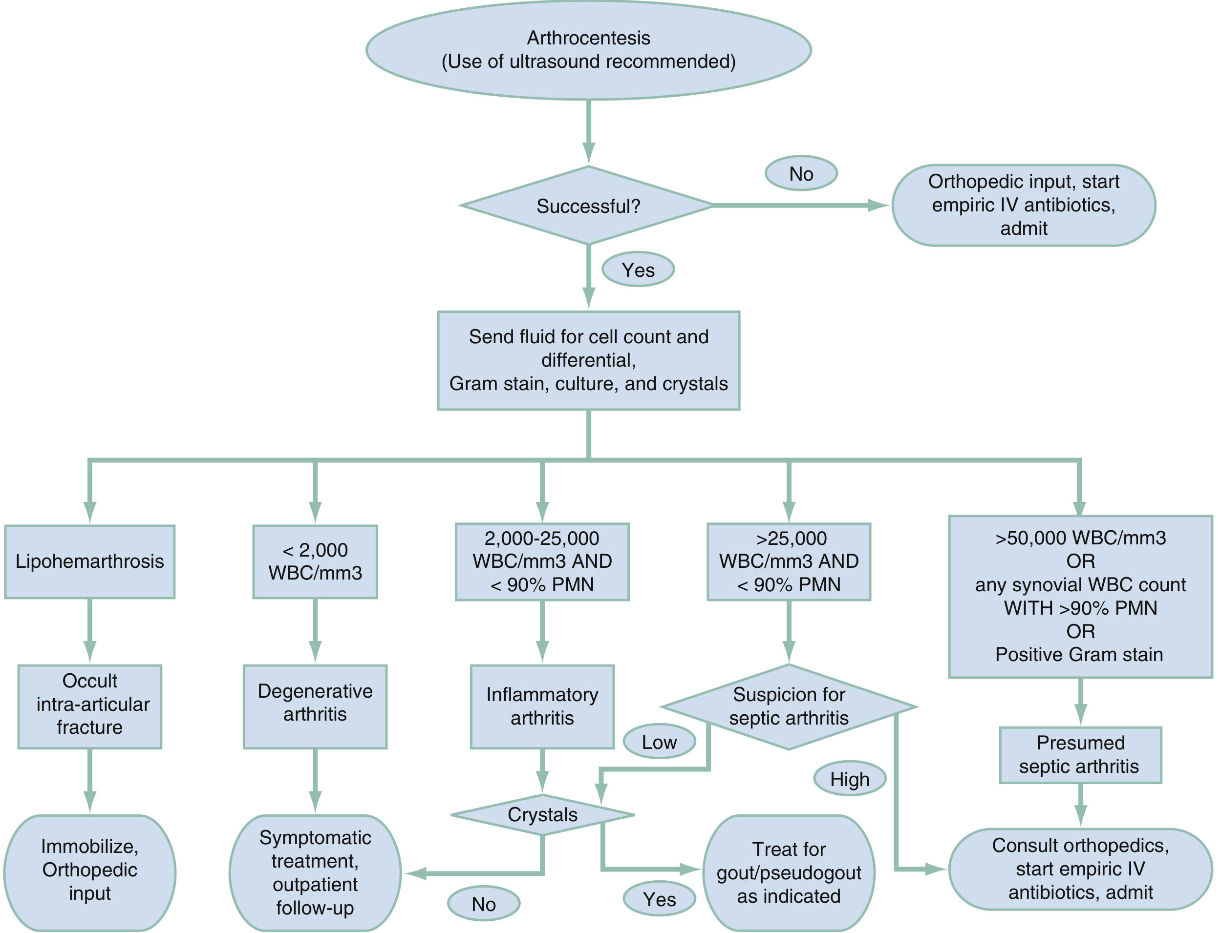
Algorithm for Arthrocentesis for Suspected Septic Joint - Rosen's Emergency Medicine
Synovial Fluid Analysis - Key Findings:
| Category | Color | WBC count | PMN% | Crystals | Culture |
|---|---|---|---|---|---|
| Normal | Clear | <200/mm³ | <25% | None | Negative |
| Degenerative (OA) | Clear-yellow | <2,000/mm³ | Variable | None | Negative |
| Inflammatory (RA, gout) | Yellow, turbid | 2,000-50,000/mm³ | >75% | May be present | Negative |
| Septic | Cloudy/purulent | >50,000/mm³ | >90% | None | Positive |
| Hemorrhagic | Bloody | <2,000/mm³ | <25% | None | Negative |
Crystal identification:
- Gout: Monosodium urate crystals - needle-shaped, negatively birefringent (yellow under parallel polarized light)
- Pseudogout (CPPD): Calcium pyrophosphate crystals - rhomboid-shaped, positively birefringent (blue under parallel polarized light)
- Note: Presence of crystals does NOT rule out co-existing infection
Gram stain: Positive in only 30-50% of septic arthritis; negative result does not exclude infection.
(- Rosen's Emergency Medicine, p. 2331-2332)
GM 7.13 - Plain Radiographs of Joints
When to Request X-rays:
- Suspected fracture or infection
- History of malignancy
- Physical examination fails to localize source of pain
- Pain persists despite conservative treatment
- Baseline before starting disease-modifying therapy
Radiographic Findings by Disease:
Rheumatoid Arthritis:
- Early: Periarticular soft tissue swelling, periarticular osteoporosis (juxta-articular)
- Late: Marginal bony erosions (at "bare areas" where synovium contacts bone), joint space narrowing, subluxation
- Distribution: Hands and feet - MCP, PIP, wrist joints; symmetric
Osteoarthritis:
- Joint space narrowing (non-uniform/asymmetric)
- Subchondral sclerosis (increased bone density under cartilage)
- Osteophytes (bone spurs at joint margins)
- Subchondral cysts
- No periarticular osteoporosis
Gout:
- Acute: Soft tissue swelling only
- Chronic tophaceous gout: "Punched-out" erosions with overhanging edges (rat-bite erosions), NO periarticular osteoporosis (bone density preserved), tophi (soft tissue calcifications), asymmetric
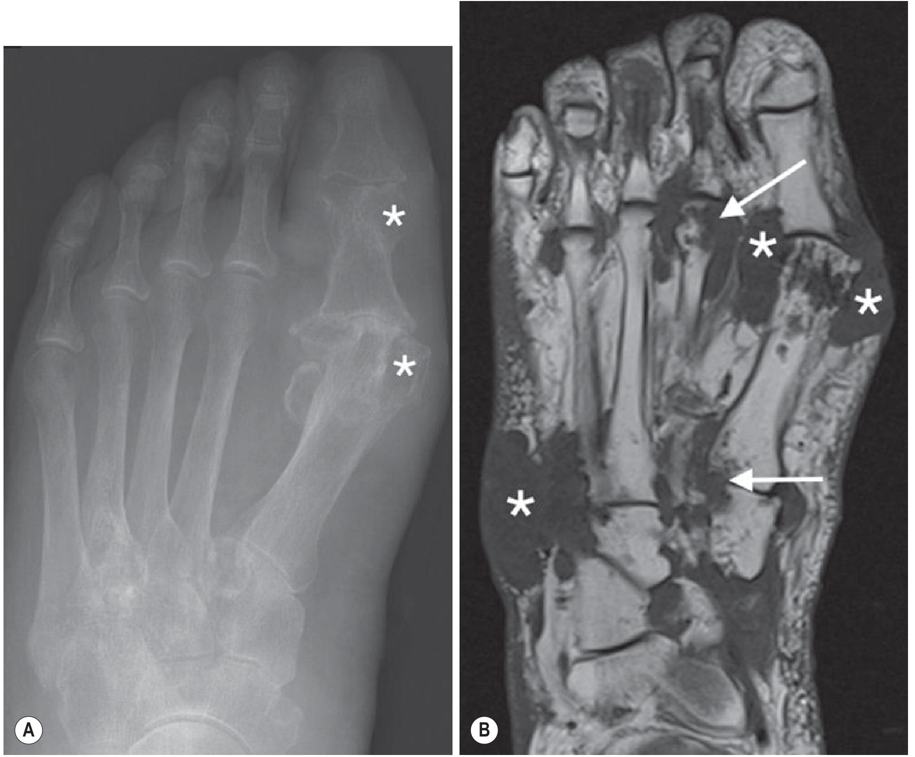
Chronic gout: (A) Plain X-ray showing erosive changes with "punched-out" erosions at 1st MTP joint. (B) MRI showing tophus deposits (low signal masses) and erosions. - Grainger & Allison's Diagnostic Radiology
Pseudogout (CPPD):
- Chondrocalcinosis: calcification in fibrocartilage (menisci of knee, triangular fibrocartilage of wrist, symphysis pubis) - pathognomonic
- Joint space narrowing
Ankylosing Spondylitis:
- Sacroiliitis: joint space widening then sclerosis then fusion
- Bamboo spine: squaring of vertebrae + syndesmophytes (vertical)
- Classic sign: "shiny corner" (Romanus lesion)
Psoriatic Arthritis:
- "Pencil-in-cup" deformity (destruction of base of phalanx, erosion of distal phalanx head)
- DIP joint involvement
- Periosteal reaction ("ivory" phalanx)
Septic Arthritis:
- Early: soft tissue swelling, joint effusion, periarticular osteoporosis
- Late (if untreated): rapid joint space narrowing, bone destruction
GM 7.15 / GM 7.17 - Treatment Plan for Rheumatologic Diseases
Rheumatoid Arthritis Treatment:
Principles:
- Early, aggressive treatment is essential to prevent joint destruction
-
70% of RF-positive polyarticular RA develops joint damage/erosion within 2 years without treatment
- Almost all patients should start a DMARD as soon as diagnosis is confirmed
Step-wise approach:
1. NSAIDs (symptom relief only, do NOT prevent joint destruction):
- Ibuprofen, naproxen, diclofenac
- Used for pain and inflammation while DMARDs take effect (weeks to months)
2. DMARDs (Disease-Modifying Antirheumatic Drugs) - first-line:
- Methotrexate (MTX): most commonly used; weekly dosing; requires folic acid supplementation; monitor LFTs, CBC
- Hydroxychloroquine (HCQ): for mild disease; monitor for retinopathy
- Sulfasalazine: for mild-moderate disease
- Leflunomide: alternative to MTX
3. Biologic DMARDs (bDMARDs) - second-line / combination:
- Anti-TNF agents: Etanercept, Infliximab, Adalimumab, Certolizumab, Golimumab
- Abatacept (T-cell co-stimulation blocker)
- Rituximab (anti-CD20, B-cell depleting)
- Tocilizumab (anti-IL-6 receptor)
4. Targeted synthetic DMARDs (tsDMARDs):
- JAK inhibitors: Tofacitinib, Baricitinib, Upadacitinib
5. Corticosteroids:
- Short-term "bridge" therapy while DMARDs take effect
- Low-dose prednisolone (<10 mg/day) for disease control
- Intra-articular injections for localized flares
Non-pharmacological:
- Patient education, joint protection
- Physical and occupational therapy
- Exercise to improve ROM and function
- Surgery (joint replacement) for severe destruction
SLE Treatment:
- Mild (skin, joints): NSAIDs + hydroxychloroquine (all SLE patients)
- Moderate: Low-dose corticosteroids + hydroxychloroquine
- Severe (nephritis, CNS, haematologic): High-dose corticosteroids + cyclophosphamide or mycophenolate mofetil
- Maintenance: Azathioprine, mycophenolate mofetil
- Biologics: Belimumab (anti-BLyS), Anifrolumab (anti-type I IFN)
- Sun protection, avoid triggers (UV, infections, certain drugs)
Ankylosing Spondylitis Treatment:
- NSAIDs (first-line, continuous use)
- Physiotherapy (essential - extension exercises, posture)
- Anti-TNF biologics (for NSAID-refractory disease)
- IL-17 inhibitors: Secukinumab, Ixekizumab
- No benefit from DMARDs for axial disease
Osteoarthritis Treatment:
- Non-pharmacological: Weight reduction, exercise, physiotherapy, assistive devices
- Topical NSAIDs (first-line for hand and knee OA)
- Oral NSAIDs / COX-2 inhibitors
- Intra-articular corticosteroids (for acute flares)
- Intra-articular hyaluronic acid (for knee OA)
- Surgery: Joint replacement for severe/refractory disease
GM 7.16 - Crystalline Arthropathies: Medications for Joint Pain and Preventive Therapy
GOUT
Pathophysiology: Monosodium urate crystal deposition due to hyperuricemia (serum uric acid >7 mg/dL men, >6 mg/dL women). 90% due to decreased renal excretion, 10% overproduction.
Risk factors: Obesity, alcohol, high-purine diet (red meat, shellfish, organ meats), diuretics, aspirin, hypertension, renal disease, metabolic syndrome
Clinical Stages:
- Asymptomatic hyperuricemia
- Acute intermittent gout
- Intercritical gout (symptom-free between attacks)
- Chronic tophaceous gout
A. Treatment of Acute Gout Attack:
1. NSAIDs (First-line):
- Indomethacin 50 mg TDS for 2 days then taper, OR
- Naproxen, ibuprofen at maximum doses
- Taper over 1-2 weeks
- Contraindicated in renal impairment, peptic ulcer disease
2. Colchicine:
- Mechanism: Inhibits microtubule polymerization; decreases neutrophil migration and phagocytosis of urate crystals
- Dose: 1 mg initially, then 0.5 mg 1 hour later (modern low-dose protocol)
- Side effects: Nausea, vomiting, diarrhoea (dose-limiting GI toxicity)
- Best when started within 12-24 hours of attack onset
- IV route now avoided (bone marrow suppression, renal/hepatic toxicity)
3. Corticosteroids (when NSAIDs and colchicine are contraindicated):
- Oral: Prednisolone 0.5 mg/kg/day, taper by 5 mg/day
- Intra-articular: Triamcinolone hexacetonide 10-40 mg (large joints), 5-20 mg (small joints) - preferred for monoarticular attack
- IM/IV ACTH: 40-80 mg every 8-12 hours (last resort)
B. Preventive / Urate-Lowering Therapy (ULT):
Indications for starting ULT:
- Recurrent attacks (≥2/year)
- Presence of tophi
- Uric acid nephrolithiasis
- Uric acid level >12 mg/dL
- Patients undergoing cancer chemotherapy
- Renal damage from gout
Note: Asymptomatic hyperuricemia does NOT need treatment.
Important: Never start ULT during an acute attack (can prolong or worsen it). Cover with colchicine prophylaxis for 3-6 months after starting ULT.
1. Allopurinol (Xanthine oxidase inhibitor - first-line ULT):
- Mechanism: Inhibits xanthine oxidase, reducing uric acid production
- Effective regardless of cause (overproduction or underexcretion)
- Start at 100 mg/day with food, increase by 100 mg/day at weekly intervals
- Target: Serum uric acid <6 mg/dL
- Usual dose: 200-300 mg/day (up to 600 mg/day)
- Adjust dose in renal impairment
- Side effects: Rash (can cause severe hypersensitivity - allopurinol hypersensitivity syndrome, especially in HLA-B*58:01 carriers), hepatotoxicity, bone marrow suppression
2. Febuxostat (Xanthine oxidase inhibitor):
- Alternative to allopurinol
- Useful in allopurinol intolerance or renal impairment
- Monitor cardiovascular events
3. Probenecid (Uricosuric agent):
- Mechanism: Blocks renal tubular reabsorption of urate, increases excretion
- Only effective if normal/good renal function
- Contraindicated with nephrolithiasis
- Adequate hydration required (>2 L/day urine output)
- Drug interactions: reduces excretion of penicillin, methotrexate
4. Colchicine (Prophylaxis):
- 0.5 mg twice daily for 3-6 months while initiating ULT
- Prevents precipitation of acute attacks when uric acid levels are fluctuating
Lifestyle measures:
- Avoid alcohol (especially beer - high in purines)
- Reduce high-purine foods (red meat, organ meats, shellfish)
- Avoid aspirin, diuretics if possible
- Maintain hydration
- Weight reduction
(- Textbook of Family Medicine, Rakel 9e, p. 887-889)
PSEUDOGOUT (Calcium Pyrophosphate Deposition Disease - CPPD)
Crystals: Calcium pyrophosphate dihydrate (CPPD) - rhomboid-shaped, positively birefringent
Clinical: Mimics gout - affects larger joints (knee most common), wrist, ankle; associated with hyperparathyroidism, haemochromatosis, hypomagnesaemia, hypothyroidism
Radiograph: Chondrocalcinosis (calcification of cartilage - menisci, triangular fibrocartilage complex)
Treatment (Acute attack):
- NSAIDs (same as gout)
- Colchicine
- Intra-articular or oral corticosteroids
- NOTE: No equivalent of allopurinol/ULT for CPPD (no drug reliably prevents CPPD crystal formation)
- Treat any underlying metabolic cause (hyperparathyroidism, haemochromatosis)
Summary Table: Key Differentiating Features
| Feature | RA | Gout | OA | SLE | AS |
|---|---|---|---|---|---|
| Sex | F > M (3:1) | M > F (20:1) | F > M (after 45) | F >> M (9:1) | M > F |
| Joint pattern | Symmetric polyarthritis | Monoarthritis (acute) | Asymmetric, weight-bearing | Variable polyarthritis | Axial + large joints |
| Key joints | MCPs, PIPs, wrist | 1st MTP, ankle, knee | Hip, knee, DIP | Variable | SI joints, spine |
| Morning stiffness | >1 hour | No | <30 min | Variable | >1 hour |
| Key serology | RF, anti-CCP | Uric acid | None | ANA, anti-dsDNA | HLA-B27 |
| X-ray | Erosions, periarticular osteoporosis | Punched-out erosions (late) | Osteophytes, sclerosis | Non-erosive usually | Sacroiliitis, bamboo spine |
| Synovial WBC | 10,000-20,000 | 20,000-100,000 | <2,000 | 5,000-15,000 | 5,000-15,000 |
Sources:
- Harrison's Principles of Internal Medicine, 22nd Edition (2025)
- Textbook of Family Medicine (Rakel, 9th Edition), Chapters 32-33
- Rosen's Emergency Medicine: Concepts and Clinical Practice, 9th Edition, pp. 2331-2335
- *Grainger & Allison's Diagnostic Radiology, p. 1125 (Crystal Arthropathies)*All 7 learning objectives are now covered. Here's a quick orientation to the full answer above:
What Was Covered
| Objective | Topic | Key Points |
|---|---|---|
| GM 7.10 | Differential Diagnosis | Mono vs. poly, inflammatory vs. non-inflammatory, pattern recognition, associated features |
| GM 7.11 | Diagnostic Workup | CBC, RF, anti-CCP, ANA, anti-dsDNA, anti-Smith, complement, uric acid - when to order and how to interpret |
| GM 7.12 | Arthrocentesis | Indications (4 main), contraindications, synovial fluid analysis table, crystal types, algorithm (with image) |
| GM 7.13 | Plain Radiographs | Disease-specific X-ray findings for RA, OA, gout, pseudogout, AS, psoriatic arthritis (with gout X-ray image) |
| GM 7.15/7.17 | Treatment Plans | RA (NSAIDs → DMARDs → biologics → JAK inhibitors), SLE, AS, OA - stepwise protocols |
| GM 7.16 | Crystalline Arthropathies | Gout acute treatment (NSAIDs, colchicine, steroids) + prophylaxis (allopurinol, probenecid, febuxostat); pseudogout management |
High-yield points to remember:
- Anti-CCP is more specific than RF for RA; anti-dsDNA is specific for SLE and tracks disease activity
- Gout crystal = negatively birefringent, needle-shaped; pseudogout = positively birefringent, rhomboid
- Never start allopurinol during an acute gout attack; cover with colchicine for 3-6 months
- Synovial WBC >50,000 = presumptive septic arthritis until proven otherwise
- Chondrocalcinosis on X-ray = pseudogout (CPPD); punched-out erosions = chronic gout
This is a shared conversation. Sign in to Orris to start your own chat.