
A patient 16 yrs old, had fever, selling over right mandible area three days back. He was treated with antibiotics. Now he has no pain but only irritation. On examination he has a hole in the gingiva over the right 1st molar, a small hole exposing th root of the teeth. What is this condition? How to proceed?
Reading File
Based on the clinical picture, this is a sinus tract (parulis/gumboil) overlying the root of the lower right first molar - a sign of a chronic periapical abscess that has drained. Let me consult the medical library for full details.
Searching sources...
Finding Sources
Finding Sources
Finding Sources
Reading File
Reading File
Finding Sources
Searching Images
dental sinus tract parulis gingival fistula periapical abscess
Finding Sources
Reading File
Reading File
Loading Image
Here is the full clinical picture and management plan for this patient.
Diagnosis: Intraoral Dental Sinus Tract (Parulis) - Secondary to Chronic Periapical Abscess
What Has Happened - The Pathological Sequence
- The lower right first molar developed pulp necrosis (likely from untreated dental caries or trauma) - the tooth became non-vital.
- Bacteria spread from the pulp canal through the root apex into the periapical space, forming an acute periapical abscess - which explains the fever and swelling over the right mandible 3 days ago.
- Antibiotics suppressed the acute phase (pain resolved, fever settled), but did not eliminate the source - the necrotic pulp and periapical infection persist.
- The infection eroded through cortical bone and gingival tissue, creating a drainage pathway - the small hole (sinus tract / parulis) you are now seeing over the root of the first molar.
This is now a chronic periapical abscess with intraoral sinus tract formation - hence only mild irritation, no pain (the pressure has been decompressed by drainage).
"A periapical abscess forms when there is a localized accumulation of inflammatory cells and bacterial debris in the periapical space of a non-vital tooth... When the infection erodes through cortical bone to create a path for drainage, a 'sinus tract' or 'parulis' forms. An intraoral dental sinus tract is characterized by a soft, non-tender, erythematous papule that develops on the alveolar process, typically in the same region as the apex of the offending non-vital tooth." - Dermatology 2-Volume Set 5e
Here is an example of a cutaneous dental sinus tract (the intraoral form looks similar but opens onto the gingiva rather than skin):
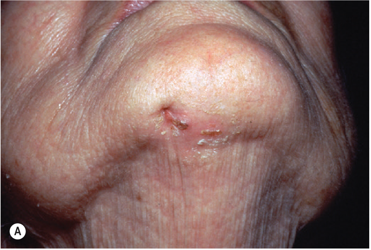
Cutaneous sinus of dental origin (Andrews' Diseases of the Skin). The intraoral form presents identically but opens onto the gingival mucosa overlying the root apex.
Why Antibiotics Alone Are Not the Answer
Antibiotics partially suppress the infection but cannot sterilize a necrotic tooth pulp - there is no blood supply to carry the antibiotic into the root canal. Without eliminating the source, the sinus tract will persist or recur, and the risk of spreading infection (cellulitis, osteomyelitis, deep space neck infection) remains. As stated by K.J. Lee's Essential Otolaryngology: treatment requires "removal of the underlying inflammatory process through endodontic treatment (root canal) or tooth extraction with enucleation and curettage; antibiotics and drainage of any soft tissue abscess."
How to Proceed - Step by Step
Step 1 - Dental Radiograph (Periapical X-ray, IOPA)
Take a periapical radiograph of the lower right first molar. You will typically see:
- A periapical radiolucency (dark halo around the root apex) indicating bone destruction
- Possible widening of the periodontal ligament space
- Any caries or previous restorations
This is both diagnostic and guides treatment planning.
Step 2 - Assess Tooth Restorability
Decide whether the tooth can be saved:
- If restorable → Root canal treatment (RCT / endodontic therapy) - the definitive treatment. The necrotic pulp is cleaned, canals shaped, disinfected, and sealed. This eliminates the source. The sinus tract will close on its own (usually within 1-2 weeks after RCT).
- If non-restorable (grossly broken down, poor prognosis, patient preference) → Extraction of the tooth, with curettage of the periapical granulation tissue.
Step 3 - Do NOT surgically excise the sinus tract separately
The sinus tract is a secondary phenomenon. Once the source (infected tooth apex) is treated, the sinus tract closes spontaneously. Surgical excision of the opening alone is futile and counterproductive.
Step 4 - Antibiotics (adjunctive only)
At 16 years old, if there are signs of systemic spread, continuing:
- Amoxicillin 500 mg TID x 5 days, or
- Amoxicillin-clavulanate if inadequate response, or
- Metronidazole added for anaerobic coverage.
However, antibiotics are adjunctive - they do NOT replace definitive dental treatment.
Step 5 - Follow-up
- Review in 1-2 weeks post-RCT to confirm sinus tract closure.
- Repeat periapical X-ray at 3-6 months to confirm periapical healing (bone fill).
Differential Diagnoses to Consider
| Condition | Distinguishing Feature |
|---|---|
| Periodontal abscess | Associated with a vital tooth, pocket depth, bone loss pattern differs |
| Actinomycosis | Sulfur granules in discharge, multiple sinus tracts, responds to long-course penicillin |
| Osteomyelitis of mandible | Fever, more extensive bone destruction on X-ray |
| Buccal bifurcation cyst | Age 5-13 years, buccal surface of erupting lower molars, foul discharge |
| Congenital fistula | Present from birth, no dental cause on X-ray |
Summary
| Feature | This Patient |
|---|---|
| Diagnosis | Chronic periapical abscess with intraoral sinus tract (parulis) |
| Causative tooth | Lower right first molar (non-vital) |
| Why pain resolved | Sinus tract decompressed the abscess |
| Why irritation persists | Chronic low-grade infection and ongoing drainage |
| Definitive treatment | Root canal treatment OR extraction |
| Role of antibiotics | Adjunctive only - not curative |
| Prognosis | Excellent with proper dental treatment |
Refer this patient to a dentist or endodontist promptly. The sinus tract will close on its own once the source is eliminated - no surgical excision of the opening is needed.
Sources: - Dermatology 2-Volume Set 5e, Andrews' Diseases of the Skin (Cutaneous Sinus of Dental Origin), K.J. Lee's Essential Otolaryngology (Odontogenic Cysts and Infections)
Ok. But the hole is on the gingiva itself not on the skin over the area.
That actually confirms the diagnosis even more precisely. The hole on the gingiva (not on the skin) is the classic intraoral sinus tract / parulis - which is exactly what was described in the textbook sources I already retrieved. Nothing changes about the diagnosis or management.
Let me clarify the distinction clearly:
The Hole IS the Intraoral Sinus Tract (Parulis)
There are two forms of dental sinus tract:
| Form | Location of opening | This patient? |
|---|---|---|
| Intraoral sinus tract (Parulis) | Gingival mucosa, overlying the root apex | YES - this is your case |
| Cutaneous sinus tract | Skin of chin/jaw/neck | No |
The image I showed earlier was the cutaneous (skin) variant - that was just for illustration. Your patient has the intraoral form, which is actually the more common presentation.
The textbook describes it exactly:
"An intraoral dental sinus tract is characterized by a soft, non-tender, erythematous papule that develops on the alveolar process, typically in the same region as the apex of the offending non-vital tooth." - Dermatology 2-Volume Set 5e
This matches perfectly:
- Hole/opening on the gingiva directly over the root of the right lower first molar
- Non-tender (patient has no pain, only mild irritation)
- 16-year-old with prior history of acute abscess (fever + swelling) treated with antibiotics
The pathological sequence is identical - the periapical infection eroded through the alveolar bone and then the overlying gingival mucosa (rather than through the skin) to create the drainage opening you are seeing.
Management - No Change
Everything in the management plan holds:
- Periapical X-ray (IOPA) - to confirm periapical radiolucency at the root apex
- Root Canal Treatment (RCT) - definitive treatment; eliminates the necrotic pulp and source of infection
- The gingival opening closes on its own within 1-2 weeks after successful RCT - no surgical excision of the parulis needed
- Extraction if the tooth is non-restorable
- Antibiotics are adjunctive only - the necrotic pulp must be physically removed
The gingival location actually makes this a textbook-perfect presentation of a chronic periapical abscess with intraoral sinus tract. Refer to a dentist/endodontist for RCT as soon as possible.
This is a shared conversation. Sign in to Orris to start your own chat.