


Can you tell me the diagnosis It has been in buttocks of female of 35 years for 2 years there is severe itching amd tell me the management too
buttock chronic pruritic verrucous papules differential lichen planus dermatitis herpetiformis clinical dermatology
chronic prurigo AND treatment
dermatitis herpetiformis grouped excoriated papules vesicles buttocks clinical photograph
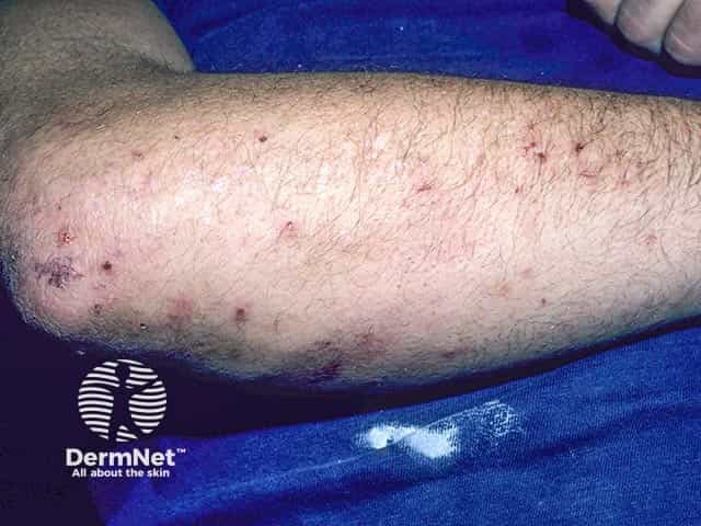
Clinical photograph of a dermatology case showing excoriated vesicles and crusted papules on the forearm, compatible with dermatitis herpetiformis (Duhring disease). The image captures an anterior (volar) view of the upper limb with multiple small erythematous papules and vesicles along the extensor surface, interspersed with crusts from scratching. A few intact vesicles are evident, confirming the vesiculopapular morphology characteristic of DH. The skin appears mildly inflamed with excoriations and postinflammatory crusting, suggesting pruritus-driven disease activity. The distribution on the forearm is typical for dermatitis herpetiformis, which often affects extensor surfaces such as elbows, knees, buttocks, and shoulders. The photograph shows clinically relevant features including clustered vesicles on an erythematous base, clear vesicular rupture sites, and secondary crusts, all consistent with DH lesions in a patient with gluten sensitivity. Visual assessment supports the diagnosis when integrated with clinical history and serology: celiac disease association, pruritic grouped vesicles, and intolerance to dietary gluten. For diagnostic confirmation, skin biopsy with direct immunofluorescence demonstrating granular IgA deposition at dermal papillae is standard; alternative tests include serology for anti-endomysial and anti-tendom gluten antibodies. This image is suitable for dermatology education, clinical case reviews, image atlases, and differential diagnosis studies of vesiculopapular pruritic eruptions.
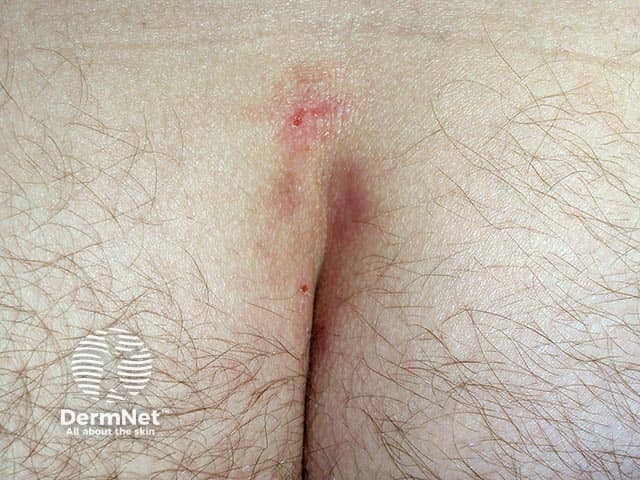
Clinical photography of the sacral skin demonstrates a pruritic vesiculopapular eruption compatible with dermatitis herpetiformis. Modality: dermatology clinical photograph; technique: close-up, posterior-oriented view with ambient lighting; no staining, no contrast agent; standard digital capture. The sacral/buttock region shows grouped vesicles and erythematous papules embedded in an excoriated, crusted base, often arranged in clustered or symmetric patterns. Lesions are intensely pruritic, with surrounding mild edema and superficial erosions; scale is minimal. Morphology includes tiny vesicles evolving to crusted erosions within an erythematous halo, sometimes with surrounding excoriations from scratching. Distribution on the buttocks and sacral area aligns with provoking factors and supports a dermatologic diagnosis in the proper clinical context. Differential considerations include contact dermatitis, papular urticaria, linear IgA disease, and various pruritic papulovesicular dermatoses. Pathophysiology is immunopathologic, with dermatitis herpetiformis characterized by granular IgA deposits at dermal papillae and neutrophilic microabscesses in histology; direct immunofluorescence confirms IgA deposition. Clinically this entity is associated with gluten sensitivity and celiac disease; management centers on a gluten-free diet and rapid symptomatic relief with dapsone or sulfapyridine. The image is educational for dermatology, medical student, and clinical decision support in vesiculopapular eruptions. This image supports educational use in dermatology curricula, case discussions, and triage.
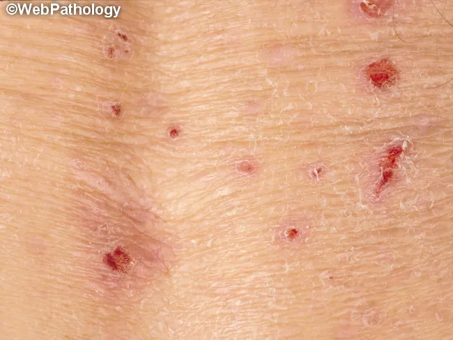
Clinical photography of the skin illustrating dermatitis herpetiformis, an intensely pruritic, grouped vesiculopapular eruption associated with gluten-sensitive enteropathy. The image demonstrates widespread, symmetrical involvement of extensor surfaces including elbows and knees, with additional lesions on the buttocks, scapular region, and sacral area; facial or scalp involvement may occur. Lesions begin as erythematous papules that rapidly evolve into firm-topped vesicles or urticarial plaques, frequently excoriated to crusts from persistent scratching. The clustered distribution and pruritus are characteristic; bullae may be present in advanced plaques. On a clinical basis, dermatitis herpetiformis correlates with underlying celiac disease and an immune response to dietary gluten. Histology (when biopsied) reveals neutrophilic microabscesses at the tips of dermal papillae with superficial dermal neutrophilic and eosinophilic infiltrates. Direct immunofluorescence classically shows granular IgA deposition at dermal papillae with accompanying C3 and sometimes C5 components. Management includes dapsone for rapid symptom control and a strict gluten-free diet to address the enteropathy. The image is a dermatology photograph suitable for educational purposes, differential diagnosis discussions, and radiology-free teaching; it can be used to illustrate skin manifestations that predate or accompany GI symptoms and to emphasize multidisciplinary care. Close clinical correlation with serology and biopsy improves diagnostic confidence in practice.
https://dermnetnz.org/topics/dermatitis-herpetiformis
- Hypertrophic lichen planus
- Lichen simplex chronicus
- Scabies or arthropod-bite reaction, especially if itching is worse at night or household contacts itch
- Dermatitis herpetiformis, if there are recurrent tiny grouped blisters or erosions on both buttocks, elbows, knees, scalp, or back. This is associated with coeliac disease and needs a different work-up and treatment.
What the dermatologist should do
- Examine the entire skin: elbows, knees, scalp, groin, hands/wrists, and check for burrows or a typical symmetrical pattern.
- Dermoscopy and a punch biopsy of a representative lesion if the diagnosis is uncertain or treatment fails. Biopsy is especially important before using prolonged potent steroids.
- Consider a cause-of-itch screen if clinically indicated: eczema/contact allergy, diabetes, thyroid, liver or kidney disease, iron deficiency, hepatitis/HIV risk factors, medication review, and sleep or anxiety factors.
- If dermatitis herpetiformis is suspected: biopsy normal-looking skin next to a fresh lesion for direct immunofluorescence, plus coeliac serology such as tissue-transglutaminase IgA and total IgA. This is the recommended confirmation approach in NIDDK guidance.
Management if it is prurigo nodularis / lichenified chronic prurigo
- Break the scratch cycle: keep nails short, avoid rubbing, use cool compresses, and cover the lesion at night with a non-adherent dressing or clothing.
- Fragrance-free moisturizer at least twice daily, especially after bathing. Avoid harsh soaps, scrubs, and tight synthetic clothing.
- A dermatologist commonly uses a short, supervised course of a potent topical corticosteroid for thick nodules, sometimes with occlusion. It should not be continued indefinitely because it can thin skin. For persistent individual nodules, clinicians may use intralesional steroid treatment. Andrews' Diseases of the Skin Clinical Dermatology, p. 71.
- Non-steroid anti-itch treatments such as tacrolimus or pimecrolimus may be used as steroid-sparing options in appropriate cases.
- A night-time sedating antihistamine may help sleep in selected people, but should be chosen by a clinician or pharmacist based on her health conditions and other medicines.
- If widespread or resistant, dermatologists may consider phototherapy or newer systemic treatments after confirming the diagnosis and evaluating causes of itch.