
What are the different functions for the right and left gastric vessels and for the right and left gastric epiploic arteries and vessels?
right left gastric vessels epiploic arteries functions anatomy differences
gastric artery blood supply stomach lesser greater curvature anatomy diagram
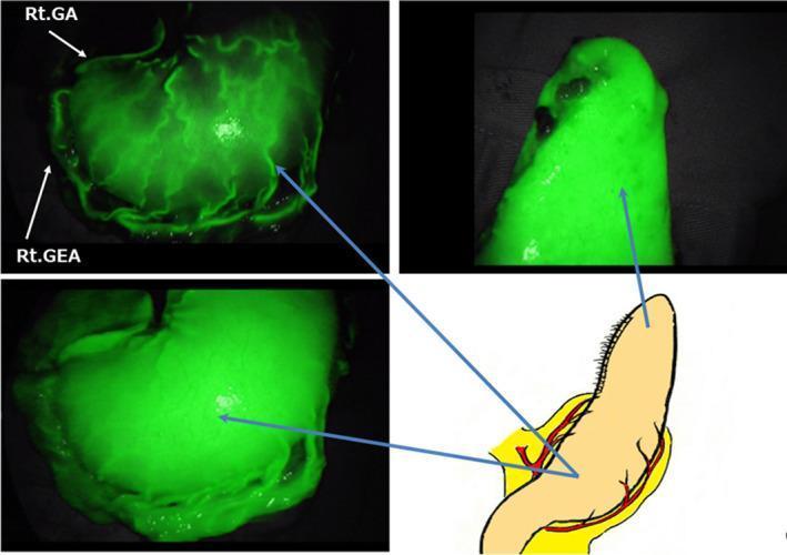
This composite figure illustrates the circulatory network of a subtotal stomach (SS) during reconstructive surgery using indocyanine green (ICG) fluorescence imaging. The panel consists of three intraoperative clinical photographs and one anatomical diagram. The ICG fluorescence images demonstrate a vibrant, dense, and interconnected vascular network beneath the gastric mucosa, indicating robust perfusion. Key arterial landmarks are labeled: the Right Gastric Artery (Rt. GA) is visible along the lesser curvature, and the Right Gastro-Epiploic Artery (Rt. GEA) is identified along the greater curvature. These primary vessels and their preserved arteriovenous arcades exhibit high-intensity fluorescence, branching into a complex capillary bed that ensures uniform tissue viability up to the tip of the gastric conduit. The anatomical diagram correlates these fluorescent findings with the surgical anatomy, highlighting the vascular supply (red) and the longitudinal orientation of the subtotal stomach. This imaging modality serves as an intraoperative tool for real-time assessment of blood flow, which is critical for minimizing anastomotic complications in esophageal or gastric reconstruction.
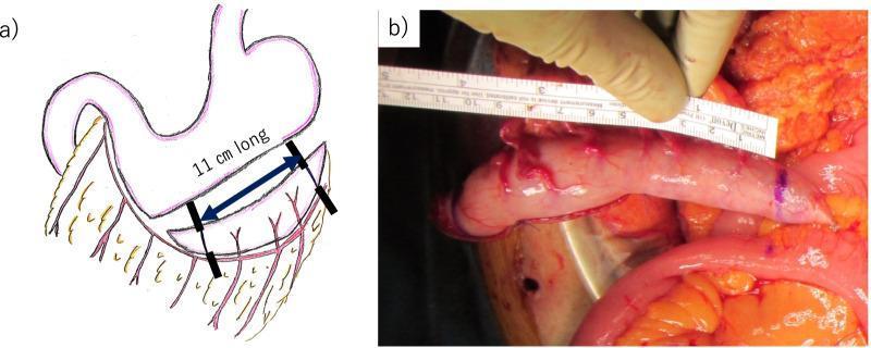
This composite figure illustrates the surgical creation of a gastric conduit for urinary diversion. Panel A is an anatomical diagram showing the stomach and the vascular supply of the greater curvature, specifically the gastroepiploic artery. A blue double-headed arrow and text indicate the 11-cm longitudinal section along the greater curvature targeted for conduit formation, starting approximately 2 cm proximal to the pylorus. Panel B is an intraoperative clinical photograph providing a surgical view of the mobilized gastric conduit. The conduit appears as a pinkish-red, vascularized tubular structure. A surgical ruler is held by a gloved hand alongside the tissue, confirming a length of approximately 11 cm. The conduit maintains its blood supply from the right gastroepiploic artery to ensure viability for subsequent ureterogastric anastomosis. This material demonstrates a specialized reconstructive technique used in complex pelvic surgeries, such as total pelvic exenteration, where standard urinary diversion methods like an ileal conduit may not be feasible.
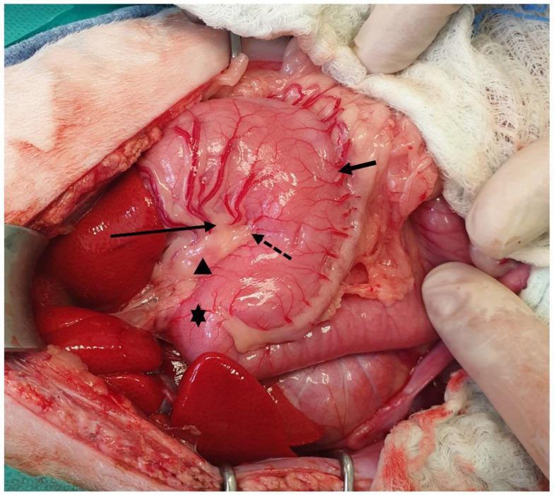
This clinical intraoperative photograph displays the macro-anatomical features of the stomach during an abdominal surgical procedure. The stomach is oriented with the cranial aspect (head) toward the left of the frame. Key anatomical landmarks are identified: the lesser curvature is indicated by a long solid arrow on the concave side, while the greater curvature is marked by a short arrow on the convex side. A distinct angular notch (incisura angularis) is visible along the lesser curvature, denoted by a dashed arrow, serving as a boundary between the gastric body and the pyloric part. The pylorus itself is marked with an asterisk (*). A gastric lymph node is localized along the lesser curvature, indicated by an arrowhead. The stomach's surface exhibits a rich vascular network of branching blood vessels. Surrounding visceral structures, including liver lobes and intestinal segments, are visible within the surgical field, which is maintained by retractors and surgical gauze. This image serves as an educational reference for gastric anatomy, vascularization, and landmark identification during laparotomy.
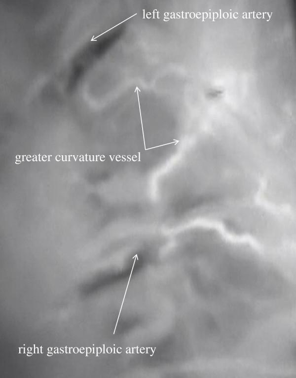
This diagnostic image shows intraoperative indocyanine green (ICG) fluorescence angiography using a photodynamic eye (PDE) system to visualize blood flow in a reconstructed stomach conduit. The image demonstrates the 'greater curvature route' of arterial supply. Visible anatomical structures include the left gastroepiploic artery in the superior field and the right gastroepiploic artery in the inferior field, both appearing as relatively thick, high-intensity curvilinear signals. Connecting these two major vessels is a network of greater curvature vessels, which exhibit a more tortuous, meandering, and complex branching pattern. The clinical significance of this imaging is to evaluate the microcirculation and perfusion of the gastric graft during esophageal cancer surgery, ensuring the viability of the conduit's fundus and anastomotic site. This technique helps surgeons identify collateral pathways and the 'greater curvature route' to prevent postoperative complications like anastomotic leakage or conduit necrosis.
Gastric Vascular Supply: Right vs. Left Vessels
Lesser Curvature: Right and Left Gastric Arteries
| Feature | Left Gastric Artery | Right Gastric Artery |
|---|---|---|
| Origin | Directly from the celiac trunk (one of its 3 main branches) | Proper hepatic artery (occasionally common hepatic or gastroduodenal artery) |
| Size | Consistently the largest artery to the stomach | Smaller caliber |
| Territory | Upper lesser curvature, cardia, lower esophagus; ascending branch supplies distal esophagus | Distal lesser curvature (antrum/pyloric region), runs proximally toward the stomach |
| Direction of flow | Descends from above (celiac trunk) toward the pylorus | Ascends from below (hepatic artery near pylorus) toward the fundus |
| Notable variant | ~20% of the time gives off an aberrant left hepatic artery traveling in the gastrohepatic (lesser) omentum - ligation can cause hepatic ischemia if this is the only supply to the left lobe | Duplications have been reported |
| Venous drainage | Left gastric vein (= coronary vein) drains into portal vein; clinically important in portal hypertension as it connects to esophageal varices | Right gastric vein drains into portal vein |
Greater Curvature: Right and Left Gastroepiploic (Gastro-omental) Arteries
| Feature | Right Gastroepiploic Artery (RGEA) | Left Gastroepiploic Artery (LGEA) |
|---|---|---|
| Origin | Gastroduodenal artery (one of its two terminal branches), behind the first part of the duodenum | Splenic artery, just proximal to the splenic hilum |
| Size | Second largest artery to the stomach overall; larger than the LGEA | Smaller |
| Direction of course | Runs right-to-left along the greater curvature, between layers of the greater omentum | Runs left-to-right within the gastrosplenic ligament, then between layers of greater omentum |
| Gastric territory | Anterior and posterior antrum and lower body of the stomach | Anterior and posterior fundus and upper/mid body of the stomach |
| Omental (epiploic) branches | Supplies right greater omentum; can occasionally branch to the final duodenal segments | Supplies middle greater omentum |
| Venous drainage | Right gastroepiploic vein joins right colic and anterior superior pancreaticoduodenal veins → Henle's trunk → superior mesenteric vein | Left gastroepiploic vein drains into the splenic vein |
| Surgical importance | Key pedicle for gastric conduit in esophageal reconstruction (stomach pulled up to the neck); also used as a free or pedicled graft in coronary artery bypass (CABG) | Vulnerable during mobilization of the colonic splenic flexure; used in some reconstructive procedures |
Key Functional Distinctions
-
Different portal system drainage pathways: Left-sided vessels (left gastric vein, left gastroepiploic vein) drain into the splenic vein or portal vein directly. Right-sided vessels (right gastroepiploic vein) drain into the superior mesenteric vein via Henle's trunk. This matters in portal hypertension - the left gastric (coronary) vein is the primary route for gastroesophageal variceal formation.
-
Anastomotic redundancy: Because of the double arcade system, the stomach can survive ligation of three of the four named arteries as long as the arcades along both curvatures are kept intact. This is exploited surgically when the stomach is used as an esophageal replacement - it can be pedicled on just the right gastric and right gastroepiploic vessels.
-
Omental supply: Only the gastroepiploic vessels (not the gastric arteries) send branches to the greater omentum itself. These omental (epiploic) branches give the omentum its blood supply for its immune and inflammatory functions.
-
Esophageal supply: Only the left gastric artery has a consistent role in supplying the distal esophagus; the right gastric artery does not.