
Give me everything available on internet about "INSULA"....but not from Wikipedia
insula brain anatomy function clinical significance
"insular cortex"[MeSH Terms]
https://pmc.ncbi.nlm.nih.gov/articles/PMC6032992
The insula (insular cortex, island of Reil)
1. What it is and where it sits
- The insula is a buried piece of cerebral cortex. It is revealed only when the margins of the lateral (Sylvian) fissure are pulled apart. It lies between the temporal and frontal lobes. - Neuroscience: Exploring the Brain, 5e, p. 660-661
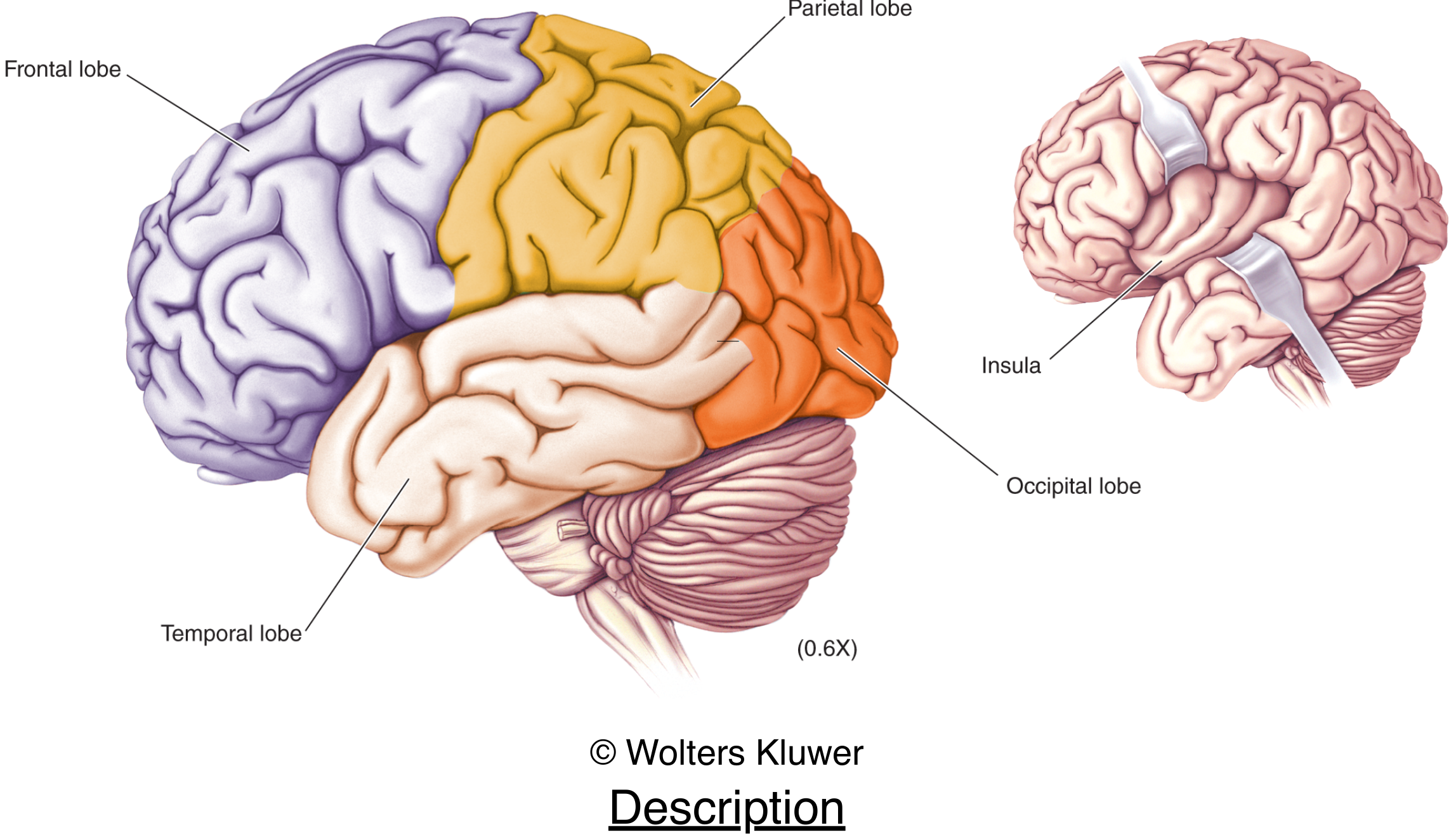
- Johann Christian Reil described it in 1809, hence "island of Reil". The PMC structure and function review describes it as traditionally paralimbic or limbic integration cortex.
- The frontal, parietal and temporal opercula ("little lids") cover it. Kenhub describes it as triangular, bounded by the anterior, superior and inferior limiting sulci.
- The limen insulae is its anteroinferior "port". It lies at the lateral edge of the anterior perforated substance, about where the middle cerebral artery (MCA) divides.
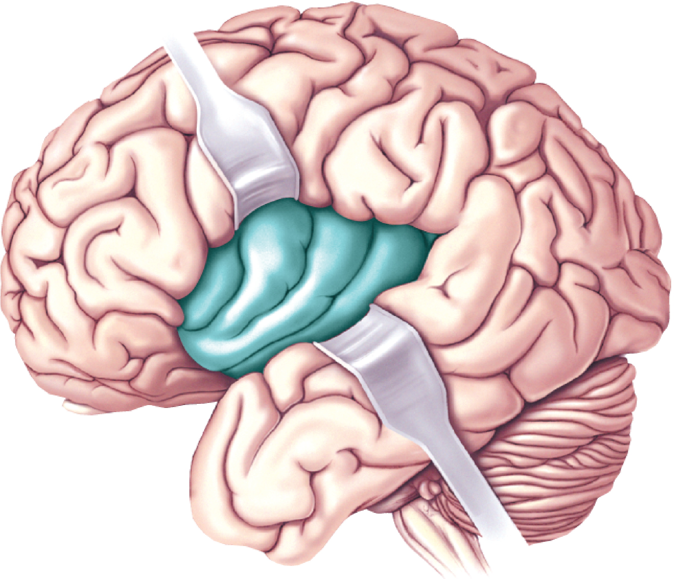
2. Gyri, microstructure and blood supply
- Gyri: three short gyri (anterior, middle, posterior) and two long gyri (anterior, posterior). These are described in Kenhub and the PMC review.
- Cytoarchitecture: granularity runs along a gradient.
- Agranular cortex lies anteriorly and centrally.
- Dysgranular cortex is intermediate.
- Granular cortex lies posteriorly, with a thick layer 4. - Neuroscience: Exploring the Brain, 5e, p. 1687
- Depending on the method, parcellations give anywhere from 2 to 13 subdivisions (PMC review).
- Arterial supply: perforating branches of the M2 segment of the MCA. The superior trunk supplies the anterior, middle and posterior short gyri, and the inferior trunk supplies the posterior long gyrus. Venous drainage is mainly to the deep middle cerebral vein. Dense vessels overlie it, which makes surgery difficult (PMC review).
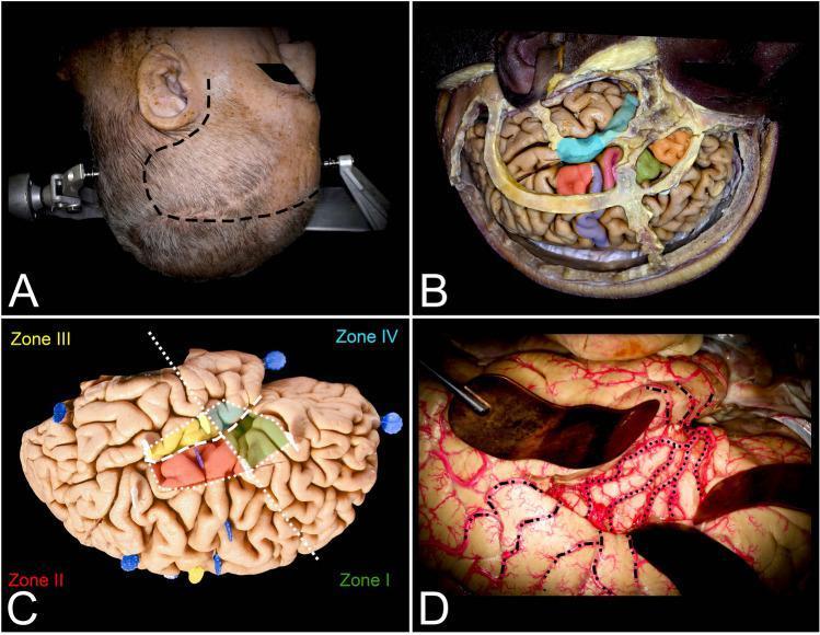
3. Connections
- It connects with the neocortex (temporal, parietal, frontal), basal ganglia, thalamus, limbic system and olfactory cortex (Kenhub).
- It projects to the central nucleus of the amygdala and to autonomic nuclei in the medulla. - Localization in Clinical Neurology, 8e, p. 1326
- The anterior insula connects to the motor and supplementary motor cortices, the thalamic VPM nucleus, and the nucleus tractus solitarius. These links are relevant to swallowing. - Localization in Clinical Neurology, 8e, p. 1326
- Anterior and posterior parts differ in connectivity. The anterior insula links to the limbic, orbitofrontal and temporopolar regions. The posterior insula links to sensorimotor and auditory regions.
4. Functions
- Interoception and visceral/autonomic control. The insula receives inputs on blood pressure, heart rate, digestion, nausea, hunger and pain. It shares this with the amygdala, thalamus, hypothalamus, cingulate and frontal cortex. - Neuroscience: Exploring the Brain, 5e, p. 1687
- Emotion and subjective feeling. The anterior insula is activated by disgust, anger and pain.
- It is also activated when we watch others in pain, which may be a neural correlate of empathy.
- The textbook describes the insula as receiving sensory input and the cingulate as sending output. - Neuroscience: Exploring the Brain, 5e, p. 1687-1689
- A PMC opinion article contrasts the two structures. The amygdala handles automatic, implicit responses. The anterior insula supports explicit, subjective feelings.
- Pain. It has neurons that respond selectively to noxious stimuli. It processes internal bodily state and the autonomic part of the pain response. - Principles of Neural Science (Kandel), 6e, p. 533
- Salience and cognitive control. With the dorsal anterior cingulate it forms the core of the salience network. This network detects the most relevant of competing internal and external stimuli. It also supports network switching (PMC review; the Menon 2024 chapter). In that model, the dorsal anterior insula supports goal-directed cognition, attention, decision-making and error detection.
- Chemosensation. Taste and olfaction are processed in the insula (Flynn, Benson and Ardila, Aphasiology 1999).
- Other roles (Flynn et al.):
- The posterior insula has vestibular and auditory roles.
- It acts as a supplementary motor and speech-articulation zone.
- It may connect language with the limbic system.
- Craving, addiction and risk-reward behavior. Functional imaging links it to cravings (Kenhub, Physiopedia).
- Social cognition. Lesion studies tie left insula damage to difficulty recognising emotions, and the right anterior insula to affective empathy (PMC review).
5. Clinical relevance
- Cardiovascular and autonomic effects of stroke.
- It is the most important cortical area for cardiovascular regulation.
- Right-sided lesions are more often linked to sympathetic dysfunction.
- Left-sided lesions are more often linked to parasympathetic effects.
- Insular strokes are associated with hypertension, arrhythmias, myocardial injury, raised catecholamines and sudden death. - Localization in Clinical Neurology, 8e, p. 1326-1327
- Isolated insular stroke. A 2021 systematic review (PMID 33575921) covers the clinical presentation. Its MeSH terms point to aphasia, dysarthria and speech deficits.
- Insular epilepsy. It is uncommon and often missed because scalp EEG can't record the insula directly.
- Typical symptoms are laryngeal discomfort, a chest or abdominal sensation, dyspnea, and perioral paresthesias.
- Signs include dysarthria or dysphonia (sometimes mutism), hypersalivation, facial and arm tonic spasm, and head and eye turning.
- Seizures can spread and mimic temporal, parietal or frontal lobe epilepsy. - Bradley and Daroff's Neurology in Clinical Practice, p. 2240
- Pain asymbolia. Insular lesions leave patients able to perceive pain and tell sharp from dull. They lack the appropriate emotional response. - Principles of Neural Science, 6e, p. 533
- Psychiatric disorders. The insula is implicated in schizophrenia, mood, panic, PTSD, OCD and addiction (Kenhub; PMC opinion article).
- Surgery. Gliomas and vascular lesions near the insula are risky because of the overlying MCA branches and nearby eloquent cortex.
6. Recent evidence (PubMed, last 5 years)
- Nani et al. 2021 (PMID 33122115): systematic review of insular morphometric "pathoconnectivity" across brain diseases.
- Di Stefano et al. 2021 (PMID 33575921): clinical presentation of strokes confined to the insula.
- Klugah-Brown et al. 2023 (PMID 35511500): structural-functional connectivity meta-analysis, supporting distinct insular subregions.
7. Don't confuse with
- "Insular thyroid carcinoma" is an unrelated tumor that shares the word. It is described in Cummings Otolaryngology.
- Insulae of Langerhans (pancreatic islets) are also unrelated.
8. Limits of this answer
- I did not fetch each web source in full. I took details from search excerpts, two page extractions, the textbooks and PubMed metadata.
- I did not read the abstracts of the PubMed papers, so I haven't summarized their findings.
?
- It is a part of the cerebral cortex hidden deep in the lateral (Sylvian) fissure, covered by the frontal, parietal and temporal opercula.
- It handles body-state sensing (interoception), emotion and empathy, pain, taste, autonomic control, and detecting what is salient.
- Clinically, damage or irritation of the insula can cause autonomic and cardiac problems after stroke, speech and swallowing deficits, "pain asymbolia", and insular epilepsy, which often mimics temporal lobe epilepsy.
What did you mean by '?' and what would help most?
The insula (insular cortex, island of Reil)
1. What it is and where it sits
- The insula is a buried piece of cerebral cortex. It is revealed only when the margins of the lateral (Sylvian) fissure are pulled apart. It lies between the temporal and frontal lobes. - Neuroscience: Exploring the Brain, 5e, p. 660-661
- Johann Christian Reil described it in 1809, hence "island of Reil". The PMC structure and function review describes it as traditionally paralimbic or limbic integration cortex.
- The frontal, parietal and temporal opercula ("little lids") cover it. Kenhub describes it as triangular, bounded by the anterior, superior and inferior limiting sulci.
- The limen insulae is its anteroinferior "port". It lies at the lateral edge of the anterior perforated substance, about where the middle cerebral artery (MCA) divides.
2. Gyri, microstructure and blood supply
- Gyri: three short gyri (anterior, middle, posterior) and two long gyri (anterior, posterior). These are described in Kenhub and the PMC review.
- Cytoarchitecture: granularity runs along a gradient.
- Agranular cortex lies anteriorly and centrally.
- Dysgranular cortex is intermediate.
- Granular cortex lies posteriorly, with a thick layer 4. - Neuroscience: Exploring the Brain, 5e, p. 1687
- Depending on the method, parcellations give anywhere from 2 to 13 subdivisions (PMC review).
- Arterial supply: perforating branches of the M2 segment of the MCA. The superior trunk supplies the anterior, middle and posterior short gyri, and the inferior trunk supplies the posterior long gyrus. Venous drainage is mainly to the deep middle cerebral vein. Dense vessels overlie it, which makes surgery difficult (PMC review).
3. Connections
- It connects with the neocortex (temporal, parietal, frontal), basal ganglia, thalamus, limbic system and olfactory cortex (Kenhub).
- It projects to the central nucleus of the amygdala and to autonomic nuclei in the medulla. - Localization in Clinical Neurology, 8e, p. 1326
- The anterior insula connects to the motor and supplementary motor cortices, the thalamic VPM nucleus, and the nucleus tractus solitarius. These links are relevant to swallowing. - Localization in Clinical Neurology, 8e, p. 1326
- Anterior and posterior parts differ in connectivity. The anterior insula links to the limbic, orbitofrontal and temporopolar regions. The posterior insula links to sensorimotor and auditory regions.
4. Functions
- Interoception and visceral/autonomic control. The insula receives inputs on blood pressure, heart rate, digestion, nausea, hunger and pain. It shares this with the amygdala, thalamus, hypothalamus, cingulate and frontal cortex. - Neuroscience: Exploring the Brain, 5e, p. 1687
- Emotion and subjective feeling. The anterior insula is activated by disgust, anger and pain.
- It is also activated when we watch others in pain, which may be a neural correlate of empathy.
- The textbook describes the insula as receiving sensory input and the cingulate as sending output. - Neuroscience: Exploring the Brain, 5e, p. 1687-1689
- A PMC opinion article contrasts the two structures. The amygdala handles automatic, implicit responses. The anterior insula supports explicit, subjective feelings.
- Pain. It has neurons that respond selectively to noxious stimuli. It processes internal bodily state and the autonomic part of the pain response. - Principles of Neural Science (Kandel), 6e, p. 533
- Salience and cognitive control. With the dorsal anterior cingulate it forms the core of the salience network. This network detects the most relevant of competing internal and external stimuli. It also supports network switching (PMC review; the Menon 2024 chapter). In that model, the dorsal anterior insula supports goal-directed cognition, attention, decision-making and error detection.
- Chemosensation. Taste and olfaction are processed in the insula (Flynn, Benson and Ardila, Aphasiology 1999).
- Other roles (Flynn et al.):
- The posterior insula has vestibular and auditory roles.
- It acts as a supplementary motor and speech-articulation zone.
- It may connect language with the limbic system.
- Craving, addiction and risk-reward behavior. Functional imaging links it to cravings (Kenhub, Physiopedia).
- Social cognition. Lesion studies tie left insula damage to difficulty recognising emotions, and the right anterior insula to affective empathy (PMC review).
5. Clinical relevance
- Cardiovascular and autonomic effects of stroke.
- It is the most important cortical area for cardiovascular regulation.
- Right-sided lesions are more often linked to sympathetic dysfunction.
- Left-sided lesions are more often linked to parasympathetic effects.
- Insular strokes are associated with hypertension, arrhythmias, myocardial injury, raised catecholamines and sudden death. - Localization in Clinical Neurology, 8e, p. 1326-1327
- Isolated insular stroke. A 2021 systematic review (PMID 33575921) covers the clinical presentation. Its MeSH terms point to aphasia, dysarthria and speech deficits.
- Insular epilepsy. It is uncommon and often missed because scalp EEG can't record the insula directly.
- Typical symptoms are laryngeal discomfort, a chest or abdominal sensation, dyspnea, and perioral paresthesias.
- Signs include dysarthria or dysphonia (sometimes mutism), hypersalivation, facial and arm tonic spasm, and head and eye turning.
- Seizures can spread and mimic temporal, parietal or frontal lobe epilepsy. - Bradley and Daroff's Neurology in Clinical Practice, p. 2240
- Pain asymbolia. Insular lesions leave patients able to perceive pain and tell sharp from dull. They lack the appropriate emotional response. - Principles of Neural Science, 6e, p. 533
- Psychiatric disorders. The insula is implicated in schizophrenia, mood, panic, PTSD, OCD and addiction (Kenhub; PMC opinion article).
- Surgery. Gliomas and vascular lesions near the insula are risky because of the overlying MCA branches and nearby eloquent cortex.
6. Recent evidence (PubMed, last 5 years)
- Nani et al. 2021 (PMID 33122115): systematic review of insular morphometric "pathoconnectivity" across brain diseases.
- Di Stefano et al. 2021 (PMID 33575921): clinical presentation of strokes confined to the insula.
- Klugah-Brown et al. 2023 (PMID 35511500): structural-functional connectivity meta-analysis, supporting distinct insular subregions.
7. Don't confuse with
- "Insular thyroid carcinoma" is an unrelated tumor that shares the word. It is described in Cummings Otolaryngology.
- Insulae of Langerhans (pancreatic islets) are also unrelated.
8. Limits of this answer
- I did not fetch each web source in full. I took details from search excerpts, two page extractions, the textbooks and PubMed metadata.
- I did not read the abstracts of the PubMed papers, so I haven't summarized their findings.
- It is a part of the cerebral cortex hidden deep in the lateral (Sylvian) fissure, covered by the frontal, parietal and temporal opercula.
- It handles body-state sensing (interoception), emotion and empathy, pain, taste, autonomic control, and detecting what is salient.
- Clinically, damage or irritation of the insula can cause autonomic and cardiac problems after stroke, speech and swallowing deficits, "pain asymbolia", and insular epilepsy, which often mimics temporal lobe epilepsy.
Give me everything available on internet about "INSULA"....but not from Wikipedia
Roman insula apartment block ancient Rome insulae
insula latest research news insular cortex 2026
"Insula" company OR product OR brand OR game OR geography meaning
https://www.nature.com/articles/d41586-026-03334-2
"Insula" on the internet, excluding Wikipedia
1. Brain: new findings from 2026 (current to Oct 29, 2026)
- 87 people with electrodes already implanted for medical reasons received small electrical currents in the insula over more than a decade. They described what they felt. (Nature news, Technology.org)
- The insula split into four functional zones.
- Posterior insula: stimulation produced pain, heat, tingling and numbness in specific body parts such as an arm or leg.
- Middle insula: stimulation produced visceral sensations such as nausea, chest pressure and fullness.
- Anterior insula: it has strong ties to the prefrontal and anterior cingulate cortex. It strongly influences other insular regions and receives weaker indirect input back.
- This gives an electrophysiological route by which cognition can modulate pain and bodily perception. The senior author Josef Parvizi says the insula is "not just reading the body. It is partly influencing the story about how the body should be felt." (abstract summary)
- Possible future use: targets for brain stimulation in anxiety and chronic pain. That is the press's speculation, not a trial result.
- Researchers recorded single human insula neurons during passive listening. The neurons responded as fast as auditory cortex neurons, though more briefly. The authors suggest a role in linking sounds to bodily sensation, with possible relevance to hearing disorders. (Iowa Neurosurgery)
- In mice, the antidepressant effect of accelerated intermittent theta-burst stimulation (aiTBS, a newer TMS protocol) depended on a prefrontal-to-insular circuit. In human patients, fMRI and EEG showed prefrontal TMS affects the insula downstream. The team plans to test whether boosting insular stimulation improves results. This is still early evidence. (Cornell Chronicle)
- This is the largest cohort of SEEG-confirmed insular epilepsy reported to date. (Consult QD summary)
- Anterior-onset seizures were more likely to have no aura (all 6 patients with no aura had anterior onset). When an aura did occur, it was nonspecific or cognitive.
- All posterior-onset patients had an aura, most often unilateral non-painful paresthesia.
- Auras ran in a gradient from front to back: none, then nonspecific or cognitive, then olfactory or gustatory, then somatosensory, auditory or abdominal. Painful auras were linked only to the dorsal insula.
- Surgical results in the surgical subgroup:
- 51.3% were seizure-free (Engel I) at about 3 years.
- Laser ablation (LITT) gave 66.6% Engel I in those treated that way.
- 46.2% had a postoperative deficit, mostly mild and transient.
- Permanent hemiparesis occurred in 5.1%.
- Practical point from the authors: reconsider the insula when semiology is atypical or earlier surgery failed.
2. Brain: other good free resources
- Structure and function of the human insula (PMC) covers anatomy, vascular supply, connectivity and lesion studies.
- The insula: an underestimated brain area (PMC) covers psychiatric and neurological roles.
- Menon 2024, insula as a hub for salience, control and interoception
- Flynn, Benson and Ardila, 1999 covers functional and clinical correlates.
- Kenhub and Physiopedia give introductory overviews.
3. Architecture: the Roman insula
- An insula was a multistorey apartment building. It was called an "island" because it rose out of the city fabric. (Britannica, Ancient Rome Live)
- Typical layout (Britannica, Common Edge):
- Shops and workshops were on the ground floor.
- Single-room units (cellae) around a light well were above.
- Access was by a shared staircase.
- Height limits were set by emperors, with 58 ft under Trajan.
- Some buildings reached 8 to 10 floors, or 20 to 25 m.
- The term also meant a city block. Most Romans lived in insulae, not in a domus (private house). A video source claims about 45,000 insulae against 2,000 houses in 4th-century Rome. I treat that figure as unverified.
- Ostia, Rome's port, has well-preserved examples with names such as Insula Bolani and Insula Vitaliana. Some had upscale apartments with up to seven rooms.
- The best-preserved example in Rome is the Capitoline Insula, 2nd century CE. It survived because it became the foundation of a later church. (UNRV overview)
4. Brands and other uses
- INSULA: a trademarked insulating technology from the golf apparel maker Galvin Green, explained by its product manager:
- The word also appears in Latin and Romance-language names and place names, including island-related ones. Searching "insula" for companies, games or places mostly returned insulation pages (R-values, thermoses), which are unrelated.
5. Caveats
- Most 2026 items are press releases or news summaries. I haven't read the primary papers, so the effect sizes and methods above are as reported by the press.
- My search budget ran out, so I did not cover less common meanings in depth (for example specific company names, medical eponyms or other languages).
- I saw one low-quality hit ("insula peptide reviews", cetaf.org). I excluded it because it looks like unreliable auto-generated content.
Make a presentation on insula anatomy and function
insula gyri short and long gyri lateral view opercula removed
insular cortex coronal MRI or functional subdivisions anterior posterior insula
/tmp/workspace
mkdir -p /tmp/workspace/insula-ppt/assets /tmp/workspace/insula-ppt/output && cd /tmp/workspace/insula-ppt && python /tmp/skills/shared/scripts/fetch_images.py "https://cdn.orris.care/cdss_images/pmc_clinical_VQA_39bacdf8d58966468741c2652baae02ebedcb6f3c059bbb4808d0153ead01079.jpg" "https://cdn.orris.care/cdss_images/pmc_clinical_VQA_4d2c22a32fbe2dc42a70976a3811fdd8f042f7239fb37bd1934c390a48294a07.jpg" "https://cdn.orris.care/cdss_images/pmc_clinical_VQA_37d79e52d61a041310c8fb5489330400c9962f3c76f37fcab375c57dc1f757c8.jpg" "https://cdn.orris.care/cdss_images/pmc_clinical_VQA_338da18b1c806374c9f7bc0e58df38692892485e752d76a1a739c9c2128fd6dc.jpg" "https://cdn.orris.care/cdss_images/pmc_clinical_VQA_4da8d41238550f757b95bc399d88756003e8c908845e7f12e9f7ad2e32bb5f08.jpg" "https://cdn.orris.care/cdss_images/4fe8b5b310080cd5588f47d7244456962d7bda7a26f02e921bdc1bd3a84ada59.png" "https://cdn.orris.care/cdss_images/25393c88775e4fdf91bb65b3f0645d5ed383aae82de99a58a2b4a1535d08c6cb.png" > assets/imgs.json; python - <<'E' import json,base64 d=json.load(open('assets/imgs.json')) names=['dissection','subregions','diagram','vascular','surgical','exposed','lobes'] from PIL import Image from io import BytesIO for n,i in zip(names,d): if i['base64']: raw=base64.b64decode(i['base64'].split(',',1)[1]) im=Image.open(BytesIO(raw)); im.save(f'assets/{n}.png'); print(n,im.size) else: print(n,i['error']) E
cd /tmp/workspace/insula-ppt && cat > build.py <<'E' from pptx import Presentation from pptx.util import Inches, Pt from pptx.dml.color import RGBColor from pptx.enum.text import PP_ALIGN, MSO_ANCHOR from pptx.enum.shapes import MSO_SHAPE from PIL import Image from lxml import etree NAVY=RGBColor(0x0B,0x1F,0x3A); TEAL=RGBColor(0x1F,0xA6,0xA0); LIGHT=RGBColor(0xF4,0xF7,0xFA) INK=RGBColor(0x1E,0x29,0x3B); GREY=RGBColor(0x5B,0x68,0x7A); WHITE=RGBColor(255,255,255); CORAL=RGBColor(0xE2,0x6D,0x5C) FONT="Calibri" prs=Presentation(); prs.slide_width=Inches(13.333); prs.slide_height=Inches(7.5) BL=prs.slide_layouts[6] W=13.333 def rect(s,x,y,w,h,color,shape=MSO_SHAPE.RECTANGLE): r=s.shapes.add_shape(shape,Inches(x),Inches(y),Inches(w),Inches(h)) r.fill.solid(); r.fill.fore_color.rgb=color; r.line.fill.background(); r.shadow.inherit=False return r def bullet(p): pPr=p._p.get_or_add_pPr() pPr.set('marL','285750'); pPr.set('indent','-285750') for t in ('a:buNone','a:buChar'): for e in pPr.findall('{http://schemas.openxmlformats.org/drawingml/2006/main}'+t.split(':')[1]): pPr.remove(e) bu=etree.SubElement(pPr,'{http://schemas.openxmlformats.org/drawingml/2006/main}buChar'); bu.set('char','\u2022') def text(s,x,y,w,h,items,size=18,color=INK,bold=False,bullets=False,align=PP_ALIGN.LEFT,anchor=MSO_ANCHOR.TOP,space=8): tb=s.shapes.add_textbox(Inches(x),Inches(y),Inches(w),Inches(h)); tf=tb.text_frame; tf.word_wrap=True tf.margin_left=tf.margin_right=Inches(0.05); tf.margin_top=tf.margin_bottom=Inches(0.03); tf.vertical_anchor=anchor if isinstance(items,str): items=[items] for i,it in enumerate(items): p=tf.paragraphs[0] if i==0 else tf.add_paragraph() p.alignment=align; p.space_after=Pt(space) if isinstance(it,tuple): head,rest=it else: head,rest=None,it if head: r=p.add_run(); r.text=head+" "; r.font.bold=True; r.font.size=Pt(size); r.font.name=FONT; r.font.color.rgb=NAVY if color==INK else color r=p.add_run(); r.text=rest; r.font.size=Pt(size); r.font.name=FONT; r.font.bold=bold; r.font.color.rgb=color if bullets: bullet(p) return tb def pic(s,path,x,y,w,h,caption=None): im=Image.open(path); iw,ih=im.size; ch=0.35 if caption else 0 bh=h-ch; sc=min(w/iw,bh/ih); pw,ph=iw*sc,ih*sc px=x+(w-pw)/2; py=y+(bh-ph)/2 s.shapes.add_picture(path,Inches(px),Inches(py),Inches(pw),Inches(ph)) if caption: text(s,x,y+h-ch,w,ch,caption,size=11,color=GREY,align=PP_ALIGN.CENTER) def slide(title,notes=None,num=[1]): s=prs.slides.add_slide(BL); num[0]+=1 bg=s.background.fill; bg.solid(); bg.fore_color.rgb=LIGHT rect(s,0,0,0.18,7.5,TEAL) text(s,0.6,0.35,12.1,0.9,title,size=32,color=NAVY,bold=True,anchor=MSO_ANCHOR.MIDDLE) text(s,12.2,7.0,0.9,0.35,str(num[0]),size=12,color=GREY,align=PP_ALIGN.RIGHT) if notes: s.notes_slide.notes_text_frame.text=notes return s def card(s,x,y,w,h,head,body,hc=TEAL,size=15): rect(s,x,y,w,h,WHITE); rect(s,x,y,w,0.08,hc) text(s,x+0.15,y+0.18,w-0.3,0.5,head,size=18,color=NAVY,bold=True) text(s,x+0.15,y+0.72,w-0.3,h-0.8,body,size=size,color=INK) # 1 Title s=prs.slides.add_slide(BL); bg=s.background.fill; bg.solid(); bg.fore_color.rgb=NAVY rect(s,0,0,0.35,7.5,TEAL) text(s,0.9,2.0,11,1.3,"The Insula",size=60,color=WHITE,bold=True) text(s,0.9,3.3,11,0.9,"Anatomy and function of the island of Reil",size=28,color=TEAL) text(s,0.9,4.6,11,1.2,["Location, structure, blood supply, connections, functions and clinical relevance","Sources: neuroscience and neurology textbooks, PubMed-indexed reviews, 2026 research reports"],size=16,color=RGBColor(0xC9,0xD6,0xE4)) s.notes_slide.notes_text_frame.text="Overview deck on insular anatomy and function. Insula means island in Latin." # 2 Location s=slide("Where is the insula?","Textbook: Neuroscience: Exploring the Brain 5e, p. 660-661.") text(s,0.6,1.5,5.6,5,[ ("Hidden cortex:","a buried piece of cerebral cortex revealed only when the lateral (Sylvian) fissure is pulled apart."), ("Name:","Latin for island. Also called the island of Reil, after Johann Christian Reil (1809)."), ("Position:","lies between the frontal and temporal lobes; some authors call it a fifth, separate lobe."), ("Classification:","traditionally paralimbic or limbic integration cortex."), ],size=18,bullets=True,space=12) pic(s,'assets/lobes.png',6.5,1.4,6.4,5.4,"Cerebral lobes and the insula (Neuroscience: Exploring the Brain, 5e)") # 3 Boundaries s=slide("Boundaries and opercula","Opercula = frontal, parietal, temporal. Source: Kenhub; Neuroscience: Exploring the Brain 5e p. 1687.") pic(s,'assets/exposed.png',0.6,1.4,6.0,5.5,"Frontal lobe lifted and temporal lobe pulled down to expose the insula") text(s,7.0,1.5,5.8,5.3,[ ("Shape:","triangular or pyramid-shaped, with its apex pointing anteriorly and inferiorly."), ("Limiting sulci:","anterior, superior and inferior (circular) sulci frame it."), ("Opercula:","the frontal, parietal and temporal lips that cover it (Latin for little lids)."), ("Limen insulae:","the anteroinferior port of the island, at the lateral edge of the anterior perforated substance, near where the middle cerebral artery divides."), ],size=17,bullets=True,space=12) # 4 Gyri s=slide("Gyri and sulci","Source: Kenhub; PMC 'Structure and function of the human insula'. Dissection image from PMC clinical image set.") pic(s,'assets/dissection.png',0.6,1.4,6.6,4.2,"Left hemisphere with opercula removed: short gyri anteriorly, long gyri posteriorly") rect(s,0.6,5.8,6.6,1.1,WHITE) text(s,0.75,5.85,6.3,1.0,"The central sulcus of the insula divides the lobe into an anterior part with short gyri and a posterior part with long gyri.",size=15,color=INK,anchor=MSO_ANCHOR.MIDDLE) card(s,7.6,1.5,2.6,2.6,"Short gyri","Anterior, middle and posterior short gyri.",size=16) card(s,10.4,1.5,2.5,2.6,"Long gyri","Anterior and posterior long gyri.",hc=NAVY,size=16) text(s,7.6,4.4,5.3,2.5,[ "Counts vary between descriptions: dissection series report 4 to 5 short and 2 to 3 long gyri.", "The inferior part near the limen is sometimes treated as its own anterior inferior area."],size=15,bullets=True,space=10) # 5 Cytoarchitecture s=slide("Cytoarchitecture: a gradient of granularity","Posterior insula has thick layer 4. Source: Neuroscience: Exploring the Brain 5e p. 1687; Kenhub.") y=1.6 for i,(h,b,c) in enumerate([("Agranular","Central and anterior. No granular layer 4. Linked with limbic and olfactory areas.",TEAL), ("Dysgranular","Intermediate zone. Some granular cells appear.",RGBColor(0x2A,0x7F,0xA8)), ("Granular","Posterior and outermost. Thick layer 4. Linked with sensorimotor and auditory areas.",NAVY)]): x=0.6+i*4.15 r=rect(s,x,y,3.95,1.0,c); text(s,x+0.1,y,3.75,1.0,h,size=24,color=WHITE,bold=True,anchor=MSO_ANCHOR.MIDDLE,align=PP_ALIGN.CENTER) text(s,x,y+1.15,3.95,1.7,b,size=16,align=PP_ALIGN.CENTER) rect(s,0.6,5.0,12.15,1.7,WHITE) text(s,0.8,5.05,11.8,1.6,[ ("Why it matters:","the anterior agranular insula joins orbitofrontal, frontal opercular and temporopolar areas into a visceral and autonomic unit; the posterior granular insula behaves more like sensory cortex."), ("Parcellation:","depending on method, the insula is split into 2 to 13 subdivisions."), ],size=16,space=8,anchor=MSO_ANCHOR.MIDDLE) # 6 Functional subdivisions s=slide("Functional subdivisions","Three-zone scheme: dAI, vAI, PI. Image from PMC clinical image set; Menon 2024 review for dAI/vAI roles.") pic(s,'assets/subregions.png',0.6,1.4,5.9,5.4,"Dorsal anterior (green), ventral anterior (red) and posterior (yellow) insula") card(s,6.9,1.5,2.9,2.55,"Dorsal anterior","Goal-directed cognition, attention, decision-making, error detection, salience network.",size=14) card(s,9.95,1.5,2.9,2.55,"Ventral anterior","Affective and chemosensory processing; emotion, taste, smell, visceral feeling.",hc=CORAL,size=14) card(s,6.9,4.25,5.95,2.5,"Posterior insula","Primary interoceptive and sensorimotor region: pain, temperature, touch, auditory and vestibular input. Feeds integrated body-state signals forward to the anterior insula.",hc=NAVY,size=15) # 7 Blood supply s=slide("Blood supply","Source: PMC 'Structure and function of the human insula'; neurosurgical anatomy literature.") pic(s,'assets/vascular.png',0.6,1.4,6.4,4.8,"Insula after opercula removal, with overlying vessels") text(s,7.4,1.5,5.4,5.3,[ ("Artery:","perforating branches of the M2 (insular) segment of the middle cerebral artery."), ("Superior trunk:","supplies the anterior, middle and posterior short gyri."), ("Inferior trunk:","supplies the posterior long gyrus."), ("Veins:","drain mainly to the deep middle cerebral vein."), ("Surgical note:","dense vessels overlie the insula, which makes operating there difficult; lenticulostriate perforators are at risk."), ],size=17,bullets=True,space=11) # 8 Connections s=slide("Connections","Sources: Localization in Clinical Neurology 8e p. 1326; Kenhub; PMC review.") cx,cy=6.67,4.2 ctr=rect(s,cx-1.1,cy-0.55,2.2,1.1,NAVY,MSO_SHAPE.ROUNDED_RECTANGLE) text(s,cx-1.1,cy-0.55,2.2,1.1,"INSULA",size=24,color=WHITE,bold=True,align=PP_ALIGN.CENTER,anchor=MSO_ANCHOR.MIDDLE) nodes=[(0.6,1.5,"Limbic","Amygdala (central nucleus), hippocampal system, cingulate"), (4.9,1.4,"Thalamus","VPM and other nuclei; spinothalamic pain and visceral relay"), (9.2,1.5,"Brainstem","Nucleus tractus solitarius and medullary autonomic nuclei"), (0.6,5.3,"Neocortex","Frontal, parietal and temporal cortex; motor and supplementary motor areas"), (4.9,5.5,"Basal ganglia","Striatal and related circuits"), (9.2,5.3,"Olfactory and gustatory","Olfactory cortex; taste pathways")] for x,y,h,b in nodes: rect(s,x,y,3.5,1.45,WHITE); rect(s,x,y,0.08,1.45,TEAL) text(s,x+0.2,y+0.05,3.2,0.45,h,size=17,color=NAVY,bold=True) text(s,x+0.2,y+0.5,3.2,0.95,b,size=13,color=INK) text(s,3.6,3.0,6.2,0.4,"Reciprocal connections with each of these",size=13,color=GREY,align=PP_ALIGN.CENTER) # 9 Functions overview s=slide("Functions at a glance","Sources: Neuroscience: Exploring the Brain 5e p. 1687-1689; Kandel 6e p. 533; Flynn, Benson and Ardila 1999; Menon 2024.") items=[("Interoception","Reads heart rate, blood pressure, digestion, nausea, hunger and pain."), ("Emotion and empathy","Anterior insula is activated by disgust, anger, pain, and by seeing others in pain."), ("Pain","Encodes sensory and autonomic aspects of noxious input."), ("Salience and control","Core of the salience network with the anterior cingulate."), ("Chemosensation","Taste and smell processing."), ("Autonomic and motor","Cardiovascular regulation, swallowing, speech articulation, vestibular input.")] for i,(h,b) in enumerate(items): x=0.6+(i%3)*4.15; y=1.5+(i//3)*2.8 card(s,x,y,3.95,2.55,h,b,hc=[TEAL,CORAL,NAVY][i%3],size=15) # 10 Interoception & emotion s=slide("Interoception, feeling and pain","Sources: Neuroscience: Exploring the Brain 5e p. 1687-1689; Kandel 6e p. 533; PMC opinion article (Namkung et al.).") text(s,0.6,1.5,6.0,5.3,[ ("Body to feeling:","the posterior insula receives body-state signals; the anterior insula integrates them into conscious feelings."), ("Amygdala vs insula:","the amygdala drives automatic, implicit responses; the anterior insula supports explicit, subjective feelings."), ("Empathy:","activation when feeling pain and when watching others in pain."), ("Anxiety:","in mice, anxiety is tied to raised heart rate and increased insular firing; inhibiting the insula reduced anxiety behavior."), ],size=17,bullets=True,space=12) rect(s,7.0,1.5,5.85,5.2,WHITE); rect(s,7.0,1.5,5.85,0.08,CORAL) text(s,7.2,1.7,5.5,0.5,"Pain: sensory vs affective",size=20,color=NAVY,bold=True) text(s,7.2,2.3,5.5,4.3,[ "Insular neurons respond selectively to noxious stimuli and drive the autonomic part of the pain response.", "Pain asymbolia: with insular lesions, patients perceive pain and tell sharp from dull, but show no appropriate emotional reaction.", "Cingulate ablation similarly reduces the affective side of pain while sparing intensity and location."],size=16,bullets=True,space=10) # 11 Salience network s=slide("The salience network","Sources: Menon 2024 chapter (Stanford); PMC 'Structure and function of the human insula'.") text(s,0.6,1.5,6.2,5.3,[ ("Members:","anterior insula, dorsal anterior cingulate, amygdala and other subcortical nodes."), ("Job:","pick out the most relevant of competing internal and external stimuli, such as an oddball sound."), ("Network switching:","insular signals help switch between the default-mode and central executive networks."), ("Consistency:","one of the most commonly activated regions across fMRI tasks."), ],size=17,bullets=True,space=12) rect(s,7.2,1.7,5.6,4.8,WHITE) cols=[("Detect","novel or relevant stimulus",TEAL),("Evaluate","body state and emotional value",RGBColor(0x2A,0x7F,0xA8)),("Switch","engage control networks",NAVY)] for i,(h,b,c) in enumerate(cols): y=1.95+i*1.5 rect(s,7.5,y,1.7,1.15,c); text(s,7.5,y,1.7,1.15,h,size=20,color=WHITE,bold=True,align=PP_ALIGN.CENTER,anchor=MSO_ANCHOR.MIDDLE) text(s,9.4,y,3.2,1.15,b,size=17,anchor=MSO_ANCHOR.MIDDLE) # 12 Clinical s=slide("Clinical relevance","Sources: Localization in Clinical Neurology 8e p. 1326-1327; Bradley and Daroff p. 2240; Kandel p. 533; Di Stefano 2021 (PMID 33575921).") card(s,0.6,1.5,6.0,2.65,"Stroke and autonomic effects","Most important cortical area for cardiovascular regulation. Right lesions favor sympathetic dysfunction, left lesions parasympathetic effects. Insular strokes link to hypertension, arrhythmia, myocardial injury, raised catecholamines.",size=14) card(s,6.8,1.5,6.0,2.65,"Insular epilepsy","Often missed on scalp EEG and mimics temporal, frontal or parietal epilepsy. Clues: laryngeal discomfort, perioral paresthesia, dyspnea, dysarthria, hypersalivation, tonic face and arm spasm.",hc=CORAL,size=14) card(s,0.6,4.3,6.0,2.5,"Isolated insular stroke","Reported presentations include aphasia, dysarthria and speech deficits; dysphagia and autonomic changes also occur.",hc=NAVY,size=14) card(s,6.8,4.3,6.0,2.5,"Psychiatric and pain disorders","Implicated in addiction and craving, depression, anxiety, PTSD, OCD, schizophrenia, and pain asymbolia after lesions.",size=14) # 13 Surgical s=slide("Surgical anatomy","Image from PMC clinical image set. Insular zones I to IV are defined by the lateral sulcus and the foramen of Monro.") pic(s,'assets/surgical.png',0.6,1.4,6.4,5.4,"Opercula, surgical zones I-IV and M2 vessels crossing the insula") text(s,7.4,1.5,5.4,5.3,[ ("Corridors:","transsylvian or transcortical routes through the opercula to reach insular gliomas and vascular lesions."), ("Hazards:","M2 branches cross the surface; lenticulostriate perforators arise deep to it; eloquent cortex (Broca area, motor cortex) sits next to it."), ("Epilepsy surgery:","SEEG localizes insular onset; laser ablation (LITT) and resection are options."), ],size=17,bullets=True,space=12) # 14 Recent research s=slide("Recent research (2026)","Press-reported summaries: Nature news Oct 2026; Iowa Neurosurgery Apr 2026; Cornell Chronicle Jun 2026; Cleveland Clinic Consult QD Mar 2026. Primary papers not independently reviewed.") card(s,0.6,1.5,6.0,2.65,"Stimulation map (Nature, Oct 2026)","87 people with implanted electrodes. Posterior insula gave pain, heat and tingling in body parts; mid insula gave nausea and chest pressure; anterior insula strongly modulates the others.",size=14) card(s,6.8,1.5,6.0,2.65,"Single neurons hear (Iowa, Apr 2026)","Human insular neurons respond to sound as fast as auditory cortex during passive listening, though more briefly.",hc=CORAL,size=14) card(s,0.6,4.3,6.0,2.5,"TMS and depression (Cell, May 2026)","Prefrontal to insular circuit mediates the effect of accelerated theta-burst TMS in mice; prefrontal TMS also affects the insula in patients.",hc=NAVY,size=14) card(s,6.8,4.3,6.0,2.5,"Insular epilepsy (Ann Neurol 2025)","Anterior onset often has no aura; posterior onset always did. About 51% Engel I outcome after surgery in the reported series.",size=14) # 15 Takeaways s=prs.slides.add_slide(BL); bg=s.background.fill; bg.solid(); bg.fore_color.rgb=NAVY; rect(s,0,0,0.35,7.5,TEAL) text(s,0.9,0.6,11.5,0.9,"Key takeaways",size=36,color=WHITE,bold=True) text(s,0.9,1.7,11.6,3.7,[ "The insula is buried cortex under the frontal, parietal and temporal opercula, supplied by M2 branches.", "Granularity runs from agranular (anterior) to granular (posterior), matching a limbic to sensory functional gradient.", "It integrates body-state signals into feelings, pain, empathy and salience detection.", "Lesions or seizures here cause autonomic, speech and pain-affect syndromes and mimic other epilepsies.", "New 2026 stimulation work maps four functional zones, but remains early and press-reported."],size=19,color=WHITE,bullets=True,space=14) text(s,0.9,5.6,11.6,1.4,["Sources: Neuroscience: Exploring the Brain 5e; Localization in Clinical Neurology 8e; Kandel Principles of Neural Science 6e; Bradley and Daroff Neurology in Clinical Practice; PMC reviews (Uddin et al.; Namkung et al.); Menon 2024; Kenhub; Flynn et al. 1999."],size=12,color=RGBColor(0xC9,0xD6,0xE4)) prs.save('output/Insula_Anatomy_and_Function.pptx') E python build.py && ls -la output
cd /tmp/workspace/insula-ppt && python - <<'E' from pptx import Presentation from pptx.util import Pt import math prs=Presentation('output/Insula_Anatomy_and_Function.pptx') for n,s in enumerate(prs.slides,1): for sh in s.shapes: if sh.has_text_frame and sh.text_frame.text.strip() and sh.shape_type==17: w=sh.width/914400-0.1; h=sh.height/914400 tot=0 for p in sh.text_frame.paragraphs: sz=max((r.font.size.pt for r in p.runs if r.font.size),default=14) txt=''.join(r.text for r in p.runs) cpl=max(1,w*72/(sz*0.48)) lines=math.ceil(len(txt)/cpl) tot+=lines*sz*1.2/72+ (p.space_after.pt/72 if p.space_after else 0) if tot>h*1.0: print(n,round(tot,2),'>',round(h,2),sh.text_frame.text[:50]) E
cd /tmp/workspace/insula-ppt && sed -i 's/size=19,color=WHITE,bullets=True,space=14)/size=18,color=WHITE,bullets=True,space=10)/' build.py && python build.py && ls -la output
Insula Anatomy and Function
Presentation · PPTX
- Title
- Location, naming and classification
- Boundaries, opercula and the limen insulae
- Gyri and sulci (short and long gyri), with a dissection image
- The agranular-to-granular gradient
- Dorsal anterior, ventral anterior and posterior subdivisions, with an image
- Blood supply (M2 branches of the middle cerebral artery), with an image
- Connections: limbic system, thalamus, brainstem, neocortex, basal ganglia and olfactory/gustatory areas
- Functions at a glance
- Interoception, feeling, empathy and pain, including pain asymbolia
- The salience network
- Clinical relevance: stroke and autonomic effects, insular epilepsy, isolated insular stroke, psychiatric and pain disorders
- Surgical anatomy, with an image
- Recent research from 2026
- Key takeaways and sources
- Sources: the facts come from the textbooks, reviews and 2026 reports I pulled earlier in this conversation. Each slide's speaker notes name its source.
- Images: two are from the textbooks. The rest come from a PMC (open-access biomedical journal) image set, with an attribution line in the slide notes.
- Not sourced directly: a few details come from my own knowledge, not from a source I read. The main ones are the lenticulostriate perforators in the surgical and blood-supply notes, and the short/long gyri counts quoted as "4 to 5" and "2 to 3". Check them if you need to cite them.
- Slide 14: the 2026 findings are as reported in press and news summaries. I didn't read the original papers.