
Forearm anatomy
forearm anatomy muscles bones cross section diagram
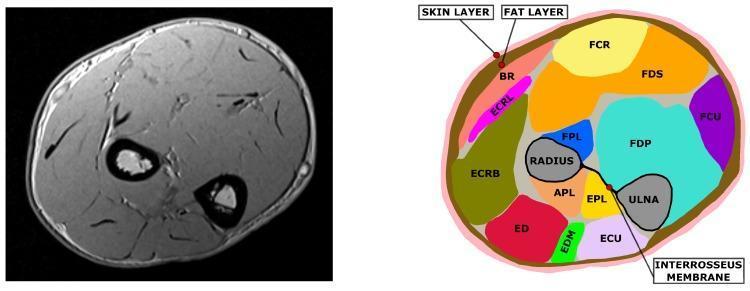
This diagnostic image and anatomical diagram comparison shows a cross-sectional MRI slice of the human forearm (left) alongside its corresponding segmented anatomical map (right). The MRI is a T1-weighted axial scan displaying cortical bone as low-signal (black) voids and muscle bundles as intermediate-signal structures. The segmented diagram identifies key anatomical structures using a color-coding system: the radius and ulna bones are in gray, the interosseous membrane is a black line connecting them, and individual muscles are uniquely colored (e.g., FCR, FDS, FDP, ECU, and ED). The interosseous membrane serves as a landmark dividing the anterior (flexor) and posterior (extensor) muscle compartments. The diagram also illustrates the superficial integumentary layers, with the skin represented in pink and the underlying subcutaneous fat layer in brown. This visual serves as an educational tool for musculoskeletal anatomy, demonstrating the spatial relationships between forearm skeletal structures, deep and superficial muscle groups, and their respective fascial boundaries.
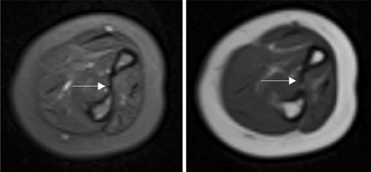
This diagnostic image displays side-by-side axial MRI scans of the human forearm, featuring a T2-weighted sequence (left) and a T1-weighted sequence (right). The cross-sectional anatomy clearly depicts the radius and ulna bones, surrounded by muscle compartments and a thick layer of subcutaneous fat. In the T1-weighted image, the bone marrow and subcutaneous fat exhibit a high (bright) signal, while the muscles appear with intermediate signal intensity. In the T2-weighted image, the fat remains bright, but fluid and certain soft tissue details show increased contrast. A white arrow in both images points specifically to the interosseous membrane, which appears as a thin, continuous, low-signal (dark) band spanning the space between the radius and ulna. The membrane shows no signs of tearing, thickening, or edema, indicating no acute injury. This comparison is used in clinical education to demonstrate normal musculoskeletal ligamentous structures and the signal characteristics of different forearm tissues in the context of orthopedic evaluation for conditions like Monteggia fractures.
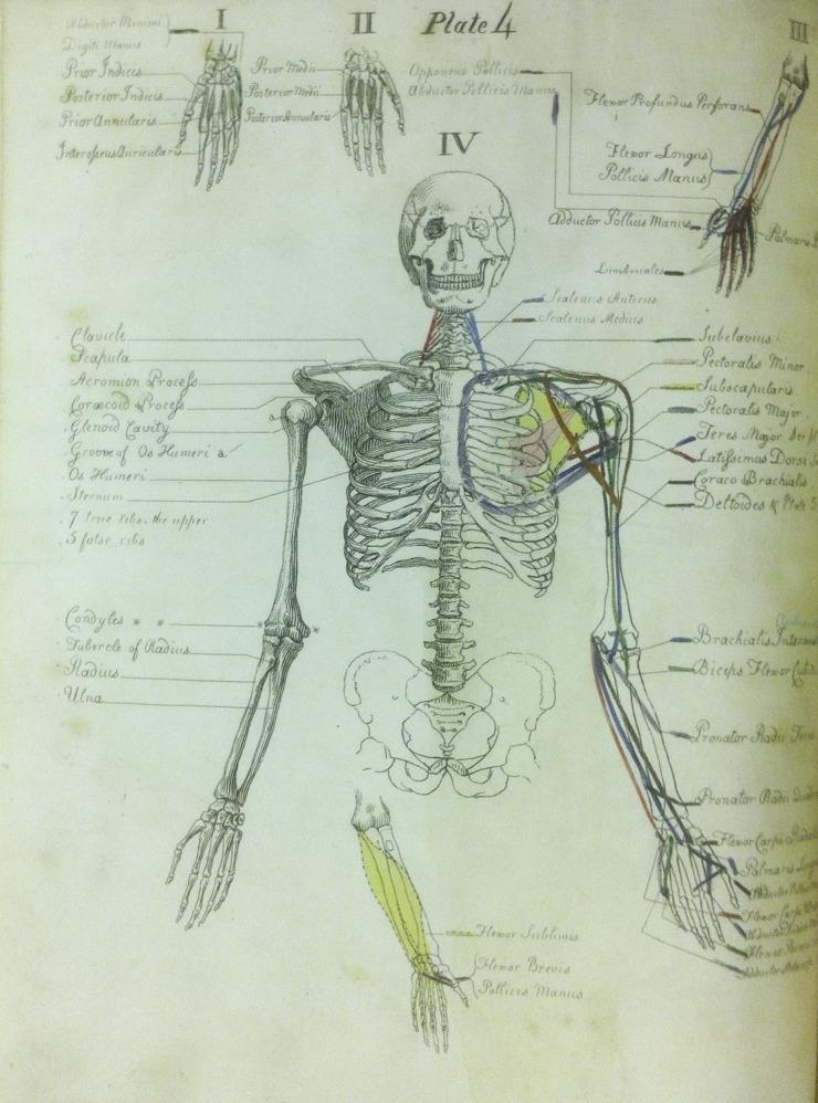
This historical hand-colored anatomical illustration, titled 'Plate 4' from Carpue's 'A Description of the Muscles of the Human Body,' serves as an educational diagram for medical students. It features a musculoskeletal depiction of the human torso and upper extremities. The central figure displays a skeletal framework including the skull, rib cage, vertebrae, pelvis, and arm bones (humerus, radius, ulna). Specific muscles and landmarks are highlighted with hand-applied pigments—predominantly yellow, red, blue, and green—to systematically represent muscle origins and insertions. Labeled structures include the scalenus medius, pectoralis minor, subscapularis, and various flexors of the forearm and hand. Multiple inset diagrams (labeled I, II, and IV) provide detailed perspectives on the osseous and muscular anatomy of the hand and wrist. The illustration demonstrates the spatial relationships and functional attachments of the upper body musculature, characteristic of early 19th-century medical education materials used for surgical preparation and anatomical dissection training.
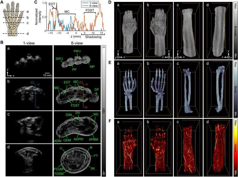
This composite educational graphic displays advanced ultrasound imaging of the human hand, wrist, and forearm. Panel A identifies anatomical planes for four cross-sectional views: (a) distal fingers, (b) distal metacarpals, (c) proximal metacarpals, and (d) distal forearm. Panel B provides a side-by-side comparison between conventional 1-view and high-fidelity 8-view cross-sections. The 8-view images clearly resolve complex anatomy including the distal interphalangeal joints (DIPJ), proximal phalanges (PP), metacarpals (MC), extensor digitorum tendons (EDT), flexor digitorum superficialis tendons (FDST), and intrinsic hand muscles (e.g., ADPM, APBM). The forearm section (d) illustrates the radius (R), ulna (U), and surrounding musculature (FDSM). Panels D–F showcase volumetric renderings from the 250 mm scan. These include grayscale B-mode images (D), blue-toned bone-specific renderings emphasizing the skeletal framework (E), and hot-colormap vascular renderings highlighting superficial and deep vessel networks (F). The figure demonstrates the clinical utility of multi-view ultrasound for improved structural clarity and tissue-specific segmentation of the musculoskeletal and vascular systems.
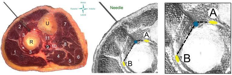
This composite educational resource illustrates a dry needling procedure targeting the supinator muscle of the forearm. The left panel shows a cadaveric cross-section of the proximal forearm with numerical labels identifying key anatomical structures: radius (R), ulna (U), brachioradialis (1), wrist extensors (2), supinator muscle (3), and various flexor compartment muscles (4–8). A needle is shown penetrating the dorsal aspect, passing through the wrist extensors into the supinator muscle belly near the radial nerve branches (*). The center and right panels provide anatomical diagrams focused on the safety profile of the procedure. They map the spatial relationship between the needle tip (blue dot) and critical neurovascular structures: the deep branch (A) and the superficial branch (B) of the radial nerve. Dashed lines represent measurement distances from the needle tip to these nerve branches, serving as a clinical guide for procedural accuracy and hazard avoidance. This material is designed for advanced musculoskeletal anatomy education and clinical training in physical therapy or sports medicine.
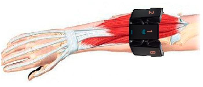
This medical illustration depicts an anatomical diagram of the human forearm and hand with a superimposed wearable surface electromyography (sEMG) device. The underlying anatomy displays the radius and ulna bones, the carpal and metacarpal structures, and the phalanges in white. The musculature of the posterior (extensor) compartment is rendered in red, with tendons highlighted in white crossing the wrist joint beneath the extensor retinaculum. A segmented black armband containing eight dry electrodes is shown positioned on the proximal third of the forearm, approximately one finger-width distal to the elbow joint. The electrodes are numbered, with electrode '1' aligned dorsally with the middle finger, while electrodes '2' and '8' are visible laterally. This visual serves as a technical and anatomical protocol for sensor placement in gesture recognition research, emphasizing the relationship between electrode orientation and underlying superficial muscles and tendons.
Forearm Anatomy
Bones
- The proximal radioulnar joint (at the elbow)
- The interosseous membrane - a fibrous sheet that transmits forces between bones, separates compartments, and provides muscle attachment
- The distal radioulnar joint (at the wrist)
Compartments
Anterior (Flexor) Compartment
| Muscle | Action |
|---|---|
| Pronator teres | Pronation, weak elbow flexion |
| Flexor carpi radialis (FCR) | Wrist flexion, radial deviation |
| Palmaris longus | Wrist flexion (absent in ~15%) |
| Flexor carpi ulnaris (FCU) | Wrist flexion, ulnar deviation |
| Flexor digitorum superficialis (FDS) | Flexion of PIP joints of digits 2-5 |
| Muscle | Action |
|---|---|
| Flexor digitorum profundus (FDP) | Flexion of DIP joints (lateral 2 digits = median; medial 2 = ulnar) |
| Flexor pollicis longus (FPL) | Flexion of thumb IP joint (anterior interosseous nerve) |
| Pronator quadratus | Pronation (anterior interosseous nerve) |
Posterior (Extensor) Compartment
| Muscle | Action |
|---|---|
| Brachioradialis | Elbow flexion (semi-pronated position) |
| Extensor carpi radialis longus (ECRL) | Wrist extension + radial deviation |
| Extensor carpi radialis brevis (ECRB) | Wrist extension |
| Extensor digitorum (ED) | Extension of digits 2-5 |
| Extensor digiti minimi (EDM) | Extension of little finger |
| Extensor carpi ulnaris (ECU) | Wrist extension + ulnar deviation |
| Muscle | Action |
|---|---|
| Supinator | Supination |
| Abductor pollicis longus (APL) | Thumb abduction |
| Extensor pollicis brevis (EPB) | Extension of thumb MCP |
| Extensor pollicis longus (EPL) | Extension of thumb IP |
| Extensor indicis (EI) | Extension of index finger |
Cross-Sectional Anatomy
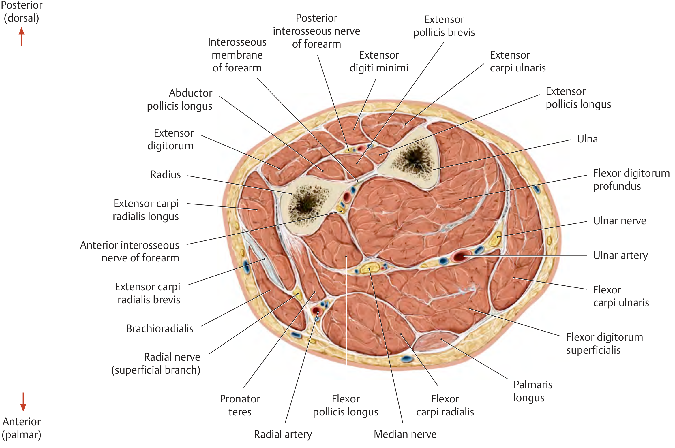
- Radius (lateral) and Ulna (medial) with the interosseous membrane between them
- Anterior compartment: flexor muscles, median nerve centrally, ulnar nerve/artery medially, radial artery laterally
- Posterior compartment: extensor muscles, posterior interosseous nerve
- Anterior interosseous nerve runs on the interosseous membrane between FDP and FPL
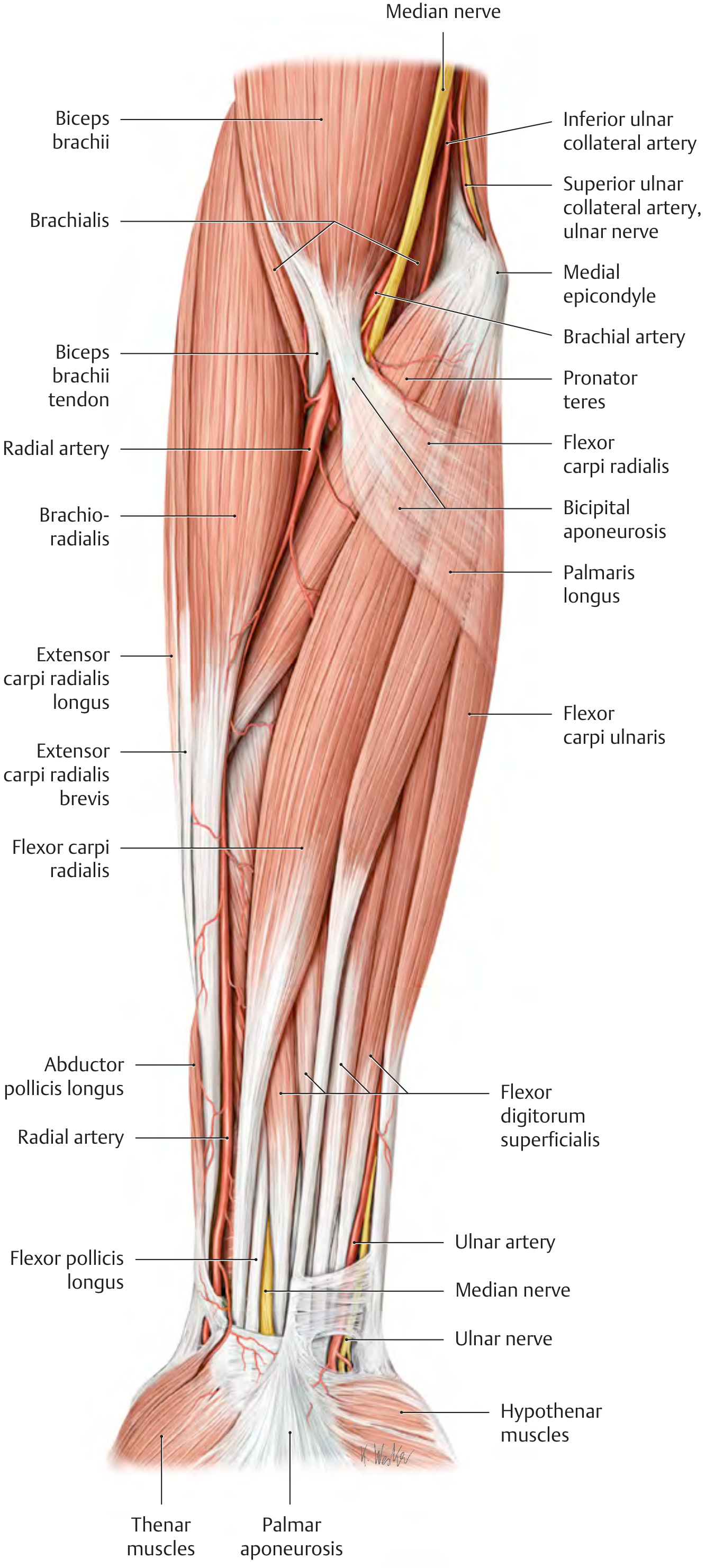
Anterior Forearm - Superficial Dissection
Nerves
Median Nerve
- Enters forearm between the two heads of pronator teres
- Runs between FDS (superficial) and FDP (deep)
- Gives off the anterior interosseous nerve (AIN) - supplies FPL, lateral FDP, pronator quadratus
- Passes through the carpal tunnel at the wrist
- Supplies all flexors except FCU and medial FDP
Ulnar Nerve
- Passes behind the medial epicondyle (through the cubital tunnel)
- Enters forearm between the two heads of FCU
- Runs medially alongside the ulnar artery in the distal forearm
- Supplies FCU and medial half of FDP
Radial Nerve
- Divides at the level of the lateral epicondyle into:
- Superficial branch - sensory, runs under brachioradialis
- Deep branch (posterior interosseous nerve, PIN) - motor, pierces through the supinator (arcade of Frohse) to supply all posterior compartment muscles
Arteries
- Anterior interosseous artery - runs on the anterior surface of the interosseous membrane
- Posterior interosseous artery - pierces the membrane to supply the extensor compartment
- Typical pattern (radius + ulna from brachial): ~84% of cases
- Persistent median artery from ulnar artery distal to common interosseous: ~8%
- Accessory superficial arteries (e.g., superficial ulnar): ~8% - clinically important during cubital fossa IV cannulation
Veins
- Cephalic vein - lateral forearm, along radial border
- Basilic vein - medial forearm, along ulnar border
- Median antebrachial vein - central, connects to median cubital vein at the elbow
Pronation / Supination
- Supination: biceps brachii (most powerful) + supinator
- Pronation: pronator teres (power) + pronator quadratus (precision at all positions)
- During pronation the radius crosses over the ulna; the ulna itself does not rotate significantly
Clinical Pearls
| Scenario | Structure at Risk |
|---|---|
| Lateral epicondyle fracture/dislocation | Posterior interosseous nerve (PIN) in radial tunnel / arcade of Frohse |
| Supracondylar fracture | Anterior interosseous nerve (AIN) and brachial artery |
| Cubital tunnel syndrome | Ulnar nerve at medial epicondyle |
| Pronator syndrome | Median nerve compressed by pronator teres |
| Monteggia fracture-dislocation | PIN injury with radial head dislocation |
| Distal radius fracture | Median nerve (acute carpal tunnel) |