
lpcb principle and morphology description
The question contains medical/microbiology terminology, so I need to invoke the medical library skill first.
Quick Answer:
LPCB (Lactophenol Cotton Blue) is a mounting medium used in mycology to examine fungal morphology. The principle relies on lactophenol killing organisms, phenol acting as a disinfectant, lactic acid preserving structures, and cotton blue (aniline blue) staining fungal cell walls (chitin) blue. It reveals characteristic morphological features like hyphae, conidia, spores, and reproductive structures.
Searching sources...
Reading File
Now let me search the library for LPCB content in parallel with a web search:
Finding Sources
Searching the Web
LPCB lactophenol cotton blue principle morphology fungal staining
Good results found. Let me now read the relevant textbook sections and search for LPCB principle content:
Reading File
Finding Sources
Reading File
Finding Sources
Excellent! I found the Tietz table entry with the principle. Let me read those sections now:
Reading File
Reading File
Reading File
Now let me read the Tietz table entry more closely for the LPCB mechanism, and also fetch some illustrative images:
Reading File
Loading Image
I need to get the actual LPCB table entry from Tietz. Let me check the line around 2519 more carefully and also fetch more images:
Reading File
Loading Image
Loading Image
I now need to find the specific LPCB row in the Tietz table. The table seems to go right to the cultures stains section. Let me search more specifically:
Finding Sources
Loading Image
Excellent! Now let me read the BOX 60.1, BOX 60.2, and Mold Morphology sections from Henry's that contain the procedure and principle details:
Reading File
Reading File
Reading File
I now have comprehensive information. Let me also check the Tietz table for the LPCB row specifically, as the initial DB query showed it was in line 3083. Let me get the exact content from Tietz:
Reading File
Let me get the actual LPCB table row from the Tietz stains table. The snippet showed it was in the cultures stains table:
Finding Sources
lpcb principle and morphology description
LPCB (Lactophenol Cotton Blue) is the standard mounting and staining medium in clinical mycology. Phenol kills organisms, lactic acid preserves structures, glycerol prevents drying, and cotton blue (aniline blue) stains fungal cell walls blue by binding chitin. It reveals hyphae, conidia, spores, and other reproductive structures used for fungal identification.
Searching sources...
Reading File
Finding Sources
Finding Sources
Reading File
Finding Sources
Finding Sources
Now let me read the key sections and fetch the best images in parallel:
Reading File
Reading File
Loading Image
Loading Image
Loading Image
Reading File
I now have all the information needed to write a comprehensive answer. Let me compile it:
Lactophenol Cotton Blue (LPCB) - Principle and Morphology
What is LPCB?
LPCB (Lactophenol Cotton Blue), also called Lactophenol Aniline Blue, is the standard mounting and staining medium used in clinical mycology for the direct microscopic examination of fungi from cultures. It simultaneously kills, preserves, and stains fungal elements in one step.
Composition and Principle
LPCB contains four active components, each with a distinct role:
| Component | Role |
|---|---|
| Phenol | Acts as a disinfectant/biocidal agent - kills (inactivates) fungal cells, making the preparation safe |
| Lactic acid | Preserves fungal structures and cell morphology |
| Glycerol | Prevents the preparation from drying out, extending shelf life of the wet mount |
| Cotton blue (aniline blue dye) | Binds to chitin in fungal cell walls, staining them blue and providing optical contrast |
As summarized in the Tietz Textbook of Laboratory Medicine (7th ed.), LPCB "preserves fungal structure, phenol inactivates the cells, and aniline (cotton blue) dye provides contrast."
The stain is used under the category "Stains Used for Fungal Cultures" - it is applied to culture isolates (not directly to patient specimens for primary diagnosis), making it ideal for the visualization of microscopic features of filamentous fungi and yeasts from cultures.
Safety Note
LPCB is fungicidal - it kills the organisms, which provides a layer of biological safety. However, when examining suspected dimorphic fungi (e.g., Histoplasma, Coccidioides), the teased preparation or cellophane tape mount must still be handled inside a certified biosafety cabinet, with the coverslip sealed with nail polish for additional protection. - Henry's Clinical Diagnosis and Management by Laboratory Methods
Methods of Preparation
Three main techniques are used to prepare LPCB mounts:
-
Cellophane (adhesive) tape mount - A piece of clear tape is briefly pressed onto the colony surface midway between the center and outer margin, then placed sticky-side down onto a drop of LPCB on a glass slide. This preserves the spatial arrangement of conidial structures.
-
Teased preparation - Mycelium is teased apart with inoculating needles, placed in a drop of LPCB on a slide, covered, and examined. The established standard method.
-
Slide culture - A square of agar (Sabouraud, potato dextrose, or potato flake) is suspended on a glass rod in a humid Petri dish and overlaid with a coverslip. After a few days of incubation, the coverslip is removed and placed in LPCB to observe undisturbed conidial structures in their original spatial relationships - used for easily disrupted reproductive structures.
Morphological Structures Visible on LPCB
All fungal elements stain blue against a pale pink-white background. Key structures identifiable include:
1. Hyphae
- Septate hyphae - hyphae divided by cross-walls (septa); seen in most pathogenic molds (Aspergillus, dermatophytes)
- Aseptate (pauciseptate) hyphae - broad, ribbon-like, mostly without septa; characteristic of the Mucorales (Rhizopus, Mucor)
2. Conidia (Asexual Spores)
Macroconidia - large, multicelled, septate conidia:
- Epidermophyton floccosum: blunt-ended, club-shaped macroconidia in aggregates along hyphae; no microconidia present
- Trichophyton mentagrophytes: elongated, pencil-shaped macroconidia lined along hyphae, with small microconidia alongside
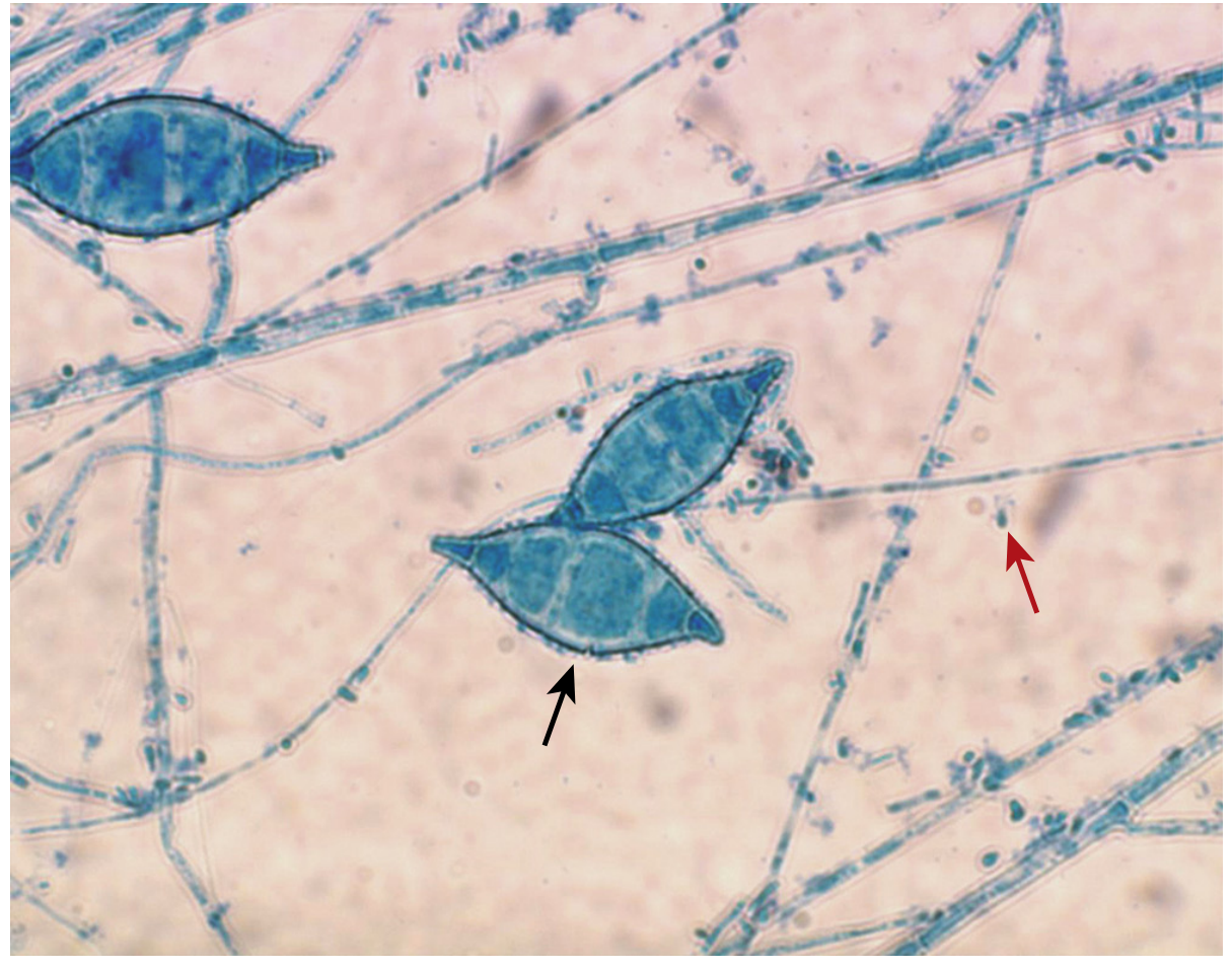
- Microsporum canis: large, spindle-shaped, thick-walled, rough-walled macroconidia (black arrow) with microconidia (red arrow) - see image below
Microconidia - small, single-celled, simple structures; the most common conidia type among the dermatophytes
Arthroconidia - barrel-shaped conidia formed by thallic conidiogenesis; alternating barrel cells separated by thin-walled empty (disjunctor) cells; seen in Coccidioides spp. - highly infectious
3. Conidiophores and Specialized Structures
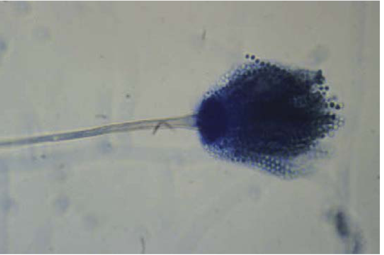
- Aspergillus spp. - conidiophore swells at the tip into a vesicle; phialides arise from the vesicle surface and bear chains of conidia parallel to the long axis; some species also have metulae (supporting sterile cells between vesicle and phialides)
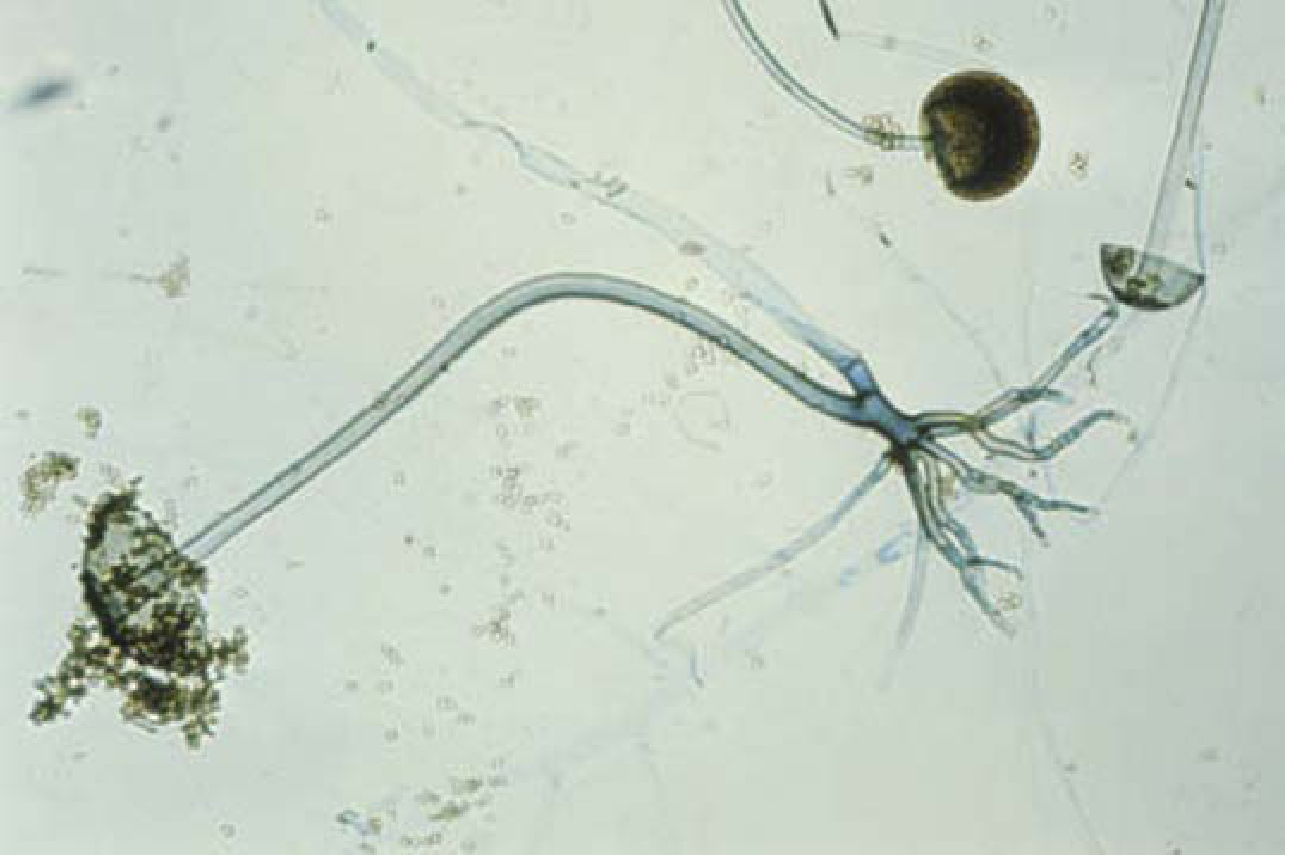
- Rhizopus spp. (Mucorales) - sporangiophores support a round sporangium containing sporangiospores; rhizoids (root-like structures) arise from stolons near the base of the sporangiophores; columella is a dome-like extension of the sporangiophore tip inside the sporangium
- Phialides - flask-shaped conidiogenous cells that release conidia from a collarette at the tip (enteroblastic conidiogenesis); seen in Aspergillus and Phialophora
- Annellides - conidiogenous cells that leave successive rings (annellations) at the apex as each conidium is released; "rings on a tree" appearance
4. Yeast Structures
- Budding yeast cells - round to oval cells with single or multiple buds
- Pseudohyphae - chains of elongated budding yeasts with constrictions at junctions (Candida spp.)
Representative LPCB Photomicrographs
Microsporum canis - smooth-walled macroconidia (black arrow = macroconidium; red arrow = microconidia), with septate hyphae in the background:
Aspergillus fumigatus - fruiting head with vesicle, phialides arising from the upper half, and parallel chains of conidia (LPCB, 400x):
Rhizopus spp. - sporangiophores supporting sporangia, with rhizoids at the base (LPCB, 100x):
Summary Table: Key LPCB Morphology by Organism
| Organism | Hyphae | Macroconidia | Microconidia | Special features |
|---|---|---|---|---|
| Epidermophyton floccosum | Septate | Blunt-ended clubs, in aggregates | Absent | Classic "beaver-tail" clubs |
| Trichophyton mentagrophytes | Septate | Pencil-shaped, elongated | Present, along hyphae | Spiral hyphae may be seen |
| Microsporum canis | Septate | Spindle-shaped, thick rough wall | Present | Echinulate (rough) macroconidial wall |
| Aspergillus fumigatus | Septate | - | - | Vesicle + phialides (uniseriate) |
| Rhizopus spp. | Aseptate/pauciseptate | - | Sporangiospores | Sporangia, rhizoids, columella |
| Coccidioides spp. | Septate | - | Arthroconidia (barrel-shaped) | Alternating with empty disjunctor cells |
Sources: Henry's Clinical Diagnosis and Management by Laboratory Methods; Tietz Textbook of Laboratory Medicine, 7th Edition; Medical Microbiology 9e (Murray)
Dermatophytes culture methods
Dermatophytes are cultured on Sabouraud Dextrose Agar (SDA) with or without cycloheximide and antibiotics. Specimens include skin scrapings, nail clippings, and hair. DTM (Dermatophyte Test Medium) is a selective/differential medium that turns red when dermatophytes grow. Colonies are identified by growth rate, surface texture, pigment, and LPCB microscopy of macro/microconidia morphology.
Searching sources...
Finding Sources
Finding Sources
Reading File
Reading File
Reading File
Finding Sources
Reading File
Finding Sources
Reading File
Finding Sources
Finding Sources
Finding Sources
Reading File
Finding Sources
Reading File
Finding Sources
Reading File
I now have comprehensive information from multiple authoritative sources. Let me compile the complete answer:
Dermatophytes - Culture Methods
Overview
The laboratory diagnosis of dermatophytosis relies on two complementary steps: direct microscopy (KOH preparation) and culture for definitive identification. Culture is essential whenever systemic treatment is planned, and is the most specific test for onychomycosis. Colonies develop within 7 to 28 days depending on the species. - Medical Microbiology 9e (Murray)
Step 1 - Specimen Collection
Correct sampling technique is critical - the wrong site or part of the lesion yields false negatives.
Hair (Tinea capitis / Tinea barbae)
- Clean the area of alopecia with alcohol (does not affect fungal viability)
- Pluck (do not cut) infected hairs from the root - cut hairs are rarely infected and often bacterial-contaminated
- Collect black dots (hair stubs in endothrix infection) or scalp scale from the area of alopecia
- For Microsporum infections, a Wood's lamp (365 nm) can be used first - fluorescing hairs (yellow-green pteridine fluorescence) are preferentially selected for culture
- Use a toothbrush, scalpel, gauze square, or cotton-tipped applicator
Skin (Tinea corporis, cruris, pedis, manuum)
- Clean the advancing edge of annular lesions with alcohol
- Scrape the active advancing margin (not the center - the center is "healed" and devoid of viable fungi)
- For intertriginous mycoses: collect from the dry scaling edge - avoid moist macerated central material (contaminated and devoid of viable fungi)
- For vesicular lesions: collect the vesicle roof - vesicular fluid and vesicle base are usually devoid of fungi
Nails (Onychomycosis)
- Distal subungual type: trim back the nail to the leading edge of infection (discard the distal portion) - then collect keratinaceous debris from beneath the remaining trimmed nail plate edge (the actual fungal reservoir)
- Proximal subungual type: collect from the active edge closest to the distal end
- White superficial type: simply scrape the surface area of involvement
- Note: Simple nail clippings and whole-removed nail plates are less ideal
Step 2 - Culture Media
Primary Isolation Media
| Medium | Composition | Purpose |
|---|---|---|
| Sabouraud Dextrose Agar (SDA) | 4% glucose, 1% neopeptone, pH 5.5-5.6 | Standard reference medium; most morphological descriptions are based on growth here |
| SDA + Cycloheximide + Chloramphenicol (Mycosel/Mycobiotic agar) | Cycloheximide inhibits saprophytic/contaminant molds; Chloramphenicol (± Gentamicin) inhibits bacteria | Selective medium of choice for dermatophyte isolation from non-sterile specimens |
| Dermatophyte Test Medium (DTM) | SDA base + cycloheximide + chloramphenicol + phenol red pH indicator | Presumptive identification + selective isolation |
| Inhibitory Mold Agar | Chloramphenicol ± gentamicin | Selective general-purpose fungal medium |
DTM - How the Color Change Works
- Dermatophyte proteolytic activity raises the pH to 8 or above → medium turns RED
- Saprophyte growth produces acidic byproducts → medium turns yellow
- Amber color with no change = most saprophytes / no dermatophyte
- Important caveat: DTM may alter colony morphology, so direct identification on DTM may not be reliable. Subculture to SDA is recommended for definitive species identification. - Fitzpatrick's Dermatology
Note on cycloheximide: Some clinically relevant non-dermatophyte nail pathogens (e.g., Scopulariopsis brevicaulis, Aspergillus spp., Fusarium spp.) do not grow on cycloheximide-containing media and may be missed if only selective media are used, especially for nail specimens. - Dermatology 5e (Bolognia)
Secondary/Identification Media (for subculture)
| Medium | Purpose |
|---|---|
| Potato Dextrose Agar (PDA) | Stimulates sporulation and pigment production; standard for differentiating Trichophyton rubrum (cherry red) vs others |
| Potato Flake Agar / Borelli Lactritmel Agar | Stimulates conidial sporulation for morphological ID |
| Cornmeal Agar + Tween 80 | Promotes chlamydoconidium and pseudohypha formation in yeasts |
| Polished Rice Agar | Differentiates M. canis (good growth) from M. audouinii (poor/no growth) |
| Trichophyton agars (1-7) | Nutritional agars to assess vitamin requirements for Trichophyton speciation |
Step 3 - Incubation Conditions
- Temperature: 20°C to 25°C (room temperature)
- Duration: Incubate for at least 4 weeks before reporting no growth
- Growth rates vary by species:
- Epidermophyton floccosum: 5-7 days
- Most Trichophyton spp.: 1-2 weeks
- Trichophyton verrucosum: up to 4 weeks (slowest grower)
Step 4 - Colony Identification
Macroscopic Features Examined
- Surface texture: powdery, granular, velvety, cottony, waxy, heaped
- Pigmentation (surface and reverse)
- Growth rate and colony size
- Radial grooves, folds, topography
Species-Specific Colony Features (Key Examples)
| Organism | Surface Colony | Reverse Pigment | Notes |
|---|---|---|---|
| Epidermophyton floccosum | Flat, feathery, central fold; yellow-green to dull gray | Yellow-brown | No microconidia |
| Microsporum audouinii | Flat, white-gray, widely spaced radial grooves; salmon reverse | Salmon-pink on PDA | No growth on polished rice |
| Microsporum canis | Flat, white-yellow, coarsely hairy, closely spaced grooves | Yellow-orange; yellow on PDA | Good growth on polished rice |
| Microsporum gypseum | Flat, granular, tan-buff | No reverse pigment | Thin-walled pickle-shaped macroconidia |
| Trichophyton rubrum | Mounded white center, maroon periphery | Maroon reverse; cherry red on PDA | Urease negative; hair perforation negative |
| Trichophyton interdigitale | White to creamy, cottony, mounded | None to light brown | Urease positive; hair perforation positive |
| Trichophyton tonsurans | Suede-like center, feathery periphery; white to yellow or maroon | Dark maroon (sometimes none) | Partial thiamine requirement |
| Trichophyton schoenleinii | Heaped/folded, whitish | Colorless to yellow-tan | Favic chandelier hyphae |
| Trichophyton verrucosum | Small, heaped, white to yellow-gray | None to yellow | Requires thiamine + inositol |
| Trichophyton violaceum | Waxy, heaped, deep purplish-red | Purple | Requires thiamine |
Step 5 - Microscopic Identification (LPCB)
A cellophane tape mount or teased preparation stained with LPCB is used. Key features:
Conidia - Genus-Level Key
| Genus | Microconidia | Macroconidia | Diagnostic Conidia |
|---|---|---|---|
| Trichophyton | Present, smooth-walled | Absent or nondiagnostic | Microconidia used for ID |
| Microsporum | Absent or nondiagnostic | Present, rough-walled | Macroconidia used for ID |
| Epidermophyton | Absent | Present, smooth-walled | Macroconidia used for ID |
Step 6 - Supplementary Biochemical Tests
| Test | Positive Result | Negative Result |
|---|---|---|
| Urease test | T. interdigitale | T. rubrum |
| Hair perforation test (in vitro) | T. interdigitale | T. rubrum |
| Growth on polished rice | M. canis (good growth) | M. audouinii, M. distortum (poor growth) |
| Nutritional requirements (Trichophyton agars) | Thiamine: T. tonsurans, T. concentricum, T. violaceum | Allows species separation |
| Thiamine + inositol: T. verrucosum | ||
| Nicotinic acid: T. equinum |
Wood's Lamp as Pre-Culture Screening (Hair)
- Wavelength: 365 nm
- Ectothrix Microsporum species (M. canis, M. audouinii): yellow-green fluorescence (pteridines) → fluorescing hairs selected for culture
- T. tonsurans (most common tinea capitis in the US): does NOT fluoresce → limits usefulness of Wood's lamp alone
- Fluorescing hairs should always be sent for culture, not relied upon solely for diagnosis
Important Caveat: Non-Dermatophyte Molds in Nails
When culture is performed for onychomycosis, at least one set of plates should be without cycloheximide because clinically relevant non-dermatophyte pathogens (Scopulariopsis brevicaulis, Fusarium, Aspergillus spp., Neoscytalidium spp.) are inhibited by cycloheximide and will be missed on selective media alone. For nail specimens specifically, PAS examination of nail clippings is the most sensitive single test and provides results faster than culture. - Fitzpatrick's Dermatology
Sources: Fitzpatrick's Dermatology (Vol 1-2); Medical Microbiology 9e (Murray); Pfenninger & Fowler's Procedures for Primary Care; Tietz Textbook of Laboratory Medicine 7e; Dermatology 5e (Bolognia); Henry's Clinical Diagnosis and Management by Laboratory Methods
Criteria for DTM spots and interpretation
DTM (Dermatophyte Test Medium) turns red when dermatophytes grow due to alkaline metabolites raising the pH, triggering the phenol red indicator. Saprophytes produce acidic byproducts and either keep the medium yellow/amber or turn it yellow. The key rule is that the color change must occur simultaneously with or before colony growth - a late color change after heavy growth suggests a saprophyte contaminant.
Searching sources...
Finding Sources
Finding Sources
Finding Sources
Finding Sources
Reading File
Finding Sources
Reading File
Finding Sources
Finding Sources
Finding Sources
Finding Sources
Finding Sources
Now let me read the full relevant sections from Fitzpatrick's and Tietz, and also search the web for more detailed DTM interpretation criteria:
Reading File
Reading File
Searching the Web
dermatophyte test medium DTM interpretation criteria color change timing simultaneous false positive saprophyte
Excellent - I now have comprehensive information from both textbooks and authoritative manufacturer/lab sources. Let me compile the full answer:
Dermatophyte Test Medium (DTM) - Criteria and Interpretation
What is DTM?
DTM (Dermatophyte Test Medium), first described by Taplin in 1969, is a selective, differential, and presumptive culture medium designed for the isolation and screening identification of dermatophytes in clinical specimens (skin scrapings, nail clippings, plucked hairs).
Composition and Rationale of Each Component
| Ingredient | Amount (per litre) | Function |
|---|---|---|
| Soy peptone (papaic digest) | 10 g | Nitrogen + carbon source for growth; protein substrate for dermatophyte proteolytic activity |
| Dextrose | 10 g | Carbohydrate energy source |
| Cycloheximide | 0.5 g | Inhibits saprophytic/contaminant molds |
| Chloramphenicol (± Gentamicin) | 0.05–0.1 g | Broad-spectrum antibacterial - suppresses gram-positive and gram-negative bacteria |
| Phenol red | 0.2 g | pH indicator (the heart of the differential system) |
| Agar | 20 g | Solidifying agent |
| Final pH | 5.6 ± 0.2 | Acidic starting pH favours dermatophyte growth; phenol red is yellow at this pH |
Principle - How the Color Change Works
The entire interpretation of DTM rests on the sequential utilization of nutrients by different organisms:
Dermatophytes (Positive Result - RED)
- Dermatophytes preferentially metabolize proteins first (proteolytic activity)
- Protein catabolism releases alkaline metabolites (ammonia/nitrogen-containing byproducts)
- pH rises from 5.6 to ≥8 (phenol red turns red at ≥8.2)
- Medium turns red simultaneously with or before colony growth
- When proteins are exhausted, dermatophytes shift to carbohydrate metabolism → acidic byproducts → medium may gradually revert to yellow if read late
Saprophytes/Contaminants (Negative - AMBER/YELLOW stays)
- Most saprophytes preferentially ferment carbohydrates first
- Carbohydrate fermentation → acidic byproducts → medium stays yellow or turns more yellow/amber
- As a contaminant uses up carbohydrates, it may eventually shift to protein metabolism → late red color change (beyond day 10-14) - this is the basis of false positives
As stated in [Fitzpatrick's Dermatology]: "The medium turns red when dermatophyte proteolytic activity increases the pH to 8 or above, and it remains amber with the growth of most saprophytes. Nondermatophyte acidic byproducts turn the medium yellow."
DTM Interpretation Criteria
POSITIVE (Dermatophyte present)
| Criterion | Details |
|---|---|
| Color | Medium turns RED around and beneath the colony |
| Timing | Color change occurs simultaneously with or before colony growth appears - this is the critical criterion |
| Colony appearance | White to buff/cream-colored aerial hyphae (fluffy, powdery, or granular surface) |
| Reading window | Within first 10-14 days of incubation |
The simultaneous or early color change with white aerial hyphae = strong presumptive positive for dermatophyte. - [Hardy Diagnostics DTM package insert]
NEGATIVE (No dermatophyte)
| Finding | Interpretation |
|---|---|
| Growth present but no color change to red | Organism is probably NOT a dermatophyte |
| Growth on control (SDA) but no growth on DTM | Organism is not a dermatophyte (may be cycloheximide-sensitive) |
| Medium remains amber/yellow-orange | No significant dermatophyte activity |
FALSE POSITIVE (Red color change but NOT a dermatophyte)
This is the most important pitfall in DTM interpretation:
| Cause | How to Recognize |
|---|---|
| Late-reading beyond 14 days | Most non-dermatophyte fungi eventually produce alkaline byproducts once carbohydrates are exhausted → rule: color change after day 14 = likely false positive |
| Saprophytic contaminant molds | Color change may occur, but colonies show dark green to black hyphae (not white/buff) - easily distinguished |
| Certain yeasts | May produce alkaline metabolites; but colonies are white/creamy and bacteria-like (not mycelial), easily distinguished from dermatophyte aerial hyphae |
| Heavy inoculum contamination | Overwhelming bacterial/fungal contamination can bypass selective agents and cause color change |
| Dormant/inactive infection site sampled | May yield false-negative result - no growth at all |
The critical differentiation rule:
Dermatophytes = white/buff aerial hyphae + early red color change (within 10-14 days, simultaneous with growth)Saprophytes = dark/colored colonies (green, black, brown) + late red color change (after day 14 if at all)Yeasts = white creamy bacterial-looking colonies ± possible red change
Step-by-Step Interpretation Protocol
Day 1-14: Check daily
Is there COLONY GROWTH?
│
├─ NO → Continue incubating up to 14-21 days
│
└─ YES
│
├─ Is the medium turning RED?
│ │
│ ├─ YES (simultaneously or before colony growth)
│ │ │
│ │ ├─ Are colonies WHITE/BUFF aerial hyphae?
│ │ │ └─ YES → PRESUMPTIVE POSITIVE for dermatophyte
│ │ │ (confirm with LPCB microscopy)
│ │ │
│ │ └─ Are colonies dark/greenish/black?
│ │ └─ YES → FALSE POSITIVE (saprophyte contaminant)
│ │
│ └─ NO red change (amber/yellow)
│ └─ Likely NOT a dermatophyte
│
└─ Is color change appearing AFTER day 14?
└─ Likely FALSE POSITIVE regardless of colony color
Important Limitations of DTM
-
Presumptive only - DTM cannot give definitive species identification. Colony morphology is often altered in DTM, making species-level ID unreliable. Always subculture to SDA or PDA for speciation.
-
Cycloheximide-sensitive pathogens missed - Clinically significant non-dermatophyte nail pathogens (Scopulariopsis brevicaulis, Aspergillus spp., Fusarium spp., Candida tropicalis, Trichosporon spp., Scedosporium spp.) are inhibited by cycloheximide and will not grow on DTM. For nail specimens especially, always culture on non-selective SDA in parallel.
-
Reading window is strict - Must read within 14 days; beyond this, false positives increase significantly as even saprophytes eventually oxidize proteins.
-
Loose cap required - The cap must be kept loosely applied during incubation to ensure proper gas exchange and optimal dermatophyte recovery. A tight cap can inhibit growth.
-
Confirm positives microscopically - All color-change positives must be confirmed by LPCB microscopic examination of the colony (tape mount or teased preparation) to identify macroconidia/microconidia morphology and confirm genus/species.
Summary Table: DTM Color Change Interpretation at a Glance
| Color of Medium | Colony Appearance | Timing | Interpretation |
|---|---|---|---|
| Red | White/buff, aerial hyphae | Day 1-14, simultaneous | ✅ Presumptive POSITIVE (dermatophyte) |
| Red | Dark green/black/brown | Any time | ❌ FALSE POSITIVE (saprophyte contaminant) |
| Red | White creamy, bacteria-like | Any time | ⚠️ Likely yeast (not dermatophyte) |
| Red | Any | After day 14 | ❌ FALSE POSITIVE (late saprophyte alkalinization) |
| Amber/Yellow | Any colony growth | Any | ❌ NEGATIVE (not a dermatophyte) |
| Yellow (more yellow) | Any colony growth | Any | ❌ NEGATIVE (acidic byproduct producer) |
| No change, no growth | - | Up to 14-21 days | Negative or cycloheximide-sensitive organism present |
Sources: Fitzpatrick's Dermatology (Vol 1-2); Tietz Textbook of Laboratory Medicine 7e; Hardy Diagnostics DTM package insert; Pfenninger & Fowler's Procedures for Primary Care; OSU CVM Veterinary Clinical and Professional Skills Center; microbeonline.com DTM review
This is a shared conversation. Sign in to Orris to start your own chat.