
pyoderma gangrenosum management
"pyoderma gangrenosum"[MeSH Terms] AND treatment
pyoderma gangrenosum ulcer clinical appearance
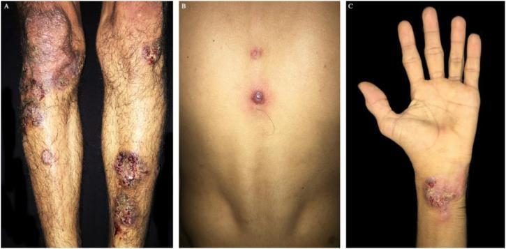
Clinical photograph series showing cutaneous manifestations of pyoderma gangrenosum. Image A displays the lower limbs with multiple erythematous nodules and deep ulcers. These lesions feature well-defined, slightly raised, violet-brownish borders and a characteristic cribriform (sieve-like) appearance in the ulcer bed, accompanied by perilesional erythema. Image B focuses on the dorsal trunk, presenting a violet papule with central vesiculation and two smaller satellite papules. Image C shows a similar ulcerated plaque on the volar aspect of the wrist/hand. The morphology is consistent with neutrophilic dermatosis often associated with systemic conditions or vasculitis. Educational focus includes the recognition of undermined borders and cribriform scarring typical of pyoderma gangrenosum. These clinical findings are relevant for dermatology and internal medicine, particularly in the context of identifying diagnostic cutaneous markers for underlying systemic inflammatory or toxicological causes.
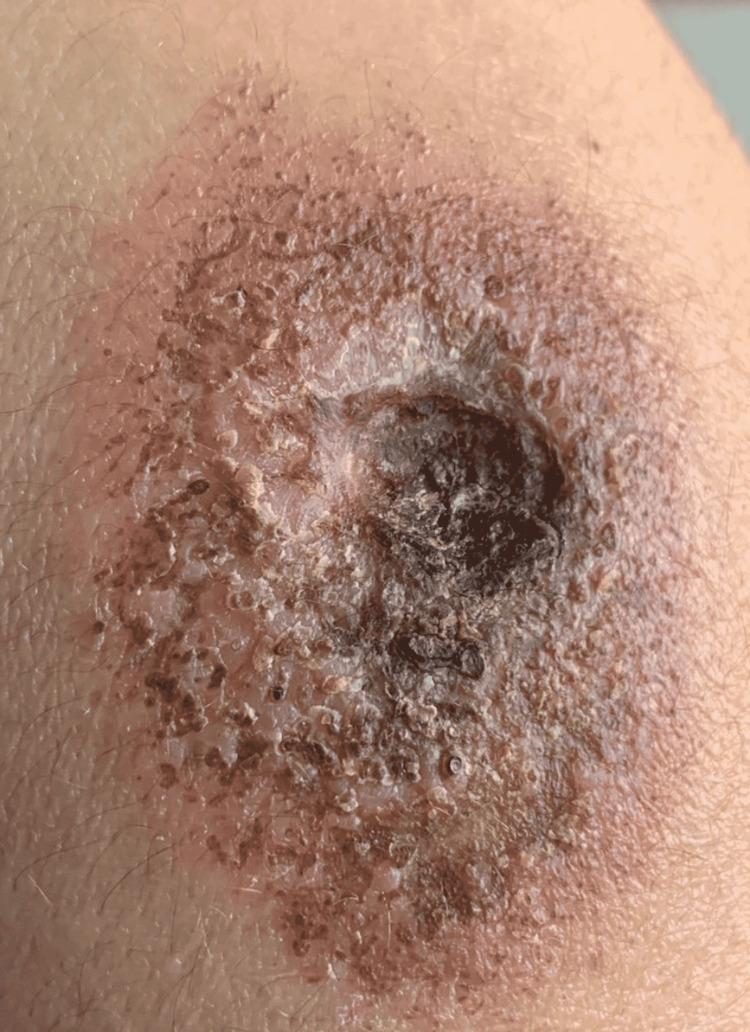
This clinical photograph displays a large, complex skin ulcer characteristic of Pyoderma Gangrenosum. The lesion features a deep central cavity with an irregular, necrotic base containing dark, devitalized tissue. The borders of the ulcer are poorly defined and notably undermined, with a violaceous or dusky hue. Surrounding the primary ulceration is a broad zone of intense erythema and inflammatory induration. The periphery of the lesion shows evidence of crusting, scaling, and small pustular elements, suggesting active progression. The overall appearance is consistent with an aggressive, inflammatory neutrophilic dermatosis, emphasizing the classic 'undermined' edge and necrotic floor used for clinical diagnosis in dermatology.
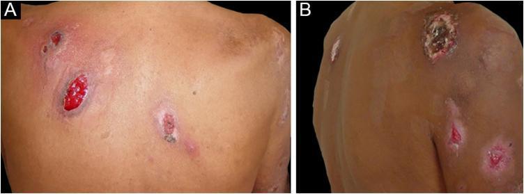
Two-panel clinical photograph demonstrating bullous pyoderma gangrenosum. Image A displays the posterior trunk (back) with multiple shallow ulcers characterized by a bright red, beefy granulating base and irregular, slightly elevated borders. The surrounding skin shows perilesional erythema and inflammation. Image B shows the upper limb with similar ulcerative lesions. A key diagnostic feature visible is the distinctive blue-gray or violaceous hue at the ulcer margins. The lesions vary in appearance from active erosions with red bases to ones with dark, hemorrhagic crusting or scabbing. This presentation illustrates a neutrophilic dermatosis often associated with underlying systemic conditions such as hematologic malignancies or inflammatory bowel disease. The educational focus is on identifying the characteristic undermined, dusky borders and the progression from bullae to shallow ulcers typical of the bullous variant of pyoderma gangrenosum.
PMID: 40740034
PMID: 39158753
Pyoderma Gangrenosum (PG): Management
Overview
Diagnostic Caveat Before Treatment
- Diagnosis is clinical and exclusionary - must rule out infection, vasculitis, malignancy, factitial disease
- Pathergy (20-30% of cases): avoid debridement and surgical intervention, as trauma worsens lesions
- Response to immunosuppression is itself a minor diagnostic criterion - failure to respond should prompt re-evaluation of the diagnosis
Step 1: Treat the Underlying Disease
| Associated Condition | Implication |
|---|---|
| IBD (Crohn's / UC) | Anti-TNF therapy overlaps; managing IBD often improves PG |
| Hematologic malignancy | Chemotherapy for underlying disease may resolve bullous PG |
| RA | DMARDs (methotrexate, anti-TNFs) serve dual purpose |
| Peristomal PG | Stoma revision or relocation may help; topical tacrolimus reported effective |
Step 2: Local / Topical Therapy
- Wound care: compresses or whirlpool baths followed by hydrophilic occlusive dressings
- Potent topical corticosteroids (e.g., clobetasol): applied directly to lesion borders
- Intralesional corticosteroid injections: caution - pathergy risk at injection sites
- Topical tacrolimus 0.1-0.3%: effective, especially peristomal PG; systemic absorption possible in large wounds
- Topical dapsone: adjunct option
- Topical timolol: isolated case reports of benefit (BMJ Case Rep, 2017)
- Topical corticosteroid powder: systematic review (PMID: 39500711) supports use in peristomal PG
Step 3: Systemic Therapy
First-Line Agents
| Drug | Dosing | Notes |
|---|---|---|
| Prednisolone / Prednisone | 1-2 mg/kg/day orally | Rapid onset; taper once controlled |
| Methylprednisolone pulse | IV, 3-5 days | For rapidly progressive or severe disease |
| Cyclosporine | 4-5 mg/kg/day | STOP-GAP RCT validated 4 mg/kg; response independent of underlying cause |
| Infliximab | 5 mg/kg IV at weeks 0, 2, 6, then every 6-8 weeks | Validated in RCT; most utilized biologic in PG |
Biologic Therapies (First- or Second-Line)
- Infliximab (anti-TNF): most used (64.8% of biologic-treated cases); average time to resolution ~161 days
- Adalimumab (anti-TNF): 16.7% of cases; 40-80 mg every other week; also effective for IBD-associated PG
- Etanercept: 11.1% of cases; used as alternative
- Canakinumab (anti-IL-1β): promising in small studies for steroid-refractory PG (Br J Dermatol, 2015)
- Ustekinumab (anti-IL-12/23): reported in refractory cases
Second-Line / Steroid-Sparing Agents
- Mycophenolate mofetil (MMF)
- Azathioprine
- Methotrexate
- Dapsone / Sulfasalazine / Sulfapyridine: less potent; useful for mild disease or contraindications to stronger agents
- Minocycline: adjunct
- Thalidomide: reserved for refractory cases
- IVIG / granulocyte apheresis: case series support; considered in refractory disease
- Systemic tacrolimus: for severe, recalcitrant cases (G Ital Dermatol Venereol, 2017)
- Alkylating agents (cyclophosphamide, chlorambucil): reserved for severe refractory disease
Step 4: Monitoring Response
- Expected response: within 1-2 weeks - reduced pain, less exudate, decreased erythema/edema
- Once inflammation is controlled, taper systemic immunosuppression
- Do not continue high-dose immunosuppression just for wound closure once inflammation is under control - the remaining wound can be managed with optimal dressings
- Ongoing wound care is essential even after achieving inflammatory control
Special Situations
Postoperative / Surgical PG
- Occurs at surgical incision sites; often misdiagnosed as wound infection
- History of sterile wound cultures and multiple failed debridements is a clue
- Avoid debridement - treat with systemic immunosuppression
Peristomal PG
- Topical tacrolimus is well supported (Lyon et al., J Dermatol)
- Anti-TNF therapy (IFX/ADA) effective when IBD is active
- Stoma relocation may be considered
Childhood PG
-
40% have IBD, ~18% have leukemia
- Treat underlying cause first
- Head/neck and genital involvement possible
Bullous PG (associated with hematologic malignancy)
- Often overlaps with bullous Sweet syndrome
- Managing the underlying hematologic disorder is central
Summary Algorithm
PG confirmed (clinical + exclusion)
     |
     +--> Identify & treat underlying disease
     |
     +--> Severity assessment
            |
            +--> Mild / solitary / vegetative
            |         --> Topical steroids / tacrolimus / wound care
            |
            +--> Moderate / progressive
            |         --> Systemic prednisolone 1-2 mg/kg/day
            |              OR cyclosporine 4 mg/kg/day (STOP-GAP)
            |              + wound care
            |
            +--> Severe / rapidly progressive
                      --> IV methylprednisolone pulse
                           + Infliximab (5 mg/kg)
                           --> Taper steroids, maintain biologic
                           --> Add steroid-sparing agent if needed
Key Evidence (Recent)
- 2025 meta-analysis (Int Wound J) - pooled healing rate 59%, recurrence 30%, treatment failure 36% - highlights significant unmet need (PMID: 40740034)
- 2024 systematic review on biologics (Arch Dermatol Res) - 88.9% resolution with infliximab/adalimumab/etanercept (PMID: 39158753)
- 2023 Japanese clinical guidance (J Dermatol) - practice guideline (PMID: 37311717)
infliximab availability in india
infliximab brands available India 2024 2025 price biosimilars
infliximab brand names India Remsima Infimab Revellex price per vial INR 2024
infliximab India CDSCO approved brands Remsima Zessly Infimab availability hospitals 2025
Infliximab Availability in India
Regulatory Status
Available Brands in India
| Brand Name | Type | Manufacturer / Marketer | Notes |
|---|---|---|---|
| Remicade | Originator | Janssen (J&J) | Reference product; available but expensive |
| Infimab (BOW015) | Similar biologic | Epirus Biopharmaceuticals / Reliance Life Sciences (manufactured in Mumbai) | First infliximab similar biologic approved in India (Sept 2014); marketed by Ranbaxy (now Sun Pharma) |
| Remsima (CT-P13) | Biosimilar | Celltrion (South Korea) | Available via importers/distributors (e.g., GNH India); most widely studied biosimilar globally |
| Zessly (PF-06438179) | Biosimilar | Sandoz / Pfizer | Available in Indian market through specialty distributors |
Note: Infliximab leads the Indian biosimilar market with 14% revenue share in 2025, driven by autoimmune disease adoption (RA, IBD, psoriasis). India is also a major manufacturer - Reliance Life Sciences produces Infimab domestically.
Approximate Cost (100 mg vial)
| Product | Approximate Price (INR/vial) |
|---|---|
| Remicade (originator) | ~₹16,000 - ₹17,500 |
| Infimab (biosimilar) | ~₹12,000 - ₹14,500 |
| Remsima (imported biosimilar) | ~₹12,000 - ₹17,000 |
Indications Approved in India
- Rheumatoid arthritis
- Ankylosing spondylitis
- Crohn's disease
- Ulcerative colitis
- Psoriatic arthritis
- Plaque psoriasis
- Behcet's disease
Practical Access Considerations
| Factor | Detail |
|---|---|
| Setting | IV infusion only; requires hospital/clinic infusion facility |
| Cold chain | Must be refrigerated (2-8°C); not available at general retail pharmacies |
| Procurement | Through specialty distributors, hospital pharmacies, or online exporters (GNH India, etc.) |
| Patient support | Ranbaxy/Sun Pharma launched RAISE (Rheumatoid Ailment Infimab Support Endeavor) for patient access assistance |
| Government schemes | Not currently on PMJAY (Ayushman Bharat) essential medicines list for PG specifically; IBD patients may access through GI specialty programs |
| Monitoring | Requires pre-infusion TB screening (Mantoux/IGRA), baseline LFTs, CBC; infusion centre with resuscitation capability |