
1. Define inflammation. Describe endothelial and leucocyte derived adhesion molecules and their role in inflammation. 2. Describe differential diagnosis of granulomatous disease in detail. 3. Discuss complement system in health and disease. 4. Define and classify granuloma. Discuss wegener’s granulomatosis.
leukocyte recruitment adhesion molecules rolling marginaton steps inflammation
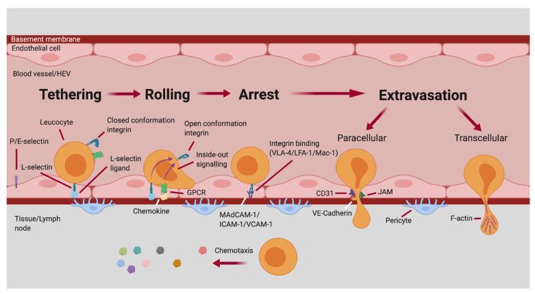
This pathophysiology diagram illustrates the sequential stages of the leukocyte adhesion cascade within a blood vessel or high endothelial venule (HEV). The process is divided into four main phases: Tethering, Rolling, Arrest, and Extravasation. 1. Tethering: Mediated by L-selectin and P/E-selectins interacting with their respective ligands. Leukocyte integrins are in a closed conformation. 2. Rolling: Leukocyte shape elongates as it interacts with chemokines via GPCRs. This triggers 'inside-out signaling,' shifting integrins to an open conformation. 3. Arrest: Firm adhesion is achieved through high-affinity integrin binding (VLA-4, LFA-1, Mac-1) to endothelial ligands (MAdCAM-1, ICAM-1, VCAM-1). 4. Extravasation: The leukocyte exits into the tissue/lymph node via two pathways: Paracellular migration (between endothelial cells, involving CD31, JAM, and VE-Cadherin) or Transcellular migration (through the endothelial cell, involving F-actin). The diagram concludes with the leukocyte passing the basement membrane and pericytes, directed by chemotaxis. This visual is designed for advanced immunology and medical students to understand cellular recruitment during inflammation or immune surveillance.
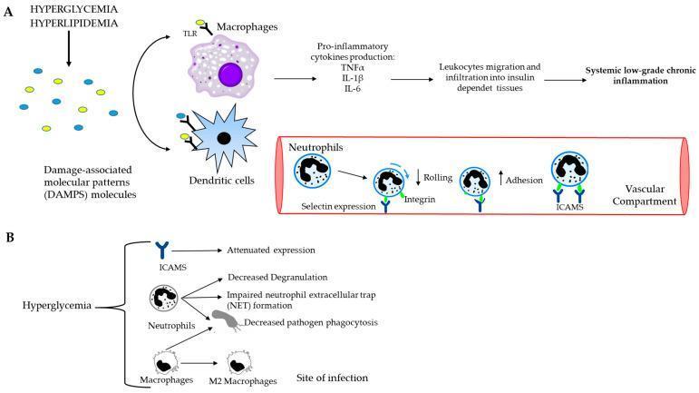
A two-part medical pathophysiology diagram illustrating the link between metabolic dysfunction and systemic inflammation. Part A depicts the cascade where hyperglycemia and hyperlipidemia generate Damage-Associated Molecular Patterns (DAMPs). These molecules bind to Toll-like receptors (TLRs) on macrophages and dendritic cells, triggering the release of pro-inflammatory cytokines (TNFͱ, IL-1̢, IL-6). This lead to leukocyte migration into insulin-dependent tissues and systemic low-grade chronic inflammation. An inset showing the vascular compartment details the neutrophil recruitment process, highlighting decreased rolling (mediated by selectin) and increased adhesion (mediated by integrins and ICAMs). Part B focuses on the cellular impact of hyperglycemia, showing attenuated ICAM expression, decreased neutrophil degranulation, impaired Neutrophil Extracellular Trap (NET) formation, and decreased phagocytosis by neutrophils and macrophages. It also notes the transition of macrophages to the M2 phenotype at sites of infection. This diagram is designed for advanced medical education regarding the immunology of metabolic diseases like diabetes and obesity.
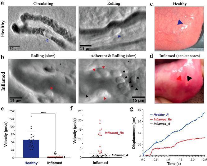
Educational panel illustrating in vivo leukocyte dynamics within human oral mucosa microvasculature under healthy and inflamed conditions, captured using oblique back-illumination microscopy (mOBM). Panels (a) and (c) depict healthy tissue with uniform mucosal appearance and leukocytes exhibiting normal circulation and rolling behavior (blue arrowheads). Panels (b) and (d) show inflamed tissue featuring a canker sore, characterized macroscopically by focal erythema and microscopically by slow-rolling (red arrowheads) and stationary, adherent leukocytes (black arrowheads) forming clusters along the endothelium. Quantitative analysis is provided in graphs (e-g): Panel (e) shows a significant reduction in mean leukocyte velocity in inflamed tissue (p < 0.0001); panel (f) distinguishes two subpopulations in inflammation—slow-rolling (Inflamed_Rs) and adherent (Inflamed_A); panel (g) tracks displacement over time, contrasting the continuous movement in healthy vessels against the 'stop-and-go' or near-zero displacement patterns in inflamed vessels. This content demonstrates the pathophysiological recruitment of leukocytes (tethering, rolling, and adhesion) during a localized inflammatory response.
granuloma epithelioid cells Langhans giant cells tuberculosis histology
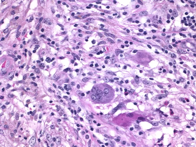
Histology of granulomatous mastitis on bright-field light microscopy. A dense inflammatory infiltrate forms granulomas centered on Langhans-type giant cells surrounded by epithelioid histiocytes, scattered lymphocytes, and occasional eosinophils. The image depicts a high-power view of the center of a granuloma within breast tissue biopsy; multinucleated giant cells with horseshoe-shaped nuclei are evident. The granulomatous architecture includes a peripheral rim of lymphocytes and histiocytes with minimal necrosis described. Staining by Hematoxylin and Eosin highlights basophilic nuclei and eosinophilic cytoplasm; pink collagenous stroma and adipose breast tissue may be visible in the background. Clinically, granulomatous mastitis can mimic infectious etiologies or neoplasia; the presence of Langhans giant cells strongly suggests a granulomatous process and warrants infectious workup for tuberculosis and fungal pathogens, as well as consideration of sarcoidosis or granulomatous reaction to foreign material. This image is diagnostic for granulomatous inflammation in breast tissue when correlated with clinical data and microbiologic studies; useful for education, differential diagnosis, and research on breast granulomatous disease. Additional educational value lies in distinguishing nonnecrotizing granulomas from caseating TB patterns, correlating histology with mammography and ultrasound, and guiding microbiologic testing and patient management in suspected granulomatous breast disease. The image supports teaching of granuloma biology and pathology.
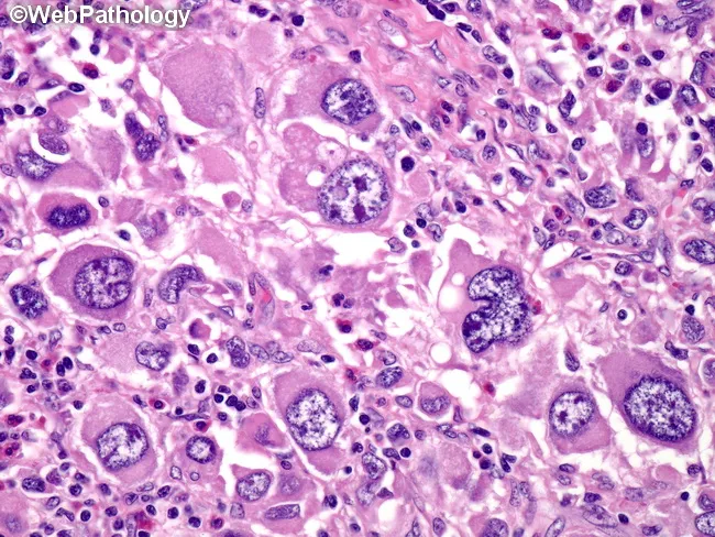
Histology image obtained with brightfield light microscopy on a hematoxylin and eosin stained tissue section. The specimen shows granulomatous inflammation characterized by aggregates of epithelioid macrophages (histiocytes) with elongated, pale to eosinophilic cytoplasm. Multinucleated giant cells, occasionally Langhans-type with peripheral, radiating nuclei, are interspersed among the epithelioid cells. Surrounding lymphocytes and a sparse fibrous stroma delineate nodular granulomas. In some fields there are crystalline or granular pink to eosinophilic deposits within the cytoplasm of histiocytes, and there may be mild necrosis at the center of larger granulomas, though necrosis is not uniformly present across the slide. The pattern is compatible with a granulomatous infectious or inflammatory process, requiring clinical correlation and microbiologic assessment. Differential diagnoses include mycobacterial infection (tuberculosis), fungal granulomas (histoplasmosis, blastomycosis), sarcoidosis, and foreign-body granulomatous reactions. The image emphasizes a macrophage-dominant infiltrate with discrete nodular architecture, preserving basic tissue planes while revealing cellular detail such as nuclear atypia is not prominent. This histologic phenotype is critical for guiding targeted tests (AFB stain, fungal stains, PCR) and therapeutic decisions, and it serves as an educational reference for recognizing granuloma formation in biopsy specimens. Correlation with clinical history and imaging is essential for diagnosis.
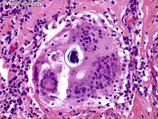
Histopathology image showing a granulomatous lesion composed of epithelioid histiocytes with a surrounding rim of lymphocytes and occasional Langhans-type multinucleated giant cells. Within a giant cell, lamellar inclusions known as Schaumann bodies are evident as concentric rings of protein and mineral material; calcium oxalate crystals may also be present. Asteroid bodies may be seen as stellate inclusions, though they are not required for diagnosis. The granuloma is noncaseating, lacking central necrosis, which favors sarcoidosis over infectious granulomatous processes such as tuberculosis. The tissue architecture shows well-formed granulomas with distinct borders, often adjacent to blood vessels in this section. The image is stained with Hematoxylin and Eosin (H&E), highlighting eosinophilic granulomatous material and basophilic nuclei within giant cells. Clinically, the presence of Schaumann bodies and Langhans giant cells supports a granulomatous inflammatory syndrome but is not pathognomonic for sarcoidosis. Differential considerations include berylliosis, hypersensitivity pneumonitis, fungal or mycobacterial infections, and foreign body reactions. Correlation with chest imaging, serum ACE levels, and clinical features is essential for diagnosis. This slide serves an educational role in recognizing granulomatous histology, giant cell inclusions, and lamellar calcific bodies, informing diagnostic workflows and teaching pathology trainees about sarcoidosis-related granulomatous disease.
Question 1: Inflammation - Definition & Adhesion Molecules
Definition of Inflammation
- Recognition of the injurious agent
- Recruitment of leukocytes
- Removal of the agent
- Regulation (control) of the response
- Repair (resolution)
Causes
Cardinal Signs (Celsus + Virchow)
- Rubor (redness) - due to vasodilation
- Calor (heat) - increased blood flow
- Tumor (swelling) - edema from vascular leakage
- Dolor (pain) - bradykinin, prostaglandins
- Functio laesa (loss of function)
Leukocyte Recruitment: The Adhesion Cascade
A. Endothelial-Derived Adhesion Molecules
1. Selectins (Endothelial)
- Stored in cytoplasmic Weibel-Palade bodies of endothelial cells (and platelets)
- Rapidly redistributed to the luminal surface within minutes in response to histamine (from mast cells) and thrombin (during coagulation)
- Mediates initial low-affinity "tethering" and rolling of leukocytes
- Synthesized de novo and expressed on the endothelial surface within 1-2 hours in response to IL-1 and TNF (produced by tissue macrophages and DCs)
- Also induced by microbial products such as LPS
- Mediates rolling of granulocytes, monocytes, and some T cells
2. Ig Superfamily Molecules (Endothelial Ligands for Integrins)
- Expressed on cytokine-activated endothelial cells, lymphocytes, macrophages, fibroblasts, and epithelial cells
- Ligand for leukocyte integrins LFA-1 (CD11a/CD18) and MAC-1 (CD11b/CD18)
- Contains extracellular Ig domains - member of the Ig superfamily
- Expressed constitutively on endothelial cells
- Also binds LFA-1
- Expressed on cytokine-activated endothelial cells in some tissues
- Ligand for VLA-4 (α4β1, CD49d/CD29) on eosinophils, lymphocytes, and monocytes
- Important in eosinophil recruitment in allergic inflammation
- Expressed on endothelial cells in intestinal mucosa
- Ligand for integrin α4β7 on gut-homing lymphocytes
B. Leukocyte-Derived Adhesion Molecules
1. Selectin (Leukocyte)
- Expressed constitutively on leukocytes (not on endothelial cells)
- Binds to sialomucins on activated endothelial cells (e.g., GlyCAM-1, CD34, MAdCAM-1)
- Recognizes sialyl 6-sulfo Lewis X on endothelial ligands
- Expression is upregulated by IL-1, TNF, and other inflammatory cytokines
2. Integrins (Leukocyte)
| Integrin | Also Known As | Chains | Endothelial Ligand | Function |
|---|---|---|---|---|
| LFA-1 | CD11a/CD18, αLβ2 | α and β2 | ICAM-1, ICAM-2, ICAM-3 | Firm adhesion; T cell-APC interaction |
| MAC-1 | CD11b/CD18, αMβ2, CR3 | α and β2 | ICAM-1, iC3b | Firm adhesion; complement receptor; phagocytosis |
| VLA-4 | CD49d/CD29, α4β1 | α4 and β1 | VCAM-1 | Adhesion of lymphocytes, monocytes, eosinophils |
| α4β7 | LPAM-1 | α4 and β7 | MAdCAM-1 | Gut-homing lymphocytes |
| αEβ7 | CD103 | αE and β7 | E-cadherin on epithelium | Mucosal T cells and DCs |
| CR4 | CD11c/CD18, αXβ2 | α and β2 | iC3b, ICAM-1 | Phagocytosis; mainly DCs |
3. Leukocyte Selectin Ligands
Steps of Leukocyte Recruitment (Summary)
| Step | Molecules Involved |
|---|---|
| 1. Margination & Rolling | E-selectin/P-selectin (endothelium) + PSGL-1/sialyl LewisX (leukocyte); L-selectin (leukocyte) + GlyCAM/CD34 |
| 2. Chemokine activation | Chemokines (IL-8/CXCL8, MCP-1, MIP-1) bind GPCR on leukocyte → inside-out signaling → integrin activation |
| 3. Firm adhesion (Arrest) | LFA-1/MAC-1 (leukocyte) + ICAM-1/ICAM-2 (endothelium); VLA-4 + VCAM-1 |
| 4. Transmigration (Diapedesis) | CD31 (PECAM-1), JAM-A/B/C, CD99 (at endothelial junctions); F-actin for transcellular migration |
| 5. Migration in tissue | Chemotaxis via C5a, LTB4, IL-8, bacterial products |
Question 2: Differential Diagnosis of Granulomatous Disease
Categories of Granulomatous Disease
I. Infectious Causes
1. Tuberculosis (Mycobacterium tuberculosis)
- Prototype of granulomatous disease - must always be excluded first
- Caseating (necrotic) granulomas = "tubercles"
- Foci of epithelioid macrophages rimmed by fibroblasts, lymphocytes, histiocytes, and occasional Langhans giant cells (nuclei arranged peripherally in horseshoe pattern)
- Central amorphous, granular, eosinophilic debris (caseous necrosis)
- Diagnosis: Acid-fast stain (Ziehl-Neelsen), culture (gold standard), PCR, tuberculin skin test, IGRA
- Key feature: AFB visible in 60-70% of cases
2. Leprosy (Mycobacterium leprae)
- Tuberculoid leprosy: Compact, well-formed noncaseating granulomas with few bacilli; nerve involvement
- Lepromatous leprosy: Poorly formed granulomas, abundant AFB in macrophages ("foam cells" or Virchow cells), no central necrosis
- Diagnosis: Slit-skin smear, biopsy, Fite-Faraco stain
3. Syphilis (Treponema pallidum)
- Gumma: microscopic to grossly visible lesion; wall of histiocytes and plasma cells; central coagulative necrosis (cellular outlines preserved - unlike TB)
- Tertiary stage manifestation; can affect liver, bone, CNS, cardiovascular system
- Diagnosis: Serology (RPR, VDRL, FTA-ABS), darkfield microscopy, biopsy with silver stain
4. Fungal Infections
- Histoplasmosis (Histoplasma capsulatum): Endemic (Ohio/Mississippi River valleys); intracellular yeast in macrophages; can cavitate; Gomori methenamine silver (GMS) stain
- Coccidioidomycosis (Coccidioides immitis): Southwestern US; large spherules with endospores; can be caseating
- Blastomycosis (Blastomyces dermatitidis): Broad-based budding yeast; mixed granulomatous + suppurative pattern
- Cryptococcosis (Cryptococcus neoformans): Immunocompromised hosts; "soap bubble" lesions in brain; India ink stain, mucicarmine for capsule
- Aspergillosis: Septate hyphae with acute-angle branching; granulomatous in immunocompetent, invasive in immunocompromised
5. Cat-Scratch Disease (Bartonella henselae)
- Gram-negative bacillus
- Rounded or stellate granuloma with central granular debris and recognizable neutrophils ("suppurative granuloma" or "stellate necrosis")
- Giant cells uncommon
- Diagnosis: Warthin-Starry silver stain, serology, PCR
6. Schistosomiasis (Parasitic)
- Granulomas form around Schistosoma egg antigens
- Associated with Th2 response and eosinophils (unlike typical Th1-mediated granulomas)
- Can lead to portal hypertension (hepatic), bladder cancer risk
7. Leishmaniasis, Toxoplasmosis, Brucellaosis
- All can cause granulomatous reactions
- Brucellosis: small, poorly formed granulomas
II. Non-Infectious Inflammatory/Immune-Mediated
1. Sarcoidosis
- Unknown etiology - possible abnormal immune response to an unidentified antigen
- Noncaseating granulomas (no central necrosis) - this is pathognomonic when combined with clinical picture
- Characteristic inclusions: Schaumann bodies (concentric calcific lamellated structures), asteroid bodies (stellate inclusions in giant cells)
- Multisystem: Lungs (bilateral hilar lymphadenopathy + pulmonary infiltrates), skin, eyes, heart, nervous system
- Diagnosis: Elevated serum ACE, hypercalcemia (macrophages produce 1-hydroxylase), Kveim test, biopsy
- Key histology: Well-formed epithelioid granulomas, no organisms on special stains
2. Crohn's Disease
- Immune reaction against intestinal bacteria/self-antigens
- Occasional noncaseating granulomas in the bowel wall (only 30-40% of biopsies show granulomas)
- Dense chronic inflammatory infiltrate; transmural inflammation; skip lesions
- Distinguishes from ulcerative colitis (which has no granulomas)
3. Granulomatosis with Polyangiitis (Wegener's Granulomatosis) - see Question 4
4. Primary Biliary Cholangitis (PBC)
- Florid duct lesion with granulomatous bile duct destruction
- Anti-mitochondrial antibody (AMA) positive
5. Hypersensitivity Pneumonitis (Extrinsic Allergic Alveolitis)
- Inhalation of organic antigens (bird proteins, fungal spores)
- Poorly formed noncaseating granulomas in interstitium
- Eosinophilia may be present; precipitating antibodies (IgG)
III. Foreign Body Reactions
- Reaction to inert, non-immunogenic foreign materials
- No T cell involvement; no specific immune response
- Materials: talc (IV drug users), silica, suture material, prosthetic implant debris, beryllium (berylliosis), silicone
- Epithelioid macrophages and giant cells apposed to foreign material
- Foreign material often identifiable in giant cells, especially with polarized light (birefringent/refractile material)
- Berylliosis: Indistinguishable from sarcoidosis histologically; occupational history is key
IV. Summary Comparison Table
| Disease | Necrosis | Giant Cells | Special Features | Key Test |
|---|---|---|---|---|
| Tuberculosis | Caseous (central) | Langhans (present) | AFB, caseation | AFB stain, culture, PCR, IGRA |
| Sarcoidosis | None | Langhans/foreign body | Asteroid/Schaumann bodies | ACE, biopsy, CXR |
| Crohn's disease | None | May be present | Skip lesions, transmural | Colonoscopy + biopsy |
| Syphilis (gumma) | Coagulative | Absent | Plasma cells, treponemes | Serology |
| Cat-scratch disease | Stellate/suppurative | Uncommon | Neutrophils in center | Warthin-Starry stain, PCR |
| Foreign body | None | Foreign body type | Birefringent material | Polarized light microscopy |
| Histoplasmosis | ± Caseous | Present | Intracellular yeast | GMS stain, culture, urine antigen |
| Wegener's (GPA) | Necrotizing | Present | Vasculitis, c-ANCA | ANCA (anti-PR3), biopsy |
| Hypersensitivity pneumonitis | None | Poorly formed | Eosinophils, IgG precipitins | Precipitating antibodies, BAL |
| Leprosy | None | Poorly defined | Foam cells | AFB stain (Fite), skin smear |
Question 3: Complement System in Health and Disease
Overview
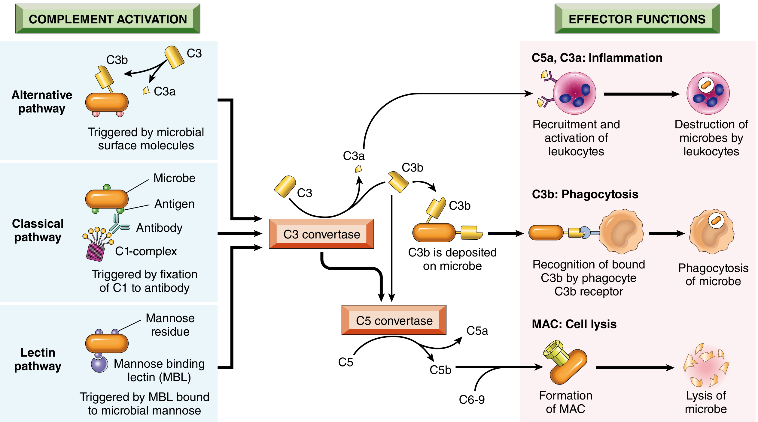
Activation Pathways
1. Classical Pathway
- Triggered by C1 binding to antibody (IgM or IgG) complexed with antigen
- Requires antigen-antibody complexes - bridges adaptive and innate immunity
- Sequence: C1 → C4 → C2 → C3 convertase (C4b2a) → C5 convertase (C4b2a3b)
2. Alternative Pathway
- Triggered by microbial surface molecules (LPS/endotoxin, complex polysaccharides, cobra venom factor) in the absence of antibody - innate immunity
- Spontaneous hydrolysis of C3 is amplified and stabilized on microbial surfaces
- Sequence: C3 spontaneous hydrolysis → Factor B + Factor D + Properdin → C3 convertase (C3bBb)
3. Lectin Pathway
- Plasma mannose-binding lectin (MBL) binds carbohydrates on microbial surfaces
- Activates MASP-1 and MASP-2 (serine proteases homologous to C1r and C1s)
- Subsequent steps mirror the classical pathway - no antibody required
Common Terminal Pathway
- C3 convertase cleaves C3 → C3a (released) + C3b (deposited on cell surface)
- C5 convertase cleaves C5 → C5a (released) + C5b (attached)
- C5b + C6 + C7 + C8 + poly-C9 → Membrane Attack Complex (MAC)
Functions (Health)
1. Inflammation (Anaphylatoxins)
- C5a, C3a, C4a stimulate histamine release from mast cells → vasodilation and increased vascular permeability
- C5a is the most potent: strong chemotactic agent for neutrophils, monocytes, eosinophils, basophils
- C5a also activates the lipoxygenase pathway in neutrophils/monocytes → further inflammatory mediators
2. Opsonization and Phagocytosis
- C3b and iC3b (inactivated C3b) coat microbial surfaces (opsonization)
- Recognized by CR1 (C3b receptor, CD35) and CR3/MAC-1 (iC3b receptor, CD11b/CD18) on neutrophils and macrophages
- Dramatically enhances phagocytosis
3. Cell Lysis (MAC)
- Deposition of MAC on thin-walled microbes (especially Neisseria species) creates membrane pores → osmotic lysis
- Particularly important against gram-negative bacteria
4. Clearance of Immune Complexes
- C3b facilitates solubilization of immune complexes and transport by erythrocytes (via CR1) to the liver and spleen for clearance
- This prevents tissue deposition of immune complexes
Regulation of Complement
| Regulator | Mechanism | Disease if Deficient |
|---|---|---|
| C1 inhibitor (C1-INH) | Blocks C1 activation; also inhibits kallikrein and Factor XII | Hereditary Angioedema (HAE) - recurrent episodes of angioedema |
| DAF (CD55) | GPI-anchored; prevents C3 convertase formation | PNH (acquired loss of GPI anchor) |
| CD59 (Protectin) | GPI-anchored; inhibits MAC formation | PNH - lysis of RBCs and platelets |
| Factor H | Promotes cleavage of C3b; accelerates alternative pathway C3 convertase decay | Atypical HUS, age-related macular degeneration (Factor H polymorphisms) |
| Factor I | Cleaves C3b to iC3b (with Factor H as cofactor) | Recurrent infections |
| C4BP (C4b-binding protein) | Inhibits classical pathway C3 convertase | - |
Complement in Disease
A. Deficiencies - Susceptibility to Infections
| Component Deficient | Consequence |
|---|---|
| C3 | Most severe - susceptibility to all encapsulated bacteria (pneumococcus, Haemophilus, meningococcus) |
| C5-C9 (terminal complement) | Susceptibility to Neisseria infections (meningococcemia, disseminated gonococcemia) |
| C1q, C2, C4 (classical pathway) | Susceptibility to infections PLUS lupus-like autoimmune disease (impaired immune complex clearance) |
| MBL | Mild immunodeficiency; recurrent upper respiratory infections in children |
| Properdin | X-linked; susceptibility to Neisseria meningitidis |
B. Complement-Mediated Tissue Injury
- Systemic Lupus Erythematosus (SLE): Classical pathway activation by DNA-anti-DNA immune complexes → nephritis, vasculitis, serositis; C3, C4 levels are LOW (consumed)
- Membranoproliferative Glomerulonephritis (MPGN): Low C3 due to alternative pathway activation
- Rheumatoid Arthritis: Complement activation by IgG immune complexes in joints
- Myasthenia Gravis and other antibody-mediated autoimmune diseases
C. Regulatory Protein Deficiencies - Excessive Activation
-
Paroxysmal Nocturnal Hemoglobinuria (PNH): Acquired somatic mutation in PIG-A gene → loss of GPI anchor → deficiency of both CD55 and CD59 → uncontrolled complement activation → intravascular hemolysis, thrombosis, cytopenias. Treatment: eculizumab (anti-C5 monoclonal antibody)
-
Hereditary Angioedema (HAE): Autosomal dominant deficiency of C1-INH → unopposed activation of contact system and complement → bradykinin overproduction → episodic non-pitting angioedema of skin, GI tract, airway (life-threatening laryngeal edema). C4 levels chronically low. Treatment: C1-INH concentrate, icatibant (bradykinin B2 receptor antagonist), lanadelumab
-
Atypical HUS (aHUS): Loss-of-function mutations in Factor H, Factor I, MCP (CD46) OR gain-of-function in C3 or Factor B → uncontrolled alternative pathway activation → endothelial damage in glomeruli → thrombotic microangiopathy. Treatment: eculizumab
-
Age-Related Macular Degeneration (AMD): Polymorphisms in Factor H (particularly Y402H variant) are the strongest genetic risk factor identified → complement deposition in Bruch's membrane → choroidal neovascularization
D. Complement Inhibitor Therapies
| Drug | Target | Indication |
|---|---|---|
| Eculizumab | Anti-C5 (blocks C5 cleavage) | PNH, aHUS, NMOSD |
| Ravulizumab | Anti-C5 (long-acting) | PNH, aHUS |
| Avacopan | C5a receptor antagonist | ANCA vasculitis (GPA, MPA) |
| C1-INH concentrate | C1 INH replacement | HAE |
Question 4: Granuloma - Definition, Classification & Wegener's Granulomatosis
Definition of Granuloma
- Granuloma formation is a cellular attempt to contain an offending agent that is difficult to eradicate
- Involves strong activation of macrophages and T lymphocytes, which can also cause injury to normal tissue
- Activated macrophages develop abundant cytoplasm resembling epithelial cells → called epithelioid cells
- Some activated macrophages fuse → multinucleate giant cells
-
- Robbins, Cotran & Kumar Pathologic Basis of Disease
Classification of Granuloma
Type 1: Foreign Body Granuloma
- Pathogenesis: Reaction to inert foreign bodies that are non-immunogenic - no T cell-mediated immune response
- Forms around materials large enough to preclude phagocytosis (talc, suture material, silicone, mineral oil, keratin, calcium deposits)
- Epithelioid cells and giant cells apposed to the surface of the foreign body
- Foreign material often identified within the giant cells, especially visible with polarized light (refractile/birefringent)
- No necrosis; giant cells are "foreign body type" with randomly scattered nuclei throughout cytoplasm
Type 2: Immune (T cell-mediated) Granuloma
- Pathogenesis: Caused by agents that induce persistent T cell-mediated immune responses - the inciting agent cannot be eliminated
- Th1 cells produce IFN-γ → activates macrophages
- In some parasitic infections (schistosomiasis), Th2 response with eosinophils predominates
- Includes: tuberculosis, fungal infections, sarcoidosis, Crohn's disease, berylliosis, hypersensitivity reactions
Histological Subtypes by Necrosis
| Subtype | Feature | Diseases |
|---|---|---|
| Caseating | Central caseous (cheese-like) necrosis; amorphous, structureless, eosinophilic debris; complete loss of cell outlines | Tuberculosis, histoplasmosis, coccidioidomycosis |
| Non-caseating | No necrosis; well-formed epithelioid granulomas | Sarcoidosis, Crohn's, foreign body, berylliosis |
| Suppurative (stellate) | Central microabscess with neutrophils within the granuloma | Cat-scratch disease, LGV, Yersinia |
| Necrotizing vasculitic | Granulomatous inflammation with vasculitis and tissue necrosis | Wegener's (GPA), Churg-Strauss |
Morphological Features of Granuloma on H&E
- Epithelioid cells: Large macrophages with pink granular cytoplasm, indistinct cell borders, oval/elongated vesicular nuclei; resemble epithelium
- Langhans Giant Cell: Multinucleated (40-50 µm); nuclei arranged in horseshoe or peripheral ring pattern; characteristic of TB but not specific
- Foreign Body Giant Cell: Nuclei randomly scattered throughout cytoplasm; forms around foreign material
- Touton Giant Cell: Wreath-like arrangement of nuclei around lipid; seen in xanthogranulomatous conditions
- Collar of lymphocytes surrounds the epithelioid aggregate
- Older granulomas have a rim of fibroblasts and connective tissue
- Healing is accompanied by fibrosis (may be extensive)
Wegener's Granulomatosis (Granulomatosis with Polyangiitis - GPA)
Definition
- Necrotizing granulomas of the upper respiratory tract (sinuses, nose, trachea)
- Necrotizing granulomas of the lower respiratory tract (lungs)
- Focal necrotizing glomerulonephritis
Epidemiology
- Rare disease: incidence ~11.8 per million person-years (UK data)
- Peak incidence: ages 55-69 years
- Slight male predominance (M:F = 1.3:1)
- Genetic risk factors: CTLA4, PTPN22, COL11A2, SERPINA1
Pathogenesis
- ~90% of patients have ANCA (Anti-Neutrophil Cytoplasmic Antibodies)
- Predominantly c-ANCA (cytoplasmic pattern by immunofluorescence), directed against PR3 (proteinase 3) in ~90% of GPA cases
- p-ANCA (perinuclear, anti-MPO) in the remaining ~10%
- Mechanism: ANCA-primed neutrophils are activated → release of PR3, ROS, and NETs (Neutrophil Extracellular Traps) → endothelial injury → necrotizing vasculitis
- NETosis is increasingly recognized as a key mediator
- An unidentified precipitating factor leads to initial ANCA production (possibly Staphylococcus aureus nasal carriage as a trigger)
Clinical Features
Upper Respiratory Tract (90% at presentation)
- Severe sinusitis and rhinorrhea
- Nasal mucosal ulcerations and epistaxis
- Nodules in nose, larynx, trachea, bronchi
- Saddle-nose deformity (destruction of nasal cartilage/bone from necrotizing granulomas)
- Subglottic stenosis (hoarseness, stridor)
- Oral involvement: necrotizing alveolar ridge, tongue ulcers, palatal perforation
- "Strawberry gums" (hypertrophic gingivitis) - biopsy of this lesion may be diagnostic
Lower Respiratory Tract (71% have radiographic infiltrates)
- Cough, dyspnea, chest pain
- Pulmonary nodules that may cavitate
- Pulmonary infiltrates, hemoptysis
- Diffuse alveolar hemorrhage (life-threatening)
Renal Involvement (85% of patients)
- Focal crescentic necrotizing glomerulonephritis
- Hematuria, proteinuria, progressive AKI
- Can be rapidly progressive (RPGN) → renal failure
- Before cyclophosphamide therapy, renal failure was the most common cause of death
Cutaneous Findings (45%)
- Palpable purpura (leukocytoclastic vasculitis)
- Nodules along extensor surfaces of extremities (may ulcerate, can mimic rheumatoid nodules)
- Petechiae, hemorrhagic pustules
- Pyoderma gangrenosum-like ulcerations
- Livedo reticularis (rare)
Other Systems
- Eyes (58%): conjunctivitis, episcleritis, proptosis (orbital granuloma)
- Joints (two thirds): arthralgia, arthritis
- CNS (22%): cranial nerve palsies, peripheral neuropathy
- Heart (12%): pericarditis, coronary vasculitis
Histopathology
- Necrotizing granulomatous vasculitis - the hallmark
- Palisaded granulomas with multinucleated giant cells and a central core of neutrophils and debris
- Leukocytoclastic vasculitis (LCV) in skin biopsies
- Renal biopsy: pauci-immune crescentic GN (little/no immune complex deposition on immunofluorescence - unlike lupus nephritis); crescents in ~90% of children with GPA
Diagnosis
| Test | Finding |
|---|---|
| c-ANCA (anti-PR3) | Positive in ~90% of active generalized GPA (almost 100% in active disease) |
| p-ANCA (anti-MPO) | Positive in ~10% - associated with MPA |
| Chest X-ray/CT | Pulmonary nodules, cavities, infiltrates |
| Urinalysis | Hematuria, red cell casts (glomerulonephritis) |
| Biopsy (lung, URT, kidney, skin) | Necrotizing granulomatous vasculitis; pauci-immune crescentic GN |
| Nasal biopsy | Granulomatous inflammation (most accessible) |
Limited vs. Generalized GPA
- Limited GPA: Upper respiratory tract only, no renal involvement; better prognosis; may respond to methotrexate + prednisone alone; TMP-SMX may be used for maintenance
- Generalized GPA: Multi-organ involvement including kidneys; requires aggressive immunosuppression
Treatment
Induction Phase
- Cyclophosphamide (2 mg/kg/day oral or 500-750 mg/m² IV every 2-4 weeks) + Prednisone (1 mg/kg/day) - traditional standard
- Rituximab (anti-CD20 monoclonal antibody) + high-dose glucocorticoids - FDA approved 2011; equivalent to cyclophosphamide for new patients; superior to cyclophosphamide for relapsing patients; preferred in PR3-ANCA positive disease
- Avacopan (C5a receptor blocker) - emerging steroid-sparing agent added to either cyclophosphamide or rituximab
- IV methylprednisolone boluses (3 days) before oral prednisone for severe disease
- Plasma exchange (PLEX): For diffuse pulmonary hemorrhage and RPGN approaching dialysis
Maintenance Phase (after remission)
- Methotrexate, azathioprine, leflunomide, MMF, or rituximab (instead of cyclophosphamide)
- Treatment for at least 1 year
- TMP-SMX long-term: reduces nasal S. aureus carriage → decreases relapse rate; useful for limited URT disease in remission
Refractory Disease
- Plasma exchange, IVIG, anti-TNF therapy (infliximab), tacrolimus
Prognosis
- Untreated GPA: Mean survival ~5 months; 90% mortality at 2 years
- With cyclophosphamide + prednisone: Complete remission in up to 93% of patients, lasting average of 4 years
- Major modern concern: treatment-related adverse effects (infection, hemorrhagic cystitis, malignancy from cyclophosphamide) now account for more deaths in year 1 than the vasculitis itself