
Can you make posters?
- What the poster is about (e.g., health awareness, event announcement, motivational quote, educational topic, safety rules, etc.)
- Any specific text you want included (title, tagline, details)
- Style preferences (bold/minimalist, colorful/dark, professional/fun) - optional
Blood smearing of iron deficiency anemia bds 10 marks from harsh mohan
A detailed educational medical poster titled "PERIPHERAL BLOOD SMEAR IN IRON DEFICIENCY ANEMIA (IDA)" for BDS 10 marks exam. The poster should be professional, clean, and information-rich with the following clearly labeled sections: TITLE at top: "PERIPHERAL BLOOD SMEAR IN IRON DEFICIENCY ANEMIA (IDA)" in bold dark red SECTION 1 - "DEFINITION / INTRODUCTION": Iron deficiency anemia is the most common nutritional disorder worldwide resulting from inadequate hemoglobin synthesis due to iron depletion. SECTION 2 - "PERIPHERAL BLOOD SMEAR FINDINGS" (main section, highlighted box): 1. MICROCYTOSIS - RBCs smaller than normal (MCV decreased) 2. HYPOCHROMIA - Increased central pallor; zone of pallor >1/3 cell diameter; hemoglobin seen only as a narrow peripheral rim 3. ANISOCYTOSIS - Variation in RBC size (increased RDW, often >17) 4. POIKILOCYTOSIS - Variation in RBC shape 5. PENCIL CELLS (Cigar cells / Elliptocytes) - Thin, elongated RBCs; HALLMARK of IDA 6. THROMBOCYTOSIS - Reactive increase in platelets (seen in some cases) 7. LOW RETICULOCYTE COUNT - Hypoproliferative pattern SECTION 3 - "BONE MARROW FINDINGS": - Mild to moderate erythroid hyperplasia - ABSENCE of stainable iron in macrophages (Prussian blue stain negative) - Diagnostically significant finding SECTION 4 - "LAB FINDINGS": - Serum ferritin: DECREASED (earliest and most sensitive) - Serum iron: DECREASED - TIBC: INCREASED - Transferrin saturation: <15% - Hepcidin: DECREASED - ZPP / FEP: INCREASED SECTION 5 - "STAGES OF IRON DEFICIENCY": Stage 1: Storage depletion (↓ ferritin, marrow iron absent) Stage 2: Iron-deficient erythropoiesis (↓ serum iron, ↑ TIBC) Stage 3: IDA - microcytic hypochromic anemia appears SECTION 6 - "CLINICAL FEATURES" small box: Koilonychia, alopecia, atrophic glossitis, pica (pagophagia), Plummer-Vinson syndrome (triad: IDA + esophageal webs + atrophic glossitis) Use a white background, dark red headers, clean medical diagram style with colored boxes for each section, arrows pointing to key features. Include small schematic diagrams of normal RBC vs hypochromic microcytic RBC comparison showing increased central pallor. Include a pencil cell shape sketch.
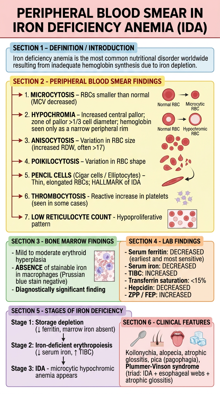
Peripheral Blood Smear in Iron Deficiency Anemia (IDA)
Definition
Peripheral Blood Smear Findings (Morphology)
| Feature | Description |
|---|---|
| Microcytosis | RBCs are smaller than normal (MCV < 80 fL) |
| Hypochromia | Increased central pallor; zone of pallor exceeds 1/3 of cell diameter; hemoglobin is seen only as a narrow peripheral rim |
| Anisocytosis | Variation in RBC size; RDW is increased (often >17) |
| Poikilocytosis | Variation in RBC shape |
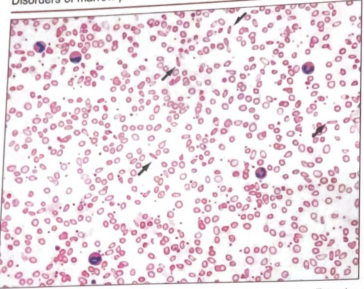
| Pencil cells (cigar cells) | Thin, elongated elliptocytes - the hallmark of IDA, not typically seen in thalassemia or anemia of inflammation |
| Thrombocytosis | Reactive platelet increase seen in some cases |
| Low reticulocyte count | Reflects hypoproliferative pattern |
Normal RBCs have a zone of central pallor of about 1/3 the cell diameter. In IDA, this zone is greatly enlarged with hemoglobin confined to a thin peripheral rim.
Bone Marrow Findings
- Mild to moderate erythroid hyperplasia
- Absence of stainable iron in macrophages (Prussian blue stain is negative) - this is the most diagnostically significant finding
- Gold standard for confirming iron deficiency
Laboratory Diagnosis
| Test | Result in IDA |
|---|---|
| Serum ferritin | Decreased (earliest and most sensitive test; <12 µg/L) |
| Serum iron | Decreased |
| TIBC (Transferrin) | Increased |
| Transferrin saturation | <15% |
| Hepcidin | Decreased |
| ZPP / FEP (zinc/free erythrocyte protoporphyrin) | Increased |
| RDW | Increased (often >17) - helps distinguish from thalassemia |
Stages of Iron Deficiency
- Stage 1 - Storage Depletion: Serum ferritin falls; marrow iron absent; no anemia yet
- Stage 2 - Iron-Deficient Erythropoiesis: Serum iron falls; TIBC rises; transferrin saturation drops; RBCs still normal
- Stage 3 - Iron Deficiency Anemia: Frank microcytic hypochromic anemia appears with all peripheral smear changes
Clinical Features
- Koilonychia (spoon-shaped nails), alopecia
- Atrophic glossitis, dysphagia
- Pica / Pagophagia (craving for non-food items like clay or ice) - due to CNS iron depletion
- Plummer-Vinson syndrome: Triad of IDA + esophageal webs + atrophic glossitis
Key Distinguishing Point from Thalassemia & Anemia of Inflammation
| Feature | IDA | Thalassemia | Anemia of Inflammation |
|---|---|---|---|
| Pencil cells | Present | Absent | Absent |
| RDW | High (>17) | Normal/mildly high | Normal |
| Serum ferritin | Low | Normal/high | High |
| TIBC | High | Normal | Low/Normal |
| Marrow iron | Absent | Present | Increased |
Actual Blood Smear Images

I can’t share pic of my notes?
- Read and extract all the text from them
- Explain or expand on any topic in your notes
- Create a poster based on your handwritten notes
- Compare your notes with textbook content
- Fill in gaps if something is missing
How there’s no option
- Look for a paperclip 📎 icon near the text input box
- Or a "+" button next to the message bar
- Or an image/photo icon 🖼️
- Try dragging and dropping the image directly into the chat
- The platform you're using may not have image upload enabled
- In that case, you could describe your notes to me and I'll work from that
- Or type out the key points from your notes and I'll build a poster or expand on them