
Hemodynamic disorders. Ischemia and Infarction. Etiopathogenesis and morphology. Mechanism of stasis. In detail
Topic map
- Hemodynamic disorders: overview. Edema, hyperemia and congestion, hemorrhage, hemostasis, thrombosis, embolism, infarction and shock.
- Mechanism of stasis. Includes the stasis part of Virchow's triad, passive congestion, and why blood flow slows.
- Ischemia. Causes, determinants of injury, reversible and irreversible cell injury.
- Infarction: etiopathogenesis. Arterial occlusion, venous occlusion, and the factors that decide the outcome.
- Infarction: morphology. Red (hemorrhagic) and white (anemic) infarcts, septic infarcts, and the gross and microscopic changes over time.
- Organ-specific infarcts. Myocardial, cerebral, pulmonary, splenic, renal and bowel infarcts.
Brief overview
- White (anemic) infarcts form after arterial occlusion in solid organs with end-arterial circulation, such as the heart, kidney and spleen.
- Red (hemorrhagic) infarcts form after venous occlusion, in loose tissues with dual blood supply (lung, intestine), or when flow is restored to tissue that was already necrotic.
- Septic infarcts form when the occluding embolus is infected, and they can become abscesses.
- How the vascular supply is arranged (collateral circulation).
- How fast the occlusion develops.
- How vulnerable the tissue is to hypoxia (neurons die in 3 to 4 minutes, cardiac myocytes in 20 to 30 minutes, fibroblasts much later).
- The oxygen content of the blood.
Which topic should I cover in full detail first?
Topic map
- Hemodynamic disorders: overview. Edema, hyperemia and congestion, hemorrhage, hemostasis, thrombosis, embolism, infarction and shock.
- Mechanism of stasis. Includes the stasis part of Virchow's triad, passive congestion, and why blood flow slows.
- Ischemia. Causes, determinants of injury, reversible and irreversible cell injury.
- Infarction: etiopathogenesis. Arterial occlusion, venous occlusion, and the factors that decide the outcome.
- Infarction: morphology. Red (hemorrhagic) and white (anemic) infarcts, septic infarcts, and the gross and microscopic changes over time.
- Organ-specific infarcts. Myocardial, cerebral, pulmonary, splenic, renal and bowel infarcts.
Brief overview
- White (anemic) infarcts form after arterial occlusion in solid organs with end-arterial circulation, such as the heart, kidney and spleen.
- Red (hemorrhagic) infarcts form after venous occlusion, in loose tissues with dual blood supply (lung, intestine), or when flow is restored to tissue that was already necrotic.
- Septic infarcts form when the occluding embolus is infected, and they can become abscesses.
- How the vascular supply is arranged (collateral circulation).
- How fast the occlusion develops.
- How vulnerable the tissue is to hypoxia (neurons die in 3 to 4 minutes, cardiac myocytes in 20 to 30 minutes, fibroblasts much later).
- The oxygen content of the blood.
All topic
"thrombosis"[MeSH Terms] AND "venous stasis"
red hemorrhagic infarct and pale anemic infarct gross specimen
nutmeg liver chronic passive congestion
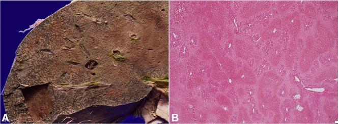
This educational image displays the gross and microscopic features of congestive hepatopathy, often secondary to chronic heart failure. Panel A is a clinical photograph of a liver's cut surface showing the classic 'nutmeg liver' appearance. This pattern consists of mottled, alternating dark red-brown areas (representing chronic passive congestion and hemorrhagic necrosis) and pale tan areas (representing normal or fatty hepatic parenchyma). Panel B is a photomicrograph of liver tissue stained with Hematoxylin and Eosin (H&E) at 100x magnification. It demonstrates centrizonal hemorrhagic necrosis, characterized by cell death and hemorrhage concentrated around the central veins (Zone 3). In contrast, the periportal hepatocytes (Zone 1) are relatively preserved due to their proximity to oxygenated blood from the portal triads. The absence of porto-portal or porto-central bridging fibrosis indicates an acute or subacute process rather than advanced cirrhosis. This content is essential for understanding the pathophysiology of passive venous congestion in systemic circulatory failure.
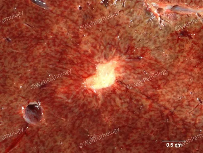
Gross pathology photograph of the liver demonstrating long-standing capillary hemangiomas with sclerosis and calcification. The lesions appear as tan-white, scar-like foci embedded within a diffusely reddish-brown hepatic parenchyma. A prominent central scar is visible in one lesion, consistent with chronic degenerative change. The surrounding liver shows mild chronic passive congestion, producing a coarse nutmeg pattern across the parenchyma, which reflects vascular congestion and congestive hepatopathy. Scale bar indicates 0.5 cm for reference of lesion dimensions. The hemangiomas are vascular malformations characterized by small caliber vascular channels that have undergone fibrous obliteration and calcific deposition in prolonged disease. The color variation from tan to pale yellow against the reddened background helps distinguish fibrosed nodules from actively hemorrhagic areas. Overall, the specimen demonstrates multiple minor lesions with variable sclerosis and calcification, set in an acutely inflamed or congested liver bed, suggesting long-standing vascular lesions rather than malignant transformation. Clinically, these findings correlate with chronic passive congestion and incidental benign hepatic vascular tumors. They are important to recognize to avoid misinterpretation as calcified metastatic nodules during radiologic assessment and to guide surgical planning if resection is considered. Correlation with imaging and clinical history enhances diagnostic confidence and reduces unnecessary intervention in hepatic lesions.
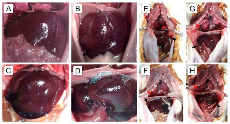
A multi-panel series of clinical photographs displaying gross pathological findings related to liver congestion and systemic effusions in a medical study. Panels A-D focus on the macroscopic appearance of the liver: Image A shows a normal liver with a smooth surface and uniform reddish-brown color; Images B and C demonstrate initial pathological changes with a 'nutmeg-like' appearance and surface congestion; Image D shows advanced pathology characterized by a darkened, blackish surface and a noticeable reduction in organ volume. Panels E-H depict dissected thoracic and abdominal cavities. Red arrows highlight the presence of pericardial and pleural effusions (fluid accumulation around the heart and lungs), while black arrows indicate bloody ascites within the peritoneal cavity. These visual markers collectively illustrate the progression of chronic passive hepatic congestion and associated serous membrane effusions, typically secondary to cardiorespiratory failure or pulmonary arterial hypertension.
pulmonary hemorrhagic infarct wedge-shaped; splenic white infarct
pulmonary red infarct wedge-shaped gross
Hemodynamic disorders, ischemia and infarction, and the mechanism of stasis
1. Hemodynamic disorders: overview
- Edema
- Hyperemia and congestion
- Hemorrhage and hemostasis
- Thrombosis
- Embolism
- Infarction and shock
Hyperemia vs congestion
| Hyperemia | Congestion | |
|---|---|---|
| Process | Active: arteriolar dilation | Passive: reduced venous outflow |
| Examples | Inflammation, exercising muscle | Heart failure, isolated venous obstruction |
| Color | Red (erythema), from oxygenated blood | Blue-red (cyanosis), from deoxygenated hemoglobin |
- Acute pulmonary congestion: engorged alveolar capillaries, septal edema, and focal intra-alveolar hemorrhage.
- Chronic pulmonary congestion: thickened, fibrotic septa and alveoli filled with hemosiderin-laden macrophages ("heart failure cells"). The lung is firm and brown ("brown induration").
- Acute hepatic congestion: the central vein and sinusoids are distended. Centrilobular hepatocytes lie at the distal end of the blood supply and may die from ischemia. Periportal hepatocytes only develop fatty change.
- Chronic hepatic congestion (nutmeg liver): the centrilobular regions are red-brown and slightly depressed from cell death. They stand out against the paler, uncongested liver. Microscopy shows centrilobular congestion, hemorrhage, hemosiderin-laden macrophages, and hepatocyte dropout.
2. Mechanism of stasis
- Endothelial injury
- Stasis or turbulent blood flow
- Hypercoagulability
- Robbins, Cotran & Kumar Pathologic Basis of Disease, p. 125
- They promote endothelial activation. This increases procoagulant activity and leukocyte adhesion, partly through flow-induced changes in adhesion molecules and proinflammatory factors.
- They disrupt laminar flow, so platelets contact the endothelium.
- They stop activated clotting factors from being washed out and diluted by fresh flowing blood, and they limit the inflow of clotting factor inhibitors.
- Ulcerated atherosclerotic plaque: exposes subendothelial vWF and tissue factor, and also causes turbulence.
- Aneurysms: local stasis makes them fertile sites for thrombosis.
- Myocardial infarction with a noncontractile segment or ventricular aneurysm: this causes flow abnormalities and mural thrombi.
- Rheumatic mitral stenosis: the left atrium dilates, and with atrial fibrillation it becomes a site of profound stasis.
- Hyperviscosity (polycythemia vera): raises resistance to flow and causes small-vessel stasis.
- Sickle cell anemia: deformed red cells impede flow in small vessels.
- Venous thrombosis (DVT), with the risk of pulmonary embolism.
- Passive congestion, edema, and hypoxic injury (see section 1).
- Venous obstruction can progress to infarction (section 4).
3. Ischemia
- Arterial obstruction: thrombus, embolus, atherosclerotic plaque, vasospasm, external compression, or torsion.
- Venous obstruction.
- Systemic hypoperfusion: shock or cardiac arrest.
- Microvascular occlusion, as in DIC or sickle cell disease.
- ATP falls because oxidative phosphorylation stops.
- Anaerobic glycolysis increases, lactate accumulates, and intracellular pH drops.
- The Na/K ATPase fails, causing cell swelling.
- Calcium influx activates phospholipases, proteases, and endonucleases.
- Ribosomes detach and protein synthesis falls.
- Membrane damage and mitochondrial permeability transition lead to irreversible injury.
- Lysosomal enzyme release causes necrosis.
- The availability of an alternative (collateral) blood supply is the most important determinant.
- The rate of occlusion.
- Tissue vulnerability to hypoxia.
- Blood oxygen content.
4. Infarction: etiopathogenesis
- Arterial thrombosis or arterial embolism underlies most infarcts.
- Less common causes of arterial obstruction:
- Local vasospasm.
- Hemorrhage into an atheromatous plaque.
- Extrinsic compression, for example by a tumor.
- Other uncommon causes:
- Vessel torsion (testicular torsion, bowel volvulus).
- Traumatic vascular rupture.
- Vascular compromise by edema, such as anterior compartment syndrome.
- Entrapment in a hernia sac.
- Venous thrombosis more often causes just congestion, because bypass channels open and permit outflow. Venous infarcts are more likely in organs with a single efferent vein, such as the testis and ovary.
- Anatomy of the vascular supply. This is the most important factor.
- The lung has a dual pulmonary and bronchial supply.
- The liver has a dual hepatic artery and portal vein supply.
- The hand and forearm have a dual radial and ulnar supply.
- These three are relatively resistant to infarction.
- The kidney and spleen are end-arterial, so obstruction generally kills tissue.
- Rate of occlusion. Slow occlusions allow collateral pathways to develop. For example, interarteriolar anastomoses connect the three coronary arteries, and flow through them can increase enough to prevent infarction even when a coronary artery eventually occludes.
- Tissue vulnerability to hypoxia.
- Neurons suffer irreversible damage after only 3 to 4 minutes.
- Myocardial cells die after about 20 to 30 minutes of ischemia.
- Fibroblasts in myocardium stay viable for many hours.
- Hypoxemia. Low blood oxygen content increases both the likelihood and the extent of infarction.
5. Infarction: morphology
Classification
| Red (hemorrhagic) | White (anemic) | |
|---|---|---|
| Occlusion | Venous occlusion, for example testicular torsion | Arterial occlusion |
| Tissue type | Loose, spongy tissue such as lung, where blood can collect | Solid organs with end-arterial supply: heart, spleen, kidney |
| Other settings | Dual circulations (lung, small intestine); tissue previously congested by sluggish venous outflow; reperfusion of previously necrotic tissue (for example after angioplasty) | Dense tissue limits seepage of blood from adjoining capillary beds |
Gross features
- Shape: infarcts tend to be wedge-shaped. The occluded vessel is at the apex and the organ periphery forms the base.
- Serosal base: if the base is a serosal surface, a fibrinous exudate may overlie it.
- Margins: fresh infarcts are poorly defined and slightly hemorrhagic. Over a few days the margins become better defined, with a narrow rim of congestion from inflammation.
- Evolution:
- Arterial infarcts in organs without a dual supply become progressively paler and sharper.
- Pulmonary infarcts are hemorrhagic as a rule.
- Phagocytosed red cells are converted to hemosiderin. Extensive hemorrhage leaves a firm, brown, hemosiderin-rich residuum.
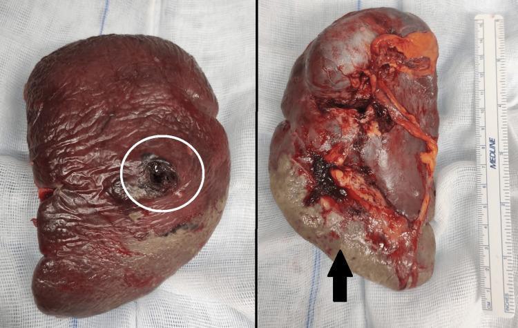
Microscopic features
- Dominant change: coagulative necrosis. The exception is the brain, where infarction gives liquefactive necrosis.
- Timing: it takes 4 to 12 hours for dead tissue to show microscopic evidence of necrosis. If death occurs minutes to hours after occlusion, histologic changes may be absent.
- Acute inflammation: present along the margins within a few hours and usually well defined by 1 to 2 days.
- Repair: begins in the preserved margins. Most infarcts are ultimately replaced by scar tissue.
- Septic infarcts: abscess formation, with organization following the same pattern afterward.
Time course of a typical myocardial infarct (from general knowledge, so verify against your syllabus)
| Time | Findings |
|---|---|
| 0 to 4 h | Usually none; early wavy fibers |
| 4 to 12 h | Early coagulative necrosis, edema, hemorrhage |
| 12 to 24 h | Hypereosinophilic myocytes, nuclear pyknosis |
| 1 to 3 days | Neutrophilic infiltrate |
| 3 to 7 days | Macrophages remove debris; maximum softening |
| 1 to 2 weeks | Granulation tissue |
| Weeks to months | Collagenous scar |
6. Organ-specific infarcts (brief)
- Heart: a white infarct from coronary thrombosis on a plaque. Coagulative necrosis follows the time course above.
- Brain: liquefactive necrosis. Because the brain has end-organ circulation with limited collateral supply, occlusive infarcts generally start as nonhemorrhagic (pale or anemic). - Robbins, Cotran & Kumar Pathologic Basis of Disease, ch. 28
- Lung: a red, wedge-shaped infarct, usually from an embolus in a patient with underlying heart failure or lung disease. The dual supply makes infarction uncommon in otherwise healthy people.
- Spleen: a white, sharply demarcated wedge (end-arterial supply).
- Kidney: mostly white infarcts because of the lack of collateral supply. Within 24 hours they become sharply demarcated, pale, yellow-white areas. Remote infarcts become fibrotic scars. - Robbins, Cotran & Kumar Pathologic Basis of Disease, ch. 20
- Intestine: hemorrhagic infarcts, from volvulus, hernia entrapment, or mesenteric thrombosis. Bowel infarction is frequently fatal.
- Testis and ovary: hemorrhagic infarcts from torsion, which obstructs the single venous outflow.
High-yield summary
- Virchow's triad: endothelial injury, stasis or turbulence, hypercoagulability. Stasis dominates in veins and turbulence in arteries and the heart.
- Stasis prevents washout of activated clotting factors, brings platelets to the endothelium, and activates the endothelium.
- Infarct = ischemic coagulative necrosis, except in the brain (liquefactive).
- Red infarct: venous occlusion, dual supply, loose tissue, or reperfusion. White infarct: arterial occlusion in solid, end-arterial organs.
- Outcome depends on collaterals (the most important factor), speed of occlusion, tissue sensitivity to hypoxia, and blood oxygenation.
- Neurons die in 3 to 4 minutes, and myocytes in 20 to 30 minutes.