
17794215649692134568791852728384.jpg
Tell task two diagram
Reading File
Searching Images
Secondary antral follicle histology H&E stained ovary antrum oocyte zona pellucida granulosa theca
Finding Sources
Loading Image
Loading Image
Loading Image
Here is what you need for Task 2: Histological Sketching — a Secondary/Antral Follicle diagram with all required labels.
Task 2: Secondary (Antral) Follicle — Labeled Diagram
Diagram 1 — Schematic cross-section of an antral follicle (with all required labels)

The diagram below (Panel A, bottom) shows the antral follicle with all five structures your task requires clearly labeled:
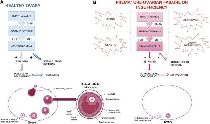
Labels visible in the diagram:
| Structure | Location in Follicle |
|---|---|
| Antrum | Large, fluid-filled central cavity (antrum with follicular fluid) |
| Oocyte | Centrally positioned egg cell, pushed to one side |
| Zona Pellucida | Clear glycoprotein layer immediately surrounding the oocyte |
| Granulosa Layer | Multi-layered epithelium surrounding the zona pellucida and antrum |
| Theca Interna | Inner vascular/endocrine layer outside the basal membrane |
| Theca Externa | Outer fibromuscular layer, outermost boundary of the follicle |
Diagram 2 — Follicular development series (progression from primordial → Graafian)
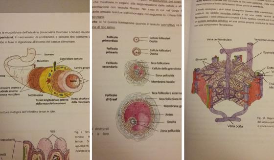
The middle panel (Follicolo secondario / Follicolo di Graaf) shows the secondary follicle transitioning to antral stage, with labeled structures including oocyte, zona pellucida, granulosa cells, theca interna/externa, and antrum.
Diagram 3 — Actual H&E photomicrograph (Graafian/antral follicle)
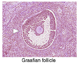
This is an actual H&E-stained slide of a Graafian (antral) follicle. You can identify:
- Antrum — large, pale, fluid-filled space (clear area)
- Oocyte — small round cell visible at the edge of the antrum
- Zona pellucida — pale halo around the oocyte
- Granulosa cells — dark-staining cells lining the follicle wall (purple nuclei)
- Theca interna/externa — outer layers of the follicle wall (arrowhead points to this region)
Key Points for Your Sketch
- Antrum = large fluid-filled space (dominant feature of a secondary/antral follicle)
- Oocyte = sits on a mound of granulosa cells called the cumulus oophorus, surrounded by the corona radiata
- Zona pellucida = eosinophilic (pink), translucent ring directly around the oocyte
- Granulosa layer = stratified cuboidal epithelium lining the antrum; no blood vessels
- Theca interna = inner layer, highly vascularized, secretes androgens → converted to estrogen by granulosa cells
- Theca externa = outer fibrous layer, structural support
This is a shared conversation. Sign in to Orris to start your own chat.