
Explain cell injury of pathology subject
Explain cell injury of pathology subject
cell injury pathology causes mechanisms reversible irreversible necrosis apoptosis
coagulative necrosis histology myocardial infarction
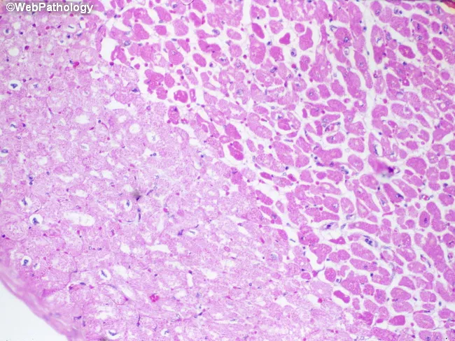
Imaging modality: Light microscopy of hematoxylin-eosin (H&E) stained cardiac tissue. Specimen is paraffin-embedded myocardial section, 5 micron thick, imaged with bright-field optics at high magnification. Anatomic region: left ventricular myocardium with an infarct in the left half of the field. The histology demonstrates pallor of necrotic cardiomyocytes (myocytolysis) produced by sarcolemmal disruption with influx of salt and water and cytoplasmic swelling. In this early post-ischemic stage, fibers lose cross-striations and display increased eosinophilia; nuclei are often absent or pyknotic. The infarct border may show waviness of fibers and mild edema; contraction bands may be present at the margin. Neutrophilic infiltration is typically minimal during the first 24 hours but may begin to appear around this window. The combination of coagulative necrosis, myocytolysis, and pallor indicates an acute myocardial infarction approximately one day old. This histologic pattern correlates with ischemia from coronary occlusion, clinical presentation of acute coronary syndrome, and aligns with diagnostic timelines (0-24 h). Clinically the image supports timing of infarct and helps distinguish acute infarction from myocarditis or chronic scar, serving as an educational reference for pathology, medical education, and research on myocardial injury dynamics. Further confirmed by clinical correlation and education.
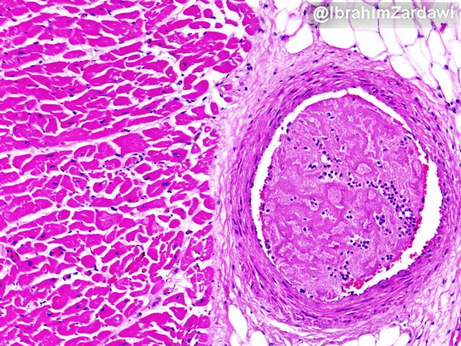
Imaging modality: Light microscopy of a hematoxylin and eosin (H&E) stained cardiac tissue section. Specimen type: paraffin-embedded myocardium with an intraluminal thrombus within a branch of a coronary artery. Imaging perspective: transverse cross-section illustrating occlusion of a coronary artery lumen by a recent thrombus and adjacent myocardial fibers. Major finding: occlusive thrombus densely eosinophilic within the arterial lumen, partially projecting into the vessel lumen and compressing flow. Vessel wall shows no striking dissection; subtle intimal thickening may reflect underlying atherosclerosis. Surrounding myocardium demonstrates preserved architectural integrity at this field of view, with no conspicuous extensive coagulative necrosis or inflammatory infiltrate evident at this early stage. The thrombus composition is suggested by a dense eosinophilic core with fibrous and platelet-rich components, typical of acute thrombosis; perivascular tissue shows mild edema. Clinical interpretation: acute coronary syndrome due to acute thrombotic occlusion of a coronary branch, likely on a preexisting atherosclerotic plaque. Diagnostic significance: provides histopathologic confirmation of thrombosis as the mechanism of ischemia; differential considerations include embolic occlusion, vasospasm with secondary thrombosis, or plaque rupture; correlate with clinical presentation and angiography. Potential educational uses: pathophysiology of myocardial ischemia, ACS, coronary thrombosis, histology of coronary arteries, and teaching images of acute myocardial infarction pathology.
reversible cell injury cellular swelling hydropic change diagram
Cell Injury (Pathology)
1. Causes of Cell Injury
- Hypoxia/Ischemia - the most common cause. Hypoxia is oxygen deficiency (from lung disease, anemia, or poor oxygen-carrying capacity of blood); ischemia is reduced blood supply, which also deprives cells of nutrients. Most hypoxia results from ischemia due to arterial blockage.
- Toxins - air pollutants, insecticides, carbon monoxide, asbestos, cigarette smoke, ethanol, and many therapeutic drugs at excessive or inappropriate doses.
- Infectious agents - viruses, bacteria, fungi, parasites - injure cells directly or via toxin release and harmful immune responses.
- Immunologic reactions - autoimmune attack on self-tissue, allergic reactions, or chronic immune responses to microbes; the resulting inflammation itself damages tissue.
- Genetic abnormalities - mutations or chromosomal defects causing loss or gain of protein function, accumulation of misfolded proteins, or damaged DNA (e.g., sickle cell anemia, inborn errors of metabolism).
- Nutritional imbalances - protein-calorie deficiency, vitamin deficiencies, or excessive intake (obesity contributing to diabetes, atherosclerosis).
- Physical agents - trauma, extremes of temperature, radiation, electric shock, sudden pressure changes.
2. Reversible vs. Irreversible Injury
- Cellular (hydropic) swelling - due to failure of ATP-dependent ion pumps, causing Na+ and water influx
- Fatty change - lipid vacuoles, especially in liver, heart, muscle
- Cytoplasmic changes: swollen ER and mitochondria, detachment of ribosomes, clumping of nuclear chromatin
- Formation of "myelin figures" from damaged membranes
- Severe mitochondrial dysfunction with inability to reverse (loss of oxidative phosphorylation even after reoxygenation)
- Marked disturbances of membrane function (plasma membrane, lysosomal, mitochondrial)
- Massive influx of calcium into the cell, activating destructive enzymes (proteases, phospholipases, endonucleases)
3. Mechanisms of Cell Injury
- Mitochondrial dysfunction - hypoxia, toxins, and radiation damage mitochondria, causing (a) failure of oxidative phosphorylation with ATP depletion, and (b) formation of the mitochondrial permeability transition pore, which can trigger necrosis, and release of cytochrome c and other proteins that activate apoptosis.
- ATP depletion - loss of energy needed for ion pumps (leading to cell swelling), protein synthesis, and lipid metabolism; anaerobic glycolysis compensates partially but causes lactic acid accumulation and intracellular acidification.
- Influx of calcium - increased cytosolic Ca2+ activates enzymes (ATPases, phospholipases, proteases, endonucleases) that damage membranes, cytoskeleton, and chromatin.
- Accumulation of reactive oxygen species (oxidative stress) - free radicals cause lipid peroxidation of membranes, oxidative modification of proteins, and DNA damage.
- Defects in membrane permeability - direct toxin/complement damage or downstream effects of the mechanisms above.
- Damage to DNA and proteins - misfolded proteins and DNA damage trigger apoptosis via checkpoint pathways.
- Autophagy - a survival response where cells "self-cannibalize" damaged organelles via lysosomal digestion during stress (e.g., nutrient deprivation, ischemia); if overwhelmed, it can also trigger cell death.
4. Patterns of Cell Death
- Coagulative necrosis - protein denaturation preserves the basic cell/tissue outline (classic in myocardial infarction, most solid organ infarcts)
- Liquefactive necrosis - enzymatic digestion produces a liquid, viscous mass (brain infarcts, bacterial abscesses)
- Caseous necrosis - "cheese-like" friable necrosis, characteristic of tuberculosis (a combination of coagulative and liquefactive features surrounded by granulomatous inflammation)
- Fat necrosis - enzymatic (lipase) destruction of fat, as in acute pancreatitis
- Fibrinoid necrosis - seen in immune-mediated vascular injury, with immune complex/protein deposition in vessel walls
- Gangrenous necrosis - not a distinct pattern but coagulative necrosis of a limb with superimposed liquefactive change from bacterial infection (wet gangrene)
Summary Table
| Feature | Reversible Injury | Irreversible Injury (Necrosis) |
|---|---|---|
| ATP | Decreased | Severely depleted |
| Membrane | Blebbing, myelin figures | Rupture, loss of integrity |
| Nucleus | Chromatin clumping | Pyknosis -> karyorrhexis -> karyolysis |
| Mitochondria | Swelling | Severe swelling, permeability transition pore |
| Outcome | Recovery if stimulus removed | Cell death, inflammation |
All
fatty liver steatosis histology hepatocytes lipid vacuoles
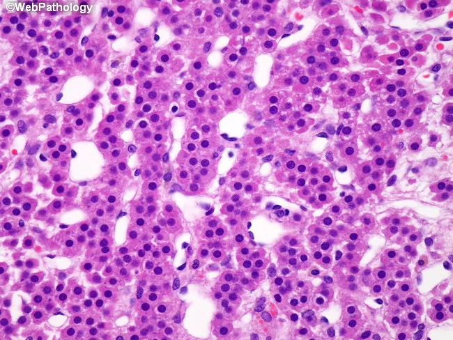
High-magnification brightfield histology of liver tissue stained with hematoxylin and eosin. The hepatic parenchyma displays preserved lobular architecture with cords of polygonal hepatocytes separated by narrow to wide sinusoids. A prominent feature is diffuse intracellular lipid accumulation, manifested as numerous clear cytoplasmic vacuoles that displace occasional nuclei to the cell margin. Both macrovesicular and microvesicular fat droplets are evident, creating a foamy to bubbly cytoplasmic appearance in many hepatocytes. The overall pattern is hepatocellular steatosis; there is no conspicuous bridging necrosis or lobular inflammation visible in this field, though mild cytoplasmic swelling of some hepatocytes can be appreciated. Bile ducts and portal tracts appear unremarkable in this view. The appearance is compatible with fatty liver changes, seen in nonalcoholic fatty liver disease (NAFLD) or alcoholic fatty liver disease, and may reflect metabolic syndrome, insulin resistance, or hepatotoxic exposure. Clinically, such steatosis can be reversible with weight reduction, glucose control, and avoidance of hepatotoxins. Pathologic significance lies in assessing the degree of steatosis and distinguishing simple steatosis from steatohepatitis, which would warrant closer clinical management. This image is suitable for educational reference in hepatology, pathology, and diagnostic imaging.
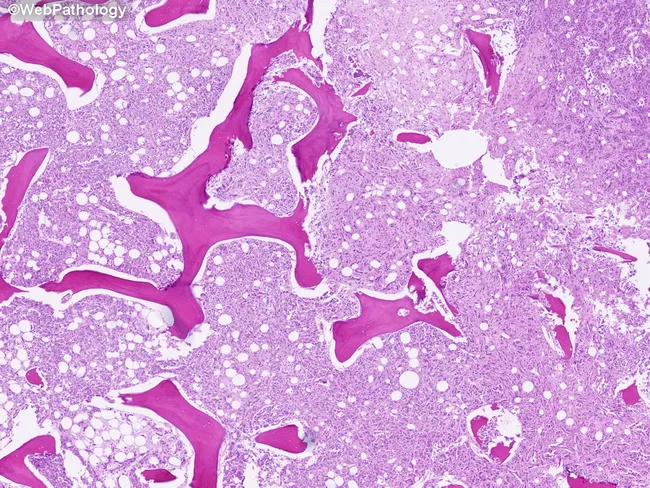
Light-microscopy histology of liver tissue demonstrates macrovesicular steatosis. Imaging modality: Histology using Hematoxylin and Eosin (H&E) stain on paraffin-embedded sections. Anatomical localization: hepatic parenchyma of the liver, with hepatocytes arranged in plates separated by sinusoids and preserved lobular architecture visible at low magnification. Visual features: numerous intracellular lipid vacuoles produce large clear droplets that displace hepatocyte nuclei to the periphery; cytoplasm otherwise eosinophilic; occasional subtle ballooning is not evident; portal tracts and central veins are discernible but not markedly inflamed. Pathological interpretation: macrovesicular steatosis consistent with fatty liver changes; most compatible with nonalcoholic fatty liver disease (NAFLD) or alcoholic fatty liver disease (AFLD) depending on patient history. In this image, steatosis is the predominant finding with minimal inflammatory infiltrate and no definitive fibrosis at this level of magnification. Clinical significance: fatty change indicates hepatocellular stress from metabolic syndrome or hepatotoxic exposure; may precede steatohepatitis, fibrosis, cirrhosis, and increased risk of hepatocellular carcinoma with ongoing injury. Potential clinical use cases: assessment of steatosis burden in NAFLD/NASH studies, biopsy-guided staging, therapeutic monitoring, and education for trainees. The image serves as an educational reference for recognizing hepatocellular lipid accumulation and distinguishing it from other hepatic pathologies such as steatohepatitis and cholestatic injury.
liquefactive necrosis brain infarct histology
caseous necrosis granuloma tuberculosis histology
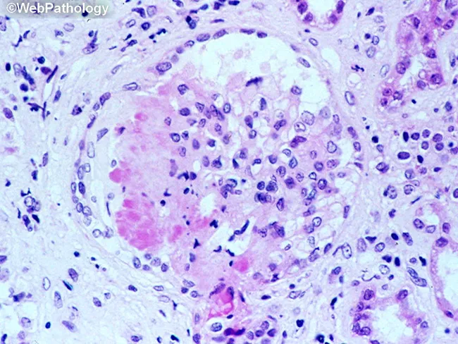
This is a light microscopy image of a formalin-fixed, paraffin-embedded tissue section stained with Hematoxylin and Eosin (H&E). The sample reveals a well-formed granulomatous lesion characterized by a surrounding cuff of lymphocytes and numerous epithelioid histiocytes with elongated, crescent-shaped nuclei. Within the granuloma, multinucleated giant cells (Langhans-type) are visible, and a central amorphous pink necrotic area (caseous necrosis) is evident. The tissue architecture demonstrates organized granulomatous inflammation with a necrotic core, consistent with infectious granulomas. The differential includes mycobacterial disease (tuberculosis) and certain fungal infections (histoplasmosis, coccioidomycosis), though non-necrotizing granulomas can occur in sarcoidosis. The staining highlights chromatin-rich nuclei and cytoplasmic eosinophilia; occasional fibroblasts and mild edema extend into the perigranulomatous region. Clinically, granulomatous tissue with caseation is highly suggestive of TB in the appropriate clinical context and warrants further diagnostic workup (acid-fast bacilli staining, fungal stains, culture, PCR). This image serves as teaching material for histopathology education, differential diagnosis of granulomatous inflammation, and correlation with radiologic and microbiologic findings in suspected infectious etiologies. Additional keywords for indexing: epithelioid macrophages, granuloma, necrosis, caseation, tuberculosis, mycobacteria, fungal infection, lymphohistiocytic rim, tissue biopsy, pathology slide, H&E, histology teaching. Clinical correlation with imaging and sputum analysis improves diagnostic yield.
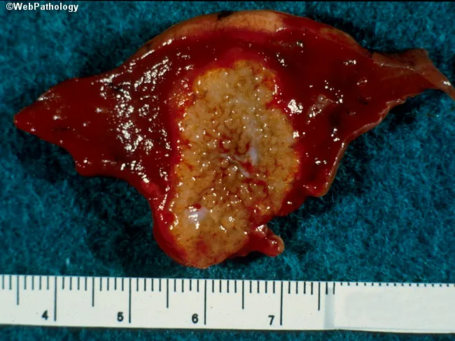
Gross pathology photograph of ex vivo hepatic tissue showing a single, lobulated lesion with a central pale-yellow to tan, crumbly necrotic core surrounded by a hyperemic, reddish-brown peripheral rind. The specimen measures approximately 5.5–6.5 cm in greatest dimension as judged by the metric ruler placed adjacent to the cut surface. The lesion appears well-demarcated from surrounding viable parenchyma, suggesting a focal process such as an infectious granuloma or abscess. The central area exhibits caseous-like necrosis with a cracked, cheese-like consistency; periphery shows congested, friable tissue with mild surface sheen. The overall sample has a smooth to slightly irregular contour. No obvious hemorrhagic stellate patterns noted beyond the margin, though diffuse vascularity is evident on the surface. The cut surface reveals a clear contrast between necrotic core and inflamed rim; there may be microcavitation within the core. These macroscopic features are compatible with granulomatous hepatic disease, notably tuberculosis or fungal infection, though bacterial abscess or parasitic granuloma cannot be excluded without microbiologic and histologic confirmation. This image is useful for education on gross-pathology correlation, differential diagnosis of hepatic necrotizing granulomas, and teaching rounds on infectious liver lesions. Correlate with histology, Ziehl-Neelsen and special stains, culture, and clinical context.
apoptosis apoptotic bodies histology cell death
Cell Injury - Complete Deep Dive
1. Apoptosis - Mechanisms
| Type | Examples |
|---|---|
| Physiologic | Embryogenesis, turnover of proliferative tissue (gut epithelium, lymphocytes), hormone withdrawal (endometrial involution), deletion of self-reactive lymphocytes, decline of leukocytes after immune response |
| Pathologic | Severe DNA damage (radiation, cytotoxic drugs), accumulation of misfolded proteins (ER stress), viral infections, killing of infected cells by cytotoxic T lymphocytes |
-
Mitochondrial (intrinsic) pathway - the dominant pathway in most physiologic and pathologic apoptosis.
- Loss of survival signals or DNA/protein damage activates BH3-only proteins, which tip the balance against anti-apoptotic proteins (BCL-2, BCL-XL).
- This increases mitochondrial outer membrane permeability, releasing cytochrome c and other pro-apoptotic proteins into the cytosol.
- Cytosolic cytochrome c binds APAF-1 to form the "apoptosome," which activates caspase-9, triggering the downstream caspase cascade (effector caspases like caspase-3) that dismantles the cell.
-
Death receptor (extrinsic) pathway - important for eliminating self-reactive lymphocytes and killing virus-infected/damaged cells.
- Death receptors of the TNF-receptor family (e.g., Fas/CD95) bind their ligand (FasL, expressed by cytotoxic T cells) or TNF.
- Receptor clustering recruits adaptor proteins (FADD) which activate caspase-8 directly, which then activates the same downstream effector caspases as the intrinsic pathway.
2. Ischemia-Reperfusion Injury
- Surge in reactive oxygen species (ROS) on reoxygenation - damaged mitochondria cannot fully reduce oxygen, generating superoxide and other radicals; cellular antioxidant defenses are also compromised by the preceding ischemia, so ROS accumulate faster than they can be neutralized. Infiltrating leukocytes contribute additional ROS.
- Continued calcium influx - reperfusion restores extracellular calcium gradients, driving further intracellular Ca2+ overload that activates destructive proteases, phospholipases, and endonucleases.
- Amplified inflammation - restored blood flow brings in more neutrophils and other leukocytes, whose activation products (proteases, ROS) cause additional tissue damage.
- Complement activation - complement proteins can deposit in previously ischemic tissue and further amplify injury.
3. Intracellular Accumulations
- Fatty change (steatosis) - abnormal triglyceride accumulation, classically in the liver (alcohol, diabetes/obesity, toxins, protein malnutrition, hypoxia), but also heart, muscle, kidney.
- Cholesterol and cholesterol esters - lipid-laden macrophages ("foam cells") accumulate cholesterol in atherosclerosis when intake/synthesis exceeds catabolism.
- Proteins - reabsorbed albumin forms pink hyaline droplets in proximal tubule cells in nephrotic syndrome (reversible); excess immunoglobulin forms Russell bodies in plasma cells; misfolded protein aggregates form alcoholic hyaline (Mallory-Denk bodies) in liver and neurofibrillary tangles in neurons.
- Glycogen - accumulates in renal tubules, cardiac myocytes, and pancreatic beta cells in poorly controlled diabetes, and in inherited glycogen storage diseases.
- Pigments:
- Carbon (exogenous) - inhaled and phagocytosed by alveolar macrophages, blackening lung parenchyma and lymph nodes (anthracosis).
- Lipofuscin ("wear-and-tear pigment," endogenous) - insoluble brown-yellow granules from lipid peroxidation, accumulating with aging, most visible in the heart, liver, and brain - a marker of prior free-radical injury, not itself harmful.
- Hemosiderin (endogenous, iron-derived) - accumulates locally after hemorrhage or systemically in iron overload states (hemosiderosis/hemochromatosis).
- Melanin (endogenous) - normal pigment, relevant pathologically in disorders of pigmentation and melanoma.
4. Necrosis Subtypes (with images)
| Type | Mechanism | Classic Example |
|---|---|---|
| Coagulative | Protein denaturation preserves cell/tissue outline for days | Infarcts in most solid organs (heart, kidney, spleen) except brain |
| Liquefactive | Enzymatic digestion by leukocytes turns tissue into a viscous liquid | Bacterial abscesses (pus); hypoxic CNS infarcts |
| Caseous | Combination pattern - "cheese-like" friable debris, tissue architecture obliterated, surrounded by granulomatous inflammation | Tuberculosis |
| Fat necrosis | Lipase-mediated digestion of fat, released fatty acids bind calcium (saponification) | Acute pancreatitis |
| Fibrinoid | Immune complexes and plasma proteins deposit in vessel walls, appearing bright pink on H&E | Vasculitis (e.g., polyarteritis nodosa), malignant hypertension |
| Gangrenous | Not a distinct microscopic pattern - coagulative necrosis of a limb; if superinfected, becomes liquefactive ("wet gangrene") | Ischemic limb, diabetic foot |