
Explain me ptyriasis versicular derma topic with real images patho diagnosis and treatment
pityriasis versicolor clinical lesions KOH microscopy spaghetti and meatballs
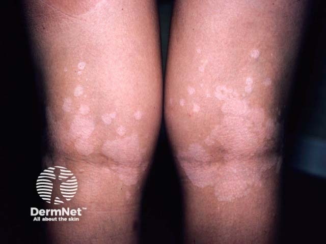
Clinical photography of bilateral popliteal fossae demonstrates multiple hypopigmented, well-demarcated macules and patches, some coalescing into larger plaques, distributed in flexural folds behind the knees. The lesions are lightly scaly on close inspection, or may show parchment-like scale in certain lighting. The overall appearance is typical for pityriasis versicolor (Malassezia) with hypopigmented variants; flexural involvement is recognized in some cases. Skin outside the lesions remains normal in tone; no erythema, atrophy, or induration is present. Images were captured with standard clinical photography setup, using diffuse neutral lighting to maximize contrast between hypopigmented patches and surrounding skin. The clinical impression is hypopigmented pityriasis versicolor; differential considerations include postinflammatory hypopigmentation, tinea versicolor in intertriginous zones, pityriasis alba, vitiligo in flexures, eczema, or hypopigmented mycosis. Confirmation may be achieved via potassium hydroxide (KOH) microscopy showing the yeast with short hyphae (spaghetti-and-meatballs), Wood’s lamp accentuation, or fungal culture. This finding guides management with topical antifungals such as azoles or allylamines; in extensive disease, systemic therapy can be considered. Emphasize recurrence risk, sun protection, and education. Relevancy to educational, diagnostic, and dermatologic research contexts is high. Images serve teaching, differential diagnosis training, and digital repository analyses for PV presentations in unusual flexural sites, for clinical practice and research.
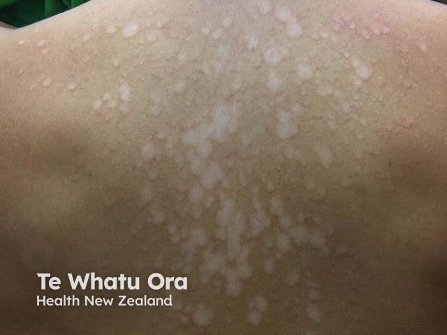
Imaging modality: Clinical photography; technique: macro close-up dermatologic image of the back under natural lighting. Precise anatomical localization: trunk, back (axillary to scapular region); dermatologic context: fine pale hypopigmented macules with fine surface scaling distributed over the upper back. Visual features: numerous small to medium-sized patches, well circumscribed, varying in size, with subtle desquamation; lesions lie on otherwise normal skin and show no surrounding erythema. Pathology: clinical diagnosis of pityriasis versicolor (tinea versicolor) caused by Malassezia species. Diagnostic adjuncts: KOH microscopy may reveal spaghetti-and-meatballs hyphae and Wood lamp examination can yield copper-orange fluorescence; these findings support diagnosis. Diagnostic significance: characteristic pigmentary changes in warm, humid climates; important differential includes vitiligo, pityriasis alba, post-inflammatory hypopigmentation; confirmation guides antifungal therapy. Therapeutic relevance: topical azoles (ketoconazole, clotrimazole) or selenium sulfide antifungal shampoos; in extensive disease, oral antifungals may be used; recurrences are common. Clinical use scenarios: dermatology atlas annotation, medical education, AI model training, image-based differential diagnosis, and longitudinal monitoring of treatment response. This image provides a classical morphology for macular hypopigmentation with fine scale on the trunk, enabling pattern recognition and educational value across clinicians, students, and researchers. Clinical utility for education.
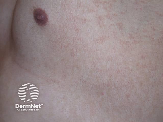
This clinical photograph shows a single, well-circumscribed pigmented patch on the skin, characteristic of pityriasis versicolor (Malassezia infection) presenting as a hypo- or hyperpigmented macule. Imaging modality: clinical photography; perspective: close-up surface view of skin. Anatomic location: cutaneous surface within the integumentary system; region not specified on this image. Visual features: circular to oval plaque with a slightly raised, well-defined border; color ranges from reddish-brown to tan relative to surrounding skin; surface may be smooth with minimal desquamation or fine scale detectable on close inspection. The lesion lacks overt edema or ulceration. In the differential diagnosis, other hypo-/hyperpigmented patches such as pityriasis alba, vitiligo, seborrheic dermatitis, postinflammatory hyperpigmentation or tinea corporis could appear similar, but the combination of color, scale, and trunk distribution often points toward Malassezia-associated disease. Notably, pityriasis versicolor commonly occurs in warm, humid climates and can fluoresce yellow-green under Wood's lamp; KOH microscopy often demonstrates the characteristic 'spaghetti and meatballs' pattern. Clinically, lesions are usually asymptomatic or mildly pruritic and can coalesce; treatment is topical antifungals (azole or allylamine) or oral antifungals in extensive disease. This image serves educational purposes for dermatology training, primary care screening, and triage in infectious dermatoses, and can be used in machine learning datasets for skin disease recognition, highlighting malassezia-related pigmentary disorders.
"Pityriasis Versicolor" AND treatment
pityriasis versicolor diagnosis treatment dermnet
https://dermnetnz.org/topics/pityriasis-versicolor
Pityriasis versicolor
Real clinical images
Cause and pathogenesis
- Organism: Lipophilic yeast Malassezia, most commonly M. globosa. It normally lives on human skin.
- It grows best in sebum-rich areas, hence involvement of the upper chest, upper back, shoulders, neck, and upper arms.
- Under favorable conditions, such as heat, humidity, sweating, or oily skin, it changes from a yeast form to a pathogenic hyphal form in the stratum corneum.
- It remains superficial and produces little inflammation.
Why do the patches become light or dark?
| Type of patch | Mechanism |
|---|---|
| Hypopigmented | Malassezia produces azelaic acid, which inhibits tyrosinase and reduces melanin production and transfer. |
| Hyperpigmented | Increased melanosomes and thickening of the stratum corneum can make lesions darker. |
| Pink/salmon | Mild inflammation and superficial scale. |
Clinical features
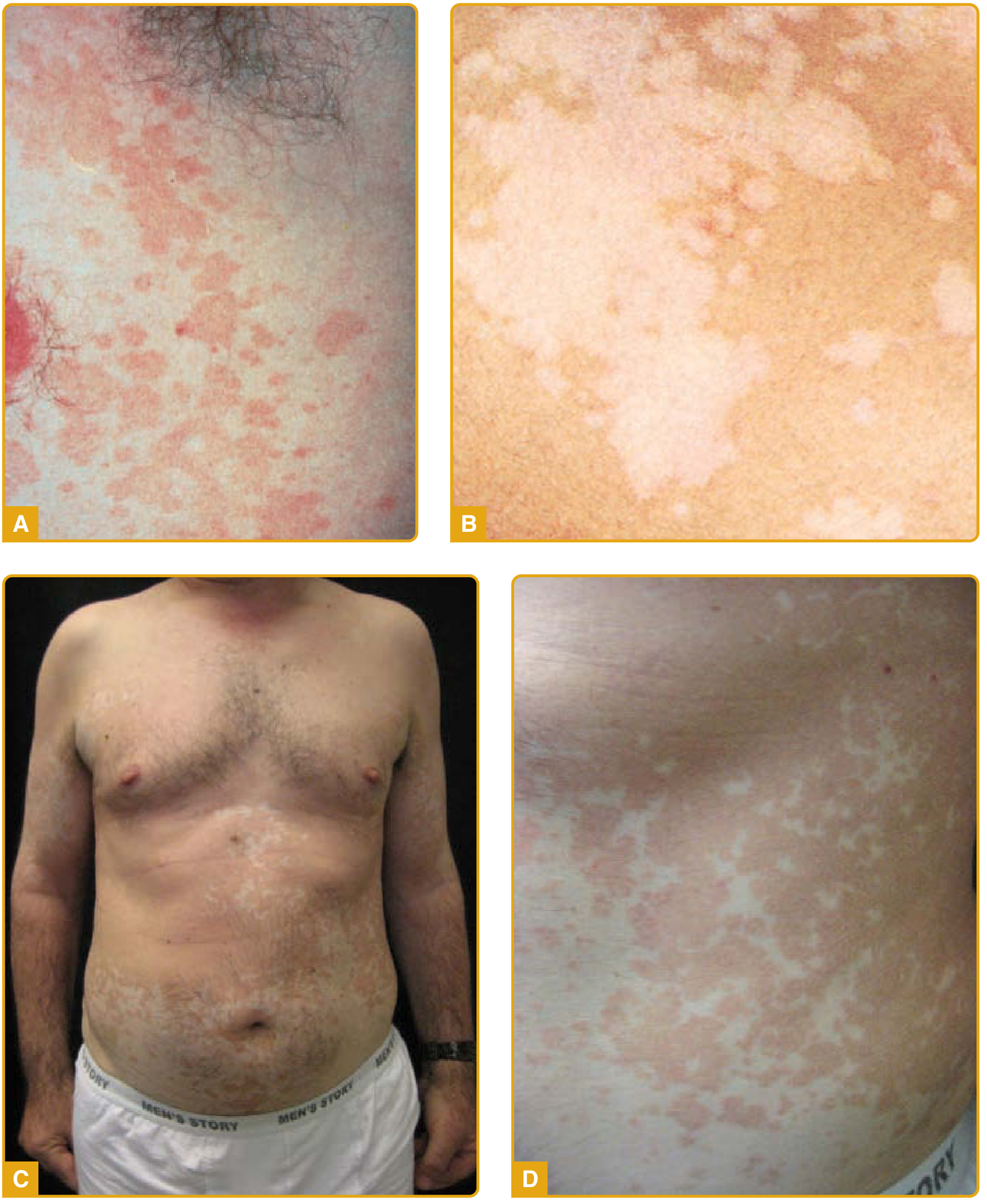
- Multiple round or oval macules, patches, or thin plaques
- Fine, dry, powdery scale, sometimes only obvious after gently scratching or stretching the skin
- Variable color:
- White or light patches
- Light brown or darker patches
- Pink or salmon-colored patches
- Lesions may merge together into large irregular patches
- Usually asymptomatic, but mild itching can occur, especially with heat/sweating
- Common distribution:
- Upper trunk and back
- Shoulders and upper arms
- Chest and neck
- Less commonly face, groin, skin folds, or scalp
Diagnosis
1. KOH examination: best office test
2. Wood lamp
- May show yellow-green or coppery fluorescence.
- Helpful as supportive evidence, but a negative Wood lamp does not rule it out.
3. Dermoscopy
- Fine white scale
- Pigment network changes
- Perifollicular or peripheral scaling
4. Fungal culture
5. Biopsy: rarely required
Pathology / histopathology
Histological findings
- Mild hyperkeratosis
- Possible mild acanthosis
- Mild superficial perivascular lymphocytic infiltrate
- Numerous spores and short “cigar-butt” hyphae in the stratum corneum
- The fungal forms produce the spaghetti-and-meatballs pattern
- PAS stain
- Grocott-Gomori methenamine silver stain, GMS
Important differential diagnoses
| Condition | How it differs |
|---|---|
| Vitiligo | Completely depigmented, sharply demarcated patches, usually no scale, KOH negative. |
| Pityriasis alba | Usually facial, poorly defined pale patches with mild eczema-like dryness, commonly in children. |
| Post-inflammatory hypopigmentation | History of previous rash, injury, or inflammation; no Malassezia on KOH. |
| Seborrheic dermatitis | Greasy yellow scale on scalp, eyebrows, nasal folds, chest. |
| Tinea corporis | Annular lesion with active scaly edge, often more inflammatory. |
| Confluent and reticulated papillomatosis | Hyperpigmented reticulated papules, usually KOH negative. |
Treatment
First-line: topical antifungal treatment
| Option | Practical use |
|---|---|
| Ketoconazole 2% shampoo | Apply to affected skin and surrounding trunk, leave for 5 to 10 minutes, then rinse. Often used daily for several days, depending on the prescribed regimen. |
| Selenium sulfide 2.5% lotion/shampoo | Apply to affected areas, leave about 10 minutes, then wash off. A commonly used regimen is daily for 7 days. |
| Topical azole cream | Ketoconazole, clotrimazole, miconazole, or econazole applied to lesions, usually once or twice daily for 2 to 4 weeks. |
| Terbinafine gel | Can be effective topically. |
| Zinc pyrithione wash/soap | A useful lower-cost option for treatment or recurrence prevention. |
Extensive, recurrent, or treatment-resistant disease
- Itraconazole
- Fluconazole
Preventing recurrence
- Keep skin dry after sweating.
- Wear loose, breathable clothing.
- Use a medicated selenium sulfide, ketoconazole, or zinc pyrithione wash intermittently during recurrent seasons, as advised by a clinician.
- Understand that loss or alteration of pigment may remain after treatment even when the scale and active infection have resolved.
Key exam points
- Cause: Malassezia yeast, especially M. globosa
- Site: upper trunk, shoulders, chest, neck
- Lesion: hypo-, hyperpigmented, or salmon-colored finely scaly macules/patches
- KOH: “spaghetti and meatballs”
- Wood lamp: yellow-green fluorescence may occur
- Histology: hyphae and spores in stratum corneum, PAS/GMS positive
- Treatment: topical selenium sulfide or azole first; oral itraconazole/fluconazole only for extensive or resistant disease
- Pigment takes time to return: persistence of color change does not equal persistent fungus