
Expansile medullary glioma with mass effects on cm cistern ,adjacent cerebellar hemispheres & pontomedullary junction understanding with images & treatment plan
medullary glioma brainstem MRI expansile mass
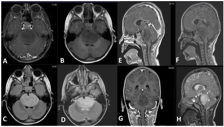
This diagnostic imaging panel consists of eight MRI slices (axial, sagittal, and coronal planes) demonstrating an expansile intra-axial mass in the brainstem, characteristic of a Diffuse Midline Glioma (DMG). The mass is primarily centered in the pons and exhibits a classic radiological profile: hypointense signal on T1-weighted pre-contrast (B, F) and post-contrast (A, E, G) sequences, indicating a lack of significant enhancement. T2-weighted (D, H) and FLAIR (C) sequences show the lesion as uniformly hyperintense. Anatomically, the tumor causes significant expansion of the pons, leading to the obliteration of the prepontine cistern and encasement of the basilar artery. Posteriorly, the mass compresses the fourth ventricle, though no proximal obstructive hydrocephalus is currently evident. Sagittal and coronal views (E, F, G, H) illustrate the longitudinal extent of the lesion, stretching from the pontomesencephalic junction superiorly to the pontomedullary junction inferiorly, with characteristically ill-defined, infiltrative borders. This series serves as a pedagogical example of the typical imaging features and local mass effect of pontine gliomas in neuro-oncology.
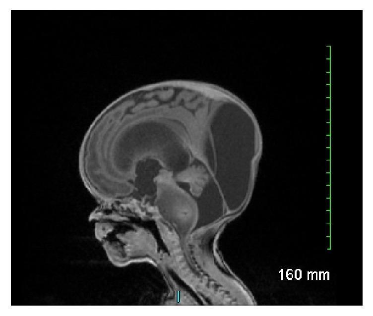
This sagittal T1-weighted fetal MRI scan demonstrates significant intracranial pathology in a developing fetus. The primary finding is a large, expansile, and poorly demarcated mass involving the brainstem, centered in the pons. The mass appears heterogeneously signal-intense and significantly expands the diameter of the pons, extending superiorly towards the midbrain and inferiorly toward the medulla. Secondary to this mass, there is evidence of severe obstructive hydrocephalus, characterized by marked dilation of the lateral ventricles and the third ventricle, with associated thinning of the overlying cerebral mantle and macrocephaly. The posterior fossa shows displacement of the cerebellar structures. This visual presentation is highly characteristic of a congenital brainstem glioma, specifically a diffuse intrinsic pontine glioma (DIPG) or similar anaplastic neoplasm, illustrating the typical obstructive complications and mass effect associated with neonatal neuro-oncology.
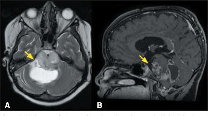
This diagnostic imaging composite displays two magnetic resonance imaging (MRI) sequences of the brain in a pediatric patient, illustrating a diffuse midline glioma affecting the brainstem. Image A is an axial T2-weighted sequence showing an expansile, infiltrative lesion located in the pons. The lesion demonstrates a heterogeneous signal intensity, with hyperintense focal areas (yellow arrow) indicative of cystic components or internal necrosis. The mass effect is evident as it expands the pontine contours. Image B is a sagittal, fat-saturated, T1-weighted sequence with gadolinium contrast enhancement. It reveals the solid portions of the tumor exhibit irregular, heterogeneous enhancement (yellow arrow), suggesting varying vascularity and blood-brain barrier disruption within the mass. The clinical presentation and imaging features are characteristic of a high-grade pediatric-type diffuse midline glioma, specifically the H3K27-altered molecular subtype. This case highlights the typical neuroradiological appearance of aggressive midline gliomas in children, focusing on expansile growth, signal heterogeneity, and variable contrast uptake.
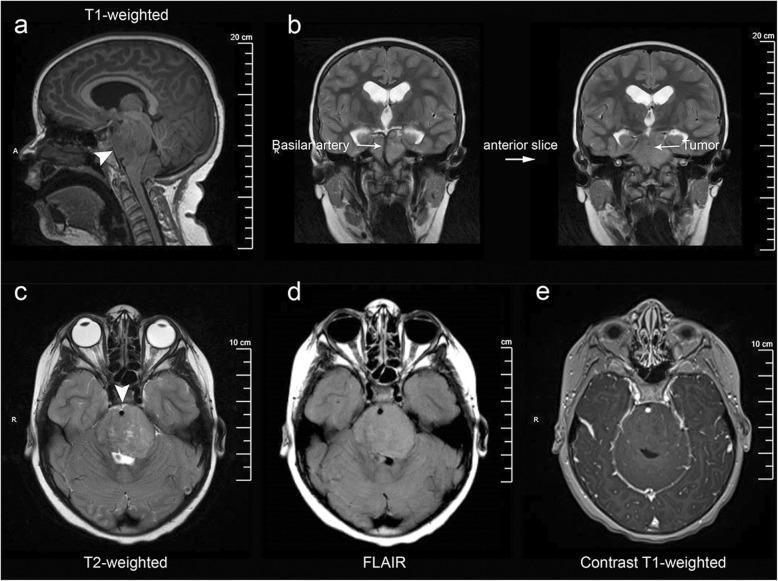
Multi-modal MRI series of the brain demonstrating a Diffuse Intrinsic Pontine Glioma (DIPG). (a) T1-weighted sagittal view shows an infiltrative, expansile mass centered within the pons. (b) Coronal T2-weighted images reveal the exophytic tumor portion extending into the prepontine and suprasellar cisterns, notably encasing the basilar artery. (c-e) Axial sequences highlight characteristic signal patterns: the lesion is hyperintense on T2-weighted (c) and Fluid-Attenuated Inversion Recovery (FLAIR) (d) sequences, indicating significant vasogenic edema or tumor infiltration. (e) Post-gadolinium T1-weighted axial sequence shows a conspicuous absence of enhancement, a typical feature of DIPG indicating an intact blood-brain barrier within the tumor. Associated secondary findings include obstructive hydrocephalus, evidenced by dilated lateral ventricles in the coronal views. This imaging series illustrates the classic neuroradiological criteria for pediatric brainstem gliomas, emphasizing anatomical distortion of the brainstem, cisternal extension, and specific signal characteristics across varying MRI sequences.
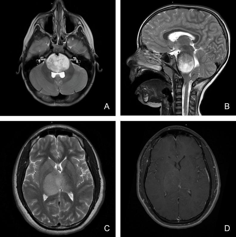
This composite of four magnetic resonance imaging (MRI) scans illustrates the diagnostic features of pediatric diffuse midline glioma in different anatomical locations. Panels A (axial) and B (sagittal) display T2-weighted sequences showing a large, hyperintense, and expansile mass involving the brainstem (pons), characteristic of a diffuse intrinsic pontine glioma (DIPG). The lesion demonstrates an infiltrative growth pattern, causing significant local mass effect and compression of adjacent structures. Panels C and D focus on a thalamic presentation. Panel C is an axial T2-weighted image showing a homogenous, hyperintense lesion within the right thalamus with poorly defined margins extending into the surrounding white matter tracts. Panel D shows the corresponding axial T1-weighted post-gadolinium sequence, revealing minimal to heterogeneous contrast enhancement within the thalamic mass. These images highlight key radiological hallmarks including the tumor's midline location, infiltrative nature, perilesional edema, and variable vascular permeability as indicated by enhancement patterns, which are critical for the clinical classification of these high-grade pediatric gliomas.
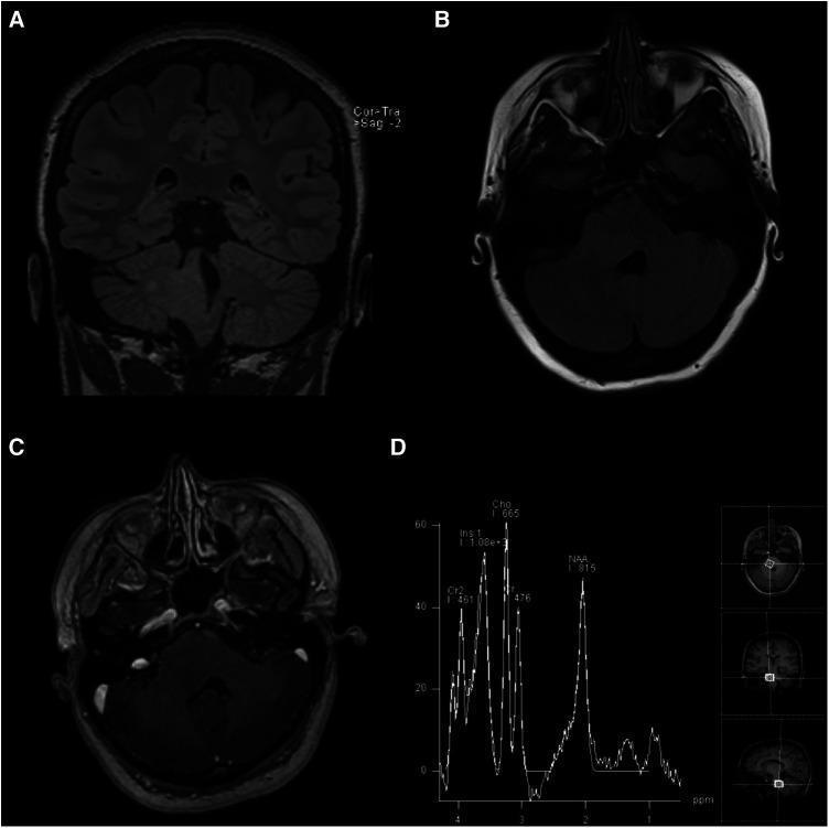
This composite of four images (A-D) presents a multi-modal MRI evaluation of a brainstem lesion. Panels (A) Coronal T2-weighted FLAIR and (B) Axial T2-weighted FLAIR demonstrate an ill-defined, hyperintense, and expansile mass within the right pons. The lesion extends into the right middle cerebellar peduncle and the right posterolateral medulla, causing partial effacement of the fourth ventricle. Panel (C) represents an axial post-gadolinium contrast T1-weighted image, which shows no contrast enhancement, indicating a preserved blood-brain barrier within the lesion. Panel (D) provides Magnetic Resonance Spectroscopy (MRS) data, showing a markedly increased choline (Cho) peak and a significantly decreased N-acetylaspartate (NAA) peak. This metabolic profile (high Cho:NAA ratio) is characteristic of increased cellular turnover and loss of neuronal integrity, typically seen in infiltrating gliomas such as Diffuse Intrinsic Pontine Glioma (DIPG). The findings are essential for neurosurgical and oncological education regarding the diagnostic imaging and metabolic characteristics of pediatric-type diffuse low-grade gliomas in adult patients.
medullary glioma treatment
"diffuse midline glioma" treatment ONC201
PMID: 41138047
PMID: 38335473
medullary brainstem anatomy pontomedullary junction cisterna magna cerebellomedullary cistern
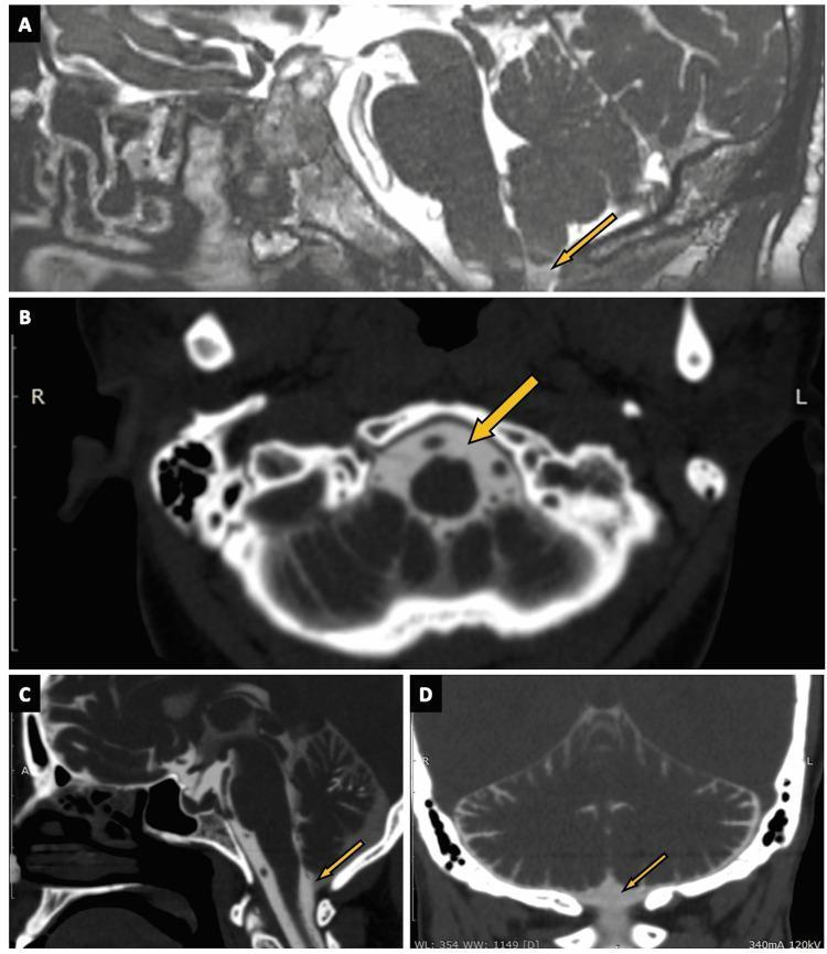
This composite diagnostic image illustrates the cisterna magna across multiple modalities and planes. Panel A shows a sagittal view using 3D constructive interference in steady state (CISS) MRI, where the cistern appears as a dark, hypointense fluid-filled space. Panels B, C, and D utilize CT cisternography in axial, sagittal, and coronal planes, respectively, to visualize the cistern as a region of low attenuation enhanced by contrast material. Anatomically, the cisterna magna is located in the posterior cranial fossa, situated inferior to the cerebellum and posterior to the medulla oblongata. It is identified by arrows in each panel. The images demonstrate the cistern's relationship to surrounding structures, including the brainstem, cerebellar vermis, and the foramen magnum. This visual guide serves as a clinical reference for identifying normal subarachnoid cistern anatomy, which is essential for diagnosing conditions such as CSF rhinorrhea or posterior fossa malformations. The target audience includes medical students and radiology residents specializing in neuroimaging.
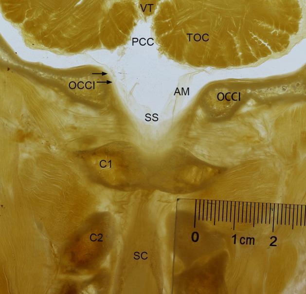
A frontal P45 plastinated anatomical section of the human occipito-cervical junction (OCJ) passing through the posterior arch of the atlas (C1). The specimen demonstrates the morphological relationship between the cerebellum and the upper cervical spine. Key structures labeled include the tuber of vermis (VT) and tonsil of cerebellum (TOC) superiorly. The posterior cerebellomedullary cistern (PCC) is visible above the foramen magnum. A prominent, rhombic-shaped subarachnoid space (SS) is observed between the cerebellum and the atlas (C1), bordered by the arachnoid membrane (AM) and the spinal dura mater (indicated by arrows). Bony landmarks identified include the occipital bone (OCCI), the atlas (C1), and the axis (C2). The spinal cord (SC) is visible descending through the vertebral canal. The image illustrates the continuity of the subarachnoid space from the posterior cranial fossa into the upper cervical vertebral canal, highlighting the anatomy of the cisterna magna and the dural sac at the craniocervical transition. A metric ruler is included for scale.
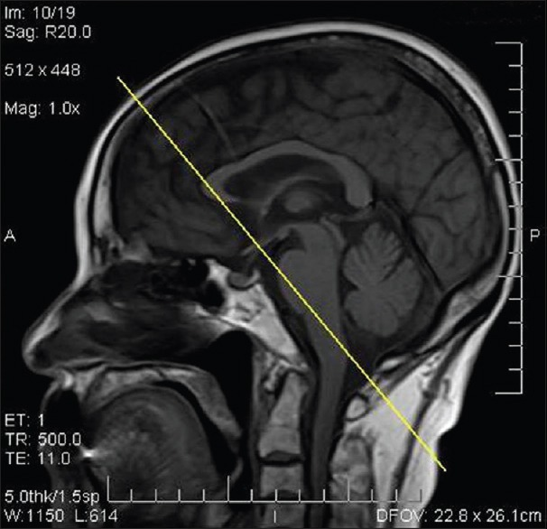
**Modality:** T1-weighted Magnetic Resonance Imaging (MRI). **Anatomical Region:** Sagittal view of the head and upper cervical spine. **Key Landmarks:** Visible structures include the cerebral cortex, corpus callosum, brainstem (midbrain, pons, medulla), cerebellum, and the upper cervical vertebrae. The cisterna magna (cerebellomedullary cistern) is clearly delineated posterior to the medulla and inferior to the cerebellum. **Procedural Context:** The image features a diagnostic overlay consisting of a straight yellow line. This line illustrates the trajectory for a suboccipital or cisternal puncture. **Characteristic Visual Features:** * **Trajectory:** The line originates from the posterior-superior aspect of the skull, passing through the brain parenchyma, and terminates in the region of the cisterna magna. * **Cisterna Magna:** Visible as a pocket of cerebrospinal fluid (CSF) located between the posterior surface of the medulla and the inferior surface of the cerebellum, superior to the foramen magnum. * **Technical Parameters:** Image annotations indicate a TR of 500.0 ms, TE of 11.0 ms, and a slice thickness of 5.0 mm. **Diagnostic Use:** This image serves as a reference for planning or demonstrating a cisternal puncture, an alternative method for CSF access when lumbar puncture is contraindicated or unsuccessful.
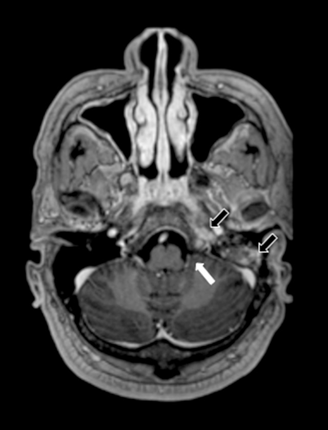
**Imaging Modality:** Magnetic Resonance Imaging (MRI), Axial plane. **Sequence:** Post-contrast T1-weighted image. **Anatomical Region:** Posterior fossa and skull base, specifically focusing on the left cerebellomedullary cistern (cisterna magna), petrous apex, and mastoid air cells. **Observed Findings:** * **Cisterna Magna:** A white arrow indicates the left cerebellomedullary cistern. There is a notable absence of a peripherally enhancing collection, signifying the resolution of a previously documented abscess. * **Skull Base and Mastoid:** Black arrows highlight persistent, though reduced, contrast enhancement within the left petrous apex and the mastoid air cells. These features are consistent with improving petrositis and mastoiditis. * **Cranial Nerves:** Normalization of contrast uptake in the vicinity of the lower cranial nerves is observed compared to previous involvement. **Diagnostic Context:** Post-treatment follow-up of a skull base infection (likely secondary to otomastoiditis) with secondary intracranial extension. The image demonstrates therapeutic response characterized by the resolution of a focal infectious collection (abscess) while showing residual inflammatory changes in the adjacent osseous structures.
Expansile Medullary Glioma: Comprehensive Understanding with Images and Treatment Plan
1. Anatomical Context - What Is the "CM Cistern"?
- Posterior and inferior to the medulla oblongata
- Inferior to the cerebellar hemispheres and vermis
- Above the foramen magnum
2. Definition and Classification
| Type | Molecular Marker | Grade | Prognosis |
|---|---|---|---|
| Diffuse Midline Glioma (DMG), H3 K27M-mutant | Histone 3 K27M | Grade 4 | Dismal (<12 months median OS) |
| Diffuse Astrocytoma (DA), IDH-wildtype | Usually IDH-WT in children | Grade 2-3 | Poor to intermediate |
| Pilocytic Astrocytoma | BRAF fusion | Grade 1 | Relatively favorable |
| Dorsally exophytic glioma | Variable | Grade 1-2 | Best surgical option |
-
It contains vital autonomic centers (cardiac, respiratory, emetic)
-
Lower cranial nerve nuclei (IX, X, XI, XII) are packed within it
-
The pontomedullary junction houses the abducens and facial nerve nuclei
-
Direct surgical access is extremely limited
-
Bradley and Daroff's Neurology in Clinical Practice, p. 1580
-
Adams and Victor's Principles of Neurology, 12th Ed., p. 691
3. Mass Effects - The Three Compartments
3a. Mass Effect on the Cisterna Magna (CM Cistern)
- Obliterates the cisterna magna, reducing the CSF reservoir
- Blocks CSF drainage from the foramen of Magendie, risking obstructive hydrocephalus
- May compress the posterior inferior cerebellar artery (PICA), risking ischemia
- In large tumors, may herniate toward the foramen magnum - a life-threatening complication
3b. Mass Effect on Adjacent Cerebellar Hemispheres
- Tumor expansion pushes laterally into the cerebellar tonsils and hemispheres
- Produces ipsilateral appendicular ataxia and dysmetria
- Large exophytic components can indent the cerebellar peduncles
- Tonsillar herniation risk increases as the cisterna magna obliterates
3c. Mass Effect on the Pontomedullary Junction
- Facial nerve (CN VII) nucleus and fascicles
- Abducens nerve (CN VI) nucleus
- Corticospinal tracts crossing regions
- Medial lemniscus fibers
- Ipsilateral CN VI/VII palsy (the classic presenting sign of brainstem glioma)
- Contralateral hemiparesis (long-tract signs)
- Internuclear ophthalmoplegia (MLF involvement)
- Pseudobulbar dysarthria and dysphagia
4. Clinical Presentation
| Symptom Domain | Specific Features |
|---|---|
| Lower CN palsies | Dysphagia, hoarseness, tongue deviation (IX, X, XII) |
| Motor | Hemiparesis, asymmetric quadriparesis |
| Cerebellar | Gait ataxia, appendicular dysmetria |
| Autonomic | Vomiting, labile BP, respiratory irregularity |
| ICP elevation (late) | Headache, papilledema, morning vomiting |
| Cervicomedullary | Neck pain/stiffness, hand numbness, upper limb weakness |
5. MRI Imaging - Characteristic Findings
MRI Sequences and Expected Findings:
| Sequence | Expected Finding |
|---|---|
| T1 (pre-contrast) | Hypointense expansion of medulla |
| T2 / FLAIR | Hyperintense infiltrative lesion |
| T1 + Gadolinium | Absent to minimal enhancement (diffuse type); rim enhancement in higher grade |
| DWI | Restricted diffusion in high-grade areas |
| MR Spectroscopy | Elevated Cho, reduced NAA (high Cho:NAA ratio) |
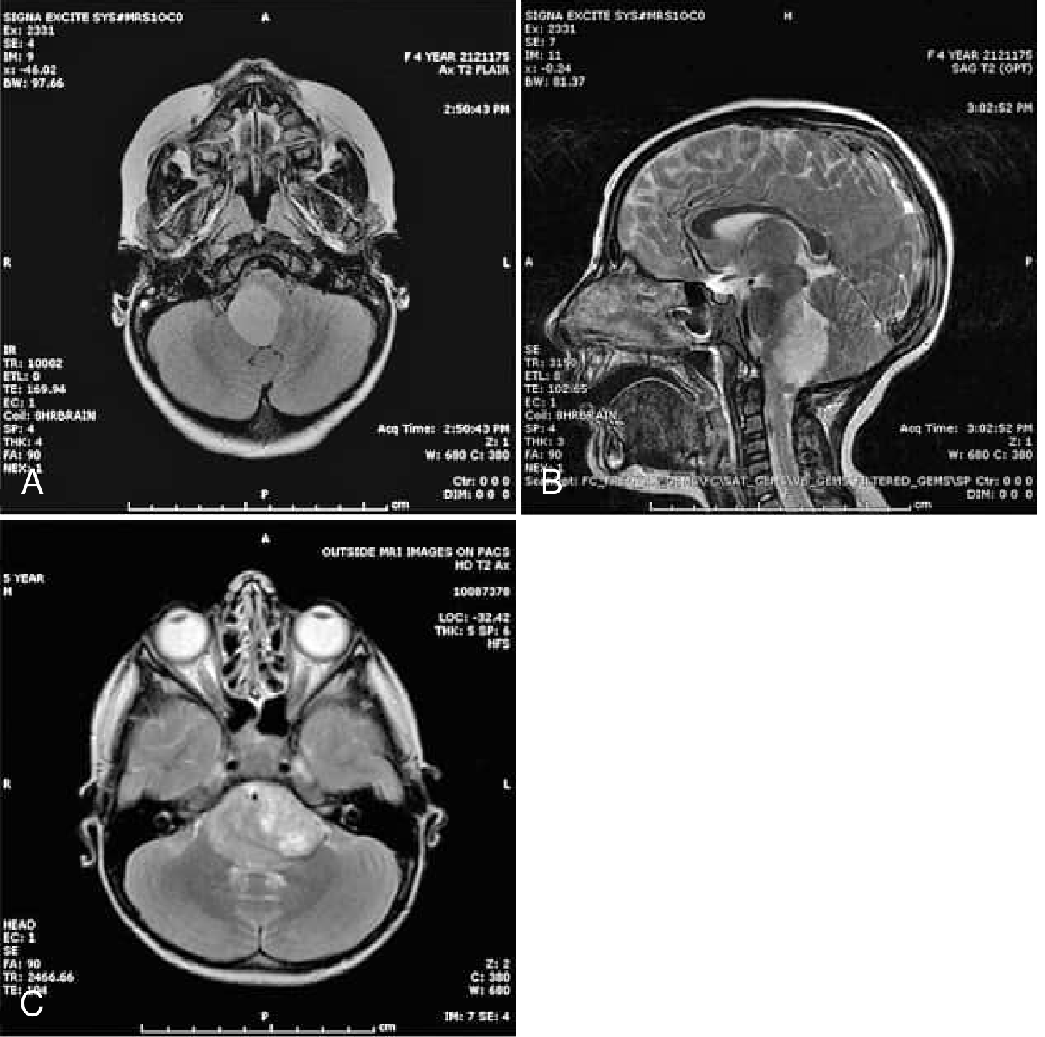
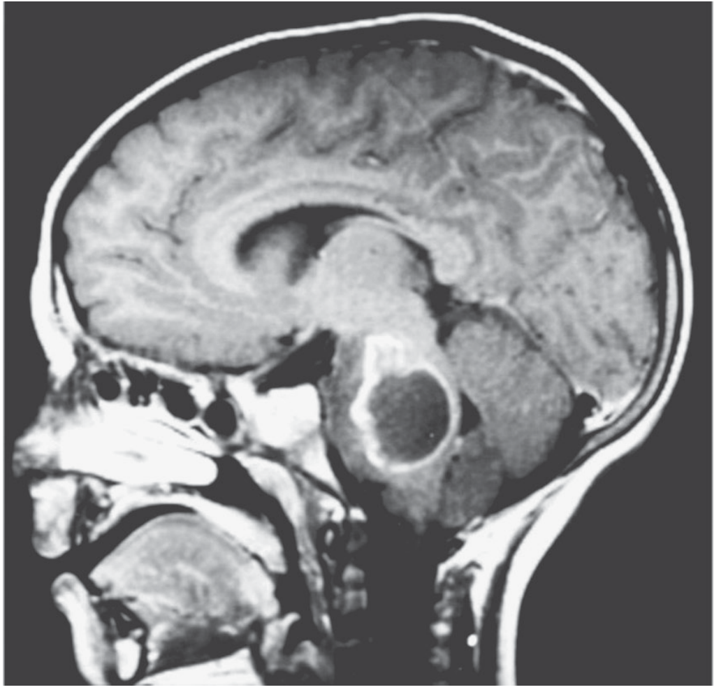
| Sagittal | Reveals craniocaudal extent - pontomedullary junction to upper cervical cord |
6. Differential Diagnosis
| Diagnosis | Distinguishing Feature |
|---|---|
| Pilocytic astrocytoma | Focal nodular/cystic, enhances brightly, dorsal exophytic |
| Pontine form of MS | Younger adult, gadolinium enhancing lesions elsewhere, CSF OCBs |
| Cavernous hemangioma | Hemosiderin "popcorn" on T2*, no expansion |
| Brainstem encephalitis | Rapid onset, fever, CSF pleocytosis, Anti-NMDA/Anti-GABA |
| Ependymoma | Arises from floor of 4th ventricle, extends via foramina |
| Embryonal tumors (ATRT) | DWI restriction, young infants |
7. Workup Protocol
- MRI brain + spine with gadolinium (staging - leptomeningeal spread present at diagnosis in some cases)
- Biopsy - now increasingly performed even in pontine/medullary location to:
- Confirm H3 K27M mutation (defines DMG, WHO grade 4)
- Search for actionable targets: PDGFRA, ACVR1, NTRK, BRAF, EGFR
- Guide clinical trial eligibility
- CSF cytology (if safely obtainable)
- MR spectroscopy - Cho:NAA ratio supports malignancy
- Ophthalmology - papilledema, ocular motility deficits
8. Treatment Plan
8a. Radiation Therapy (Standard of Care)
- Dose: 54-59.4 Gy in 1.8 Gy fractions (conventional fractionation)
- Hypofractionation: 39 Gy in 13 fractions (3 Gy/fraction) used for very young children or palliation
- Produces temporary clinical improvement in ~80% of DIPG patients
- Does NOT cure; median time to progression 6-9 months
- Re-irradiation can be considered at progression for palliation
8b. Targeted Therapy - Dordaviprone (ONC201) - FDA Approved 2025
Adult and pediatric patients ≥1 year with diffuse midline glioma harboring H3 K27M mutation, with progressive disease following prior therapy.
- ORR (RANO-HGG): 20% (10-33.7%)
- ORR (combined RANO-HGG/LGG): 30%
- Median duration of response: 11.2 months
- Corticosteroid reduction ≥50%: 47% of evaluable patients
- Tolerability: No grade 4 adverse events; fatigue most common grade 3 event (10%)
- Mechanism: imipridone compound - DRD2 antagonist + mitochondrial ClpP agonist - disrupts integrated metabolic and epigenetic pathways in H3K27M-mutant cells (Venneti et al., Cancer Discov 2023, PMID 37584601)
8c. Chemotherapy
- Temozolomide, bevacizumab, carboplatin: None have shown survival benefit over radiation alone in DMG
- BRAF/MEK inhibitors (dabrafenib + trametinib): Indicated if BRAF V600E mutation is present (more common in non-H3K27M pilocytic subtypes)
- NTRK inhibitors (larotrectinib, entrectinib): If NTRK fusion is identified on molecular profiling
8d. Neurosurgical Role
| Indication | Intervention |
|---|---|
| Obstructive hydrocephalus | Endoscopic third ventriculostomy (ETV) or VP shunt |
| Focal/nodular, dorsally exophytic | Attempted resection of mural nodule (pilocytic type) |
| Cystic medullary glioma | Drainage of cyst ± nodule resection |
| Diffuse type | Surgery NOT indicated for tumor control; risks are prohibitive |
| Biopsy | Stereotactic biopsy for molecular profiling - now routinely performed |
8e. Management of Specific Mass Effects
| Mass Effect | Management |
|---|---|
| CM cistern obliteration + hydrocephalus | ETV or VP shunt; avoid LP (herniation risk) |
| Brainstem edema | Dexamethasone (0.1-0.15 mg/kg/day); taper after radiation |
| Respiratory compromise | Early ICU involvement; consider elective intubation |
| Dysphagia | PEG/NG tube; speech therapy; aspiration precautions |
| Cerebellar dysfunction | Physio/OT rehabilitation |
8f. Palliative and Supportive Care
- Goals of care discussion with family early
- Corticosteroid management for symptomatic relief
- Symptom control: anti-emetics, analgesics, anti-epileptics if seizures
- Nutritional support
- Psychological support for patient and caregivers
9. Prognosis Summary
| Tumor Type | Median OS |
|---|---|
| DMG H3K27M-mutant (DIPG/medullary) | <12 months |
| Diffuse astrocytoma (IDH-wildtype, brainstem) | 12-24 months |
| Pilocytic astrocytoma (focal, exophytic) | >10 years (good prognosis) |
| Cervicomedullary astrocytoma (focal) | Intermediate - surgical outcome determines prognosis |
Key Takeaway
- Radiation as the backbone (54-59.4 Gy focal field)
- Dordaviprone (ONC201) for H3K27M-mutant tumors at progression - the first FDA-approved targeted agent (August 2025)
- Molecular profiling by biopsy to identify actionable targets
- CSF diversion if hydrocephalus develops
- Integrated palliative support from day one