
What is the treatment of celiac disease
"celiac disease"[MeSH Terms] AND "gluten-free diet"[MeSH Terms]
celiac disease small intestinal villous atrophy histology
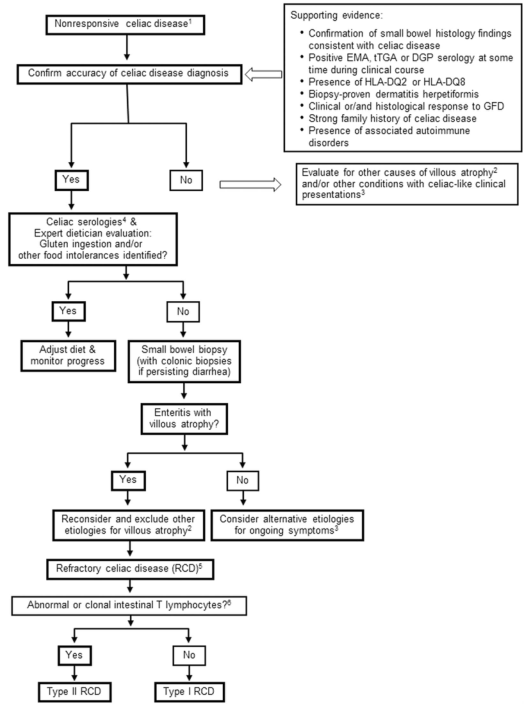
Summary : This flowchart outlines the diagnostic and management pathway for nonresponsive celiac disease, including confirmation of diagnosis, evaluation for gluten ingestion or other food intolerances, consideration of alternative causes of villous atrophy, and classification of refractory celiac disease (RCD) into Type I or Type II based on intestinal T lymphocyte findings.
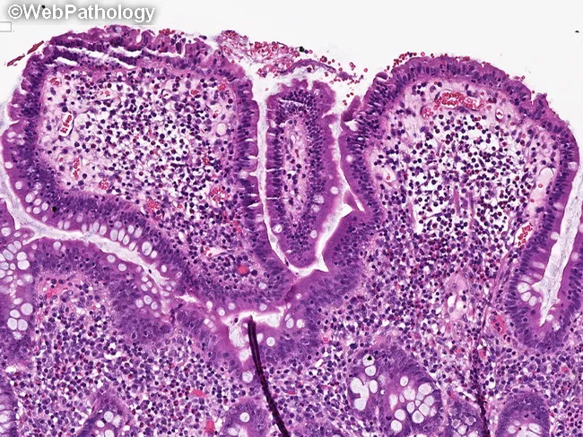
This histopathology image depicts small intestinal mucosa with celiac disease-type changes in tissue adjacent to Enteropathy-associated T-cell lymphoma (EATL). Acquired by light microscopy from a mucosal biopsy of the jejunum/ileum, the slide shows pronounced villous blunting with crypt hyperplasia, producing a columnar, shortened villi profile. The surface epithelium reveals increased intraepithelial lymphocytes arranged along the villous axis; lamina propria is chronically inflamed with lymphocytes and plasma cells. These features are classic for gluten-sensitive enteropathy and can precede or accompany overt lymphomatous transformation in EATL. The mucosal architecture is distorted, with a spectrum from mild to severe villous atrophy; goblet cells are reduced; enterocytes may display reactive changes. The image captures the inflammatory milieu and barrier dysfunction characteristic of celiac disease, reflecting malabsorption risk and nutritional compromise. In the context of suspected or known EATL, this histology underscores the background gluten-inducible enteropathy that predisposes to malignant T-cell transformation. Clinically, such specimens guide diagnosis, prognosis, and therapeutic planning, including dietary gluten exclusion and surveillance for lymphoma. This histologic pattern should be correlated with serology, imaging, and molecular studies to differentiate celiac disease from refractory celiac disease and to assess lymphoma risk. Additionally, this pattern has educational utility.
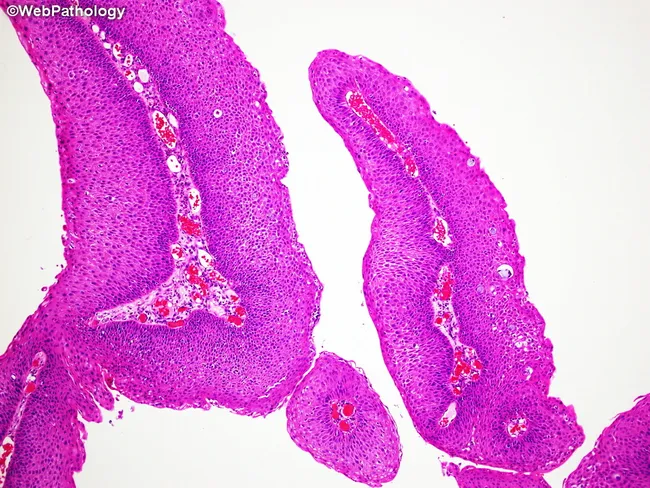
High-resolution bright-field histology image of gastrointestinal mucosa stained with hematoxylin and eosin (H&E). The specimen is a small intestinal biopsy or mucosal patch showing tall finger-like villi with slender, bulbous cores of lamina propria and a lined epithelium. The tissue architecture emphasizes the mucosal surface with exuberant villous projections, smooth luminal interface, and intact surface epithelium. Hematoxylin stains nuclei a deep blue, while eosin imparts pink–red cytoplasmic and extracellular matrix contrast, highlighting enterocyte basophilic nuclei, goblet cells, and vascular channels within the lamina propria. Goblet cells appear as pale, mucus-filled pockets along the villus stalks. There is no obvious villous blunting, crypt hyperplasia, inflammatory infiltrate, or architectural distortion evident in this field. The image demonstrates features typical of normal or near-normal small intestinal mucosa, displaying orderly cellular polarity and well-differentiated enterocytes. Clinically, this pattern is relevant for differential diagnosis of malabsorption syndromes, celiac disease, infectious enteritis, and inflammatory bowel conditions, where villous atrophy, crypt hyperplasia, or inflammatory infiltration would be expected. The slide is suitable for educational reference, histology teaching, and comparative pathology, providing a baseline for recognizing standard villous architecture in gastrointestinal mucosa and for contrast with pathological specimens. This image supports educational objectives in anatomy, histology, and diagnostics.
Treatment of Celiac Disease
1. Gluten-Free Diet (GFD) - The Cornerstone
- Wheat (including spelt, kamut, triticale, durum) - contains gliadin
- Rye - contains secalin
- Barley (including barley malt) - contains hordein
- Protein-containing derivatives of these grains (wheat bran, wheat germ)
Hidden Sources of Gluten
- Bread, crackers, cereals, pasta, baking mixes (obvious sources)
- Candy, soy sauce, soups, seasoned rice mixes, salad dressings, gravies (hidden sources)
- Communion wafers, medications, and nutritional supplements
- Labels must be re-read each time a product is purchased, as manufacturers can change ingredients without warning
Oats
2. Dietary Supplementation
| Nutrient | Reason for Deficiency |
|---|---|
| Iron | Duodenal malabsorption |
| Folate | Jejunal malabsorption |
| Vitamin B12 | Less common, terminal ileal involvement |
| Calcium & Vitamin D | Impaired Ca absorption, bone disease risk |
| Zinc, Magnesium | Generalized small bowel malabsorption |
3. Multidisciplinary Management
- Consultation with a skilled registered dietitian (RD) experienced in celiac disease
- Patient education about the disease and the diet
- Life-long adherence to the GFD
- Identification and treatment of nutritional deficiencies
- Access to an advocacy/support group
- Continuous long-term follow-up by a multidisciplinary team
4. Monitoring of Patients on Treatment
- Serology: Tissue transglutaminase IgA (tTG-IgA) should fall gradually on a successful GFD. Persistently elevated tTG-IgA suggests ongoing gluten exposure.
- Repeat biopsy: Advocated in some centers to confirm mucosal healing (endpoint), especially to reduce risk of lymphoproliferative malignancy and osteoporotic fracture.
- Direct-to-consumer gluten detection tools (handheld sensors, urine/stool gluten assays) are available to detect recent gluten exposure and may improve quality of life, though their role has not been fully validated in prospective studies.
5. Nonresponsive Celiac Disease (NRCD)
- Primary: Failure to respond from the outset
- Secondary: Relapse after initial normalization
Common Causes of NRCD (in order of frequency):
- Ongoing inadvertent gluten ingestion (most common cause)
- Disaccharide intolerance (lactose, fructose)
- Small intestinal bacterial overgrowth (SIBO)
- Microscopic colitis
- Irritable bowel syndrome
- Pancreatic exocrine insufficiency
- Refractory celiac disease (rare)
6. Refractory Celiac Disease (RCD)
Type I RCD
- Lymphocyte infiltration similar to untreated celiac disease
- Treated with glucocorticoids (e.g., budesonide or prednisone) and/or immunosuppressants (azathioprine, 6-mercaptopurine)
Type II RCD
- Abnormal T lymphocytes with clonal expansion (CD3+/CD8- intraepithelial lymphocytes)
- Associated with serious complications: ulcerative jejunoileitis, enteropathy-associated T-cell lymphoma (EATL)
- Poor prognosis
- May be treated with cladribine, stem cell transplant, or biologics in specialized centers
7. Emerging / Future Treatments
- Glutenases (oral enzymes that digest gluten peptides)
- Tight junction modulators (e.g., larazotide acetate - reduces intestinal permeability)
- Transglutaminase 2 inhibitors
- HLA-DQ2/DQ8 blockers
- Regulatory T-cell therapies / desensitization approaches
- Nexvax2 (a vaccine approach targeting gluten-reactive T cells)
Summary Table
| Situation | Treatment |
|---|---|
| All diagnosed patients | Strict lifelong GFD |
| Nutritional deficiency | Targeted supplementation (Fe, folate, Ca, D, B12) |
| Positive serology at diagnosis | Dietitian consult, repeat serology at 6-12 months |
| NRCD (most cases) | Identify cause (usually hidden gluten); correct accordingly |
| RCD Type I | Glucocorticoids ± immunosuppressants |
| RCD Type II | Specialist care; consider cladribine, stem cell transplant |