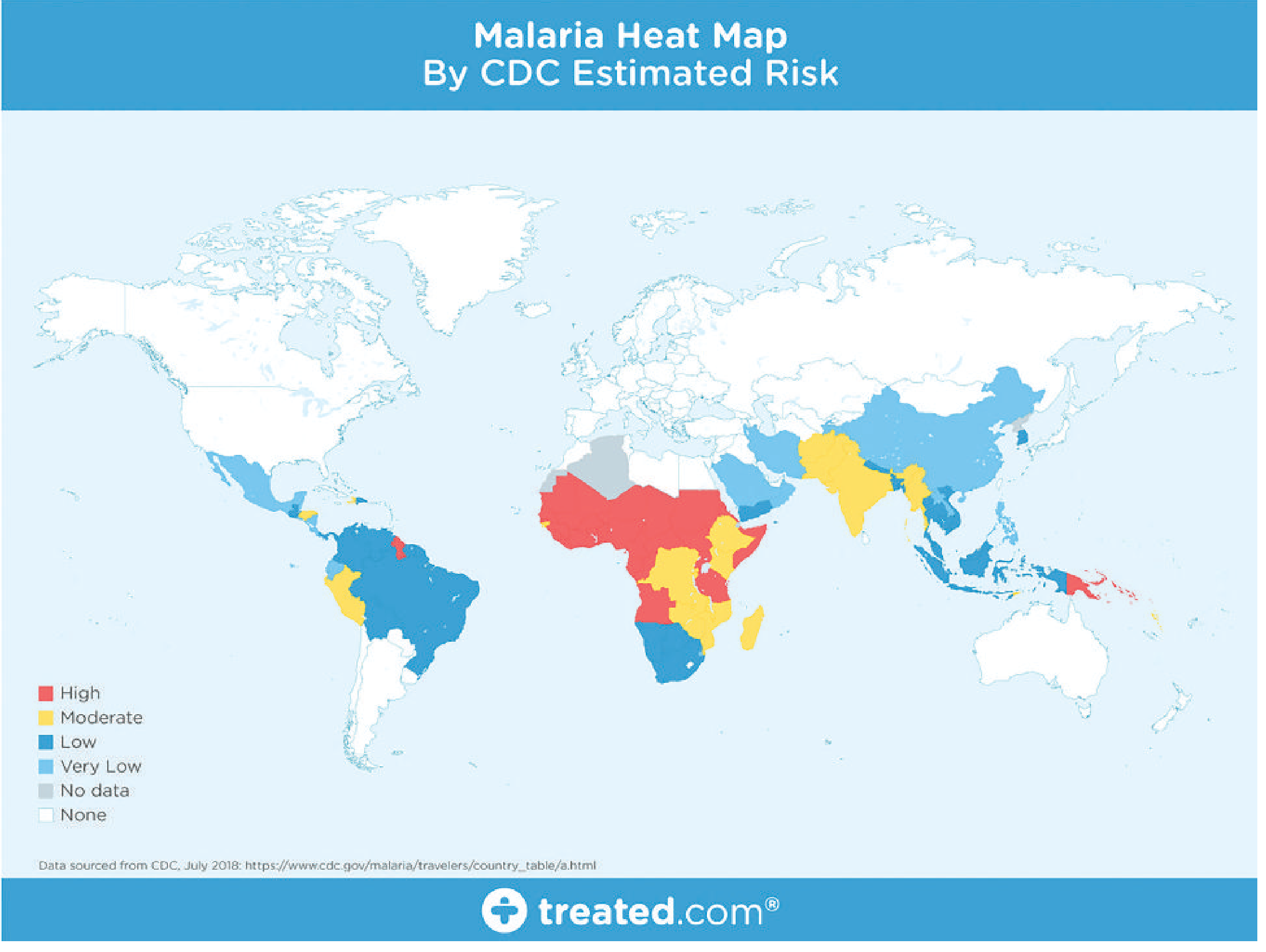
The protozoa of blood and tissues are closely related to the intestinal protozoan parasites in practically all aspects except for their sites of infection (Box 74-1). Malaria parasites (Plasmodium spp.) infect both blood and tissues. • Plasmodium Species Plasmodia are coccidian or sporozoan parasites of red blood cells, and as seen with other coccidia, they require two hosts: the mosquito for the sexual reproductive stages and humans and other animals for the asexual reproductive stages. Infection with Plasmodium spp. (i.e., malaria) accounts for 1 to 5 billion febrile episodes and 1 to 3 million deaths annually, 85% of which are in Africa (Clinical Case 74-1). The five species of plasmodia that infect humans are P. falciparum, P. knowlesi, P. vivax, P. ovale, and P. malariae (Table 74-1). These species share a common life cycle, as illustrated in Figure 74-1. Human infection is initiated by the bite of an Anopheles mosquito, which introduces infectious plasmodia sporozoites via its saliva into the circulatory system. The sporozoites are carried to the parenchymal cells of the liver, where asexual reproduction (schizogony) occurs. This phase of growth is termed the exoerythrocytic cycle and lasts 8 to 25 days, depending on the plasmodial species. Some species (e.g., P. vivax, P. ovale) can establish a dormant hepatic phase in which the sporozoites (called hypnozoites or sleeping forms) do not divide. The presence of these viable plasmodia can lead to relapse of infections months to years after the initial clinical disease (relapsing malaria). The hepatocytes eventually rupture, liberating the plasmodia (termed merozoites at this stage), which in turn attach to specific receptors on the surface of erythrocytes and enter the cells, thus initiating the erythrocytic cycle. Asexual replication progresses through a series of stages (ring, trophozoite, schizont) that culminates in rupture of the erythrocyte, releasing up to 24 merozoites, which initiates another cycle of replication by infecting other erythrocytes. Some merozoites also develop within erythrocytes into male and female gametocytes. If a mosquito ingests mature male and female gametocytes during a blood meal, the sexual reproductive cycle of malaria can be initiated, with eventual production of sporozoites infectious for humans. This sexual reproductive stage within the mosquito is necessary for the maintenance of malaria within a population. Most malaria seen in the United States is acquired by visitors or residents of countries with endemic disease (imported malaria). However, the appropriate vector, the Anopheles mosquito, is found in several sections of the United States, and domestic transmission of disease has been Table 74-1 Human Malarial Parasites Parasite Disease Plasmodium vivax Benign tertian malaria P. ovale Benign tertian or ovale malaria P. malariae Quartan or malarial malaria P. falciparum Malignant tertian malaria P. knowlesi Simian malaria or quotidian malariaobserved (introduced malaria). In addition to transmission by mosquitos, malaria can be acquired by blood transfusions from an infected donor (transfusion malaria). This type of transmission can also occur among injection drug users who share needles and syringes (“mainline” malaria). Congenital acquisition, although rare, is also a possible mode of transmission (congenital malaria). Plasmodium falciparum Physiology and Structure P. falciparum demonstrates no selectivity in host erythrocytes and invades any red blood cell (RBC) at any stage in its existence. Also, multiple merozoites can infect a single erythrocyte. Thus three or even four small rings may be seen in an infected cell (Figure 74-2). P. falciparum is often seen in the host cell at the very edge or periphery of the cell membrane, appearing almost as if it were “stuck” on the outside of the cell (see Figure 74-2). This is called the appliqué or accolé position and is distinctive for this species. Growing trophozoite stages and schizonts of P. falciparum are rarely seen in blood films because their forms are sequestered in the liver and spleen. Only in very heavy infections are they found in the peripheral circulation. Thus peripheral blood smears from patients with P. falciparum malaria characteristically contain only young ring forms and occasionally gametocytes. The typical crescentic gametocytes are diagnostic for the species (Figure 74-3). Infected RBCs do not enlarge and become distorted as they do with P. vivax and P. ovale. Occasionally, reddish granules known as Maurer dots are observed in P. falciparum. P. falciparum, similar to P. knowlesi and P. malariae, does not produce hypnozoites in the liver. Relapses from the liver are not known to occur. Epidemiology P. falciparum occurs almost exclusively in tropical and subtropical regions. Co-infection with human immunodeficiency virus (HIV) is common in these regions and may pose a risk factor for severe malaria. Clinical Syndromes The incubation period of P. falciparum is the shortest of all the plasmodia, ranging from 7 to 10 days, and does not extend for months to years. After the early influenza-like symptoms, P. falciparum rapidly produces daily (quotidian) chills and fever as well as severe nausea, vomiting, and diarrhea. The periodicity of the attacks then becomes tertian (36 to 48 hours), and fulminating disease develops. The term malignant tertian malaria is appropriate for this infection. Because the symptoms of this type of malaria are similar to those of intestinal infections, the nausea, vomiting, and diar-rhea have led to the observation that malaria is “the malignant mimic.” Although any malarial infection may be fatal, P. falciparum is the most likely to result in death if left untreated. The increased numbers of erythrocytes infected and destroyed result in toxic cellular debris, adherence of RBCs to vascular endothelium and adjacent RBCs, and formation of capillary plugging by masses of RBCs, platelets, leukocytes, and malarial pigment. Involvement of the brain (cerebral malaria) is most often seen in P. falciparum infection. Capillary plugging from an accumulation of malarial pigment and masses of cells can result in coma and death. Kidney damage is also associated with P. falciparum malaria, resulting in an illness called blackwater fever. Intravascular hemolysis with rapid destruction of RBCs produces a marked hemoglobinuria and can result in acute renal failure, tubular necrosis, nephrotic syndrome, anLiver involvement is characterized by abdominal pain, vomiting of bile, severe diarrhea, and rapid dehydration. Laboratory Diagnosis Thick and thin blood films are searched for the characteristic rings of P. falciparum, which frequently occur in multiples within a single cell, as well as in the accolé position (see Figure 74-2). Also diagnostic are the distinctive crescentic gametocytes (see Figure 74-3). A high-grade parasitemia (>10% of RBCs infected) consisting only of ring forms is suggestive of P. falciparum infection even if no gametocytes are observed. Laboratory personnel must perform a thorough search of the blood films because mixed infections can occur with any combination of the four species, but most often the combination is P. falciparum and P. vivax. Detection and proper reporting of a mixed infection directly affect the treatment chosen. Increasingly, antigen detection using a rapid diagnostic test (RDT) is being used both in the field and in diagnostic laboratories as an adjunct to conventional microscopic diagnosis. RDTs use immunochromatographic lateral-flow strip technology and use monoclonal antibodies directed at either species-specific or pan-Plasmodium targets. These tests are simple, rapid (results in < 20 minutes), and inexpensive. P. falciparum–specific monoclonal antibodies have been developed for histidine-rich protein 2 (HRP-2) and P. falciparum lactate dehydrogenase. Targets conserved across all human malarias (panmalarial antigens) have been identified on Plasmodium lactate dehydrogenase (PLDH) and aldolase enzymes. Thus far, one RDT has been approved by the U.S. Food and Drug Administration (FDA): the BinaxNOW (Binax, Scarborough, Maine) Malaria test kit, based on the antigens HRP-2 and aldolase. The sensitivity and specificity of this test for detection of P. falciparum are 95% and 94%, respectively. Treatment, Prevention, and Control Treatment of malaria is based on the history regarding travel to endemic areas, prompt clinical review and differential diagnosis, accurate and rapid laboratory work, and correct use of antimalarial drugs. Because chloroquine-resistant strains of P. falciparum are present in all areas of endemicity (Africa, Southeast Asia, South America), with the exception of Central America and the Caribbean, physicians must review all current protocols for proper treatment of P. falciparum infections, noting particularly where chloroquine resistance is known to occur. If the patient’s history indicates that the origin is not from a chloroquine-resistant area, the drug of choice is either chloroquine or parenteral quinine. Patients infected with chloroquine-resistant P. falciparum (or P. vivax) may be treated with other agents, including mefloquine ± artesunate, artemether-lumefantrine, atovaquone-proguanil (Malarone), quinine, quinidine, pyrimethamine-sulfadoxine (Fansidar), and doxycycline. Because quinine and pyrimethamine-sulfadoxine are potentially toxic, they are used more often for treatment than prophylaxis. Amodiaquine, an analog of chloroquine, is effective against chloroquine-resistant P. falciparum; however, toxicity limits its use. Newer agents with excellent activity against multidrugresistant strains of P. falciparum include the phenanthrenmethanols, halofantrine and lumefantrine, and the artemisinins, artemether and artesunate, both sesquiterpene derivatives (see Chapter 72). Combinations of the rapid-acting artemisinins with an existing or newly introduced antimalarial compound have been shown to be highly effective in both treatment and control of malaria caused by P. falciparum. The rapid reduction in parasite biomass (≈108 -fold within 3 days) produced by the artemisinins leaves a relatively small number of organisms for the second agent (usually mefloquine or lumefantrine) to clear. This reduces considerably the exposure of the parasite population to mefloquine or lumefantrine, thus reducing the chance of an escape-resistant mutant arising from the infection. Combinations of artesunate and mefloquine and of artemether and lumefantrine have both been well tolerated and highly efficacious in the treatment of multidrug-resistant falciparum malaria in semiimmune and nonimmune individuals. Of concern are reports of prolonged parasite clearance times that have been observed in artesunate-treated patients in Western Cambodia, suggesting the possible emergence of resistance to this class of agents. Although the rationale for red cell exchange transfusion in severe malaria is compelling, there are no prospective clinical trials comparing this therapy with others. Nonetheless, red cell exchange (or whole-blood exchange), if available, should be considered in cases of severe malaria complicated by clinical signs of cerebral malaria, acute lung injury, severe hemolysis with acidemia, shock, or a high or rising level of parasitemia despite adequate intravenous antimicrobial therapy. The use of anticonvulsants (phenobarbatone) and dexamethasone in cerebral malaria is likely to be ineffective or harmful and is not recommended. When there is uncertainty whether the P. falciparum is chloroquine resistant, it is advisable to assume the strain is resistant and treat the patient accordingly. If the laboratory reports a mixed infection involving P. falciparum and P. vivax, the treatment must eradicate not only P. falciparum from the erythrocytes but also the liver stages of P. vivax to avoid relapses. Failure on the part of the laboratory to detect and report such a mixed infection can result in inappropriate treatment and unnecessary delay in accomplishing a complete cure. Chemoprophylaxis and prompt eradication of infections are critical in breaking the mosquito-human transmission cycle. Control of mosquito breeding and protection of individuals by screening, netting, protective clothing, and insect repellents are also essential. Chloroquine resistance complicates the management of these patients but can be overcome by the physician’s awareness of appropriate regimens. Immigrants from and travelers to endemic areas must be carefully screened using blood films or serologic tests to detect possible infection. The development of vaccines to protect persons living in or traveling to endemic areas is under investigation. Plasmodium knowlesi Physiology and Structure Plasmodium knowlesi is a malaria parasite of Old World monkeys (long-tailed [Macaca fasicularis] and pig-tailed [Macaca nemestrina] macaques). P. knowlesi is transmimosquitoes that resides in the upper canopy of the forests and has infrequent contact with humans. Unlike other primate malarias, P. knowlesi exhibits a relaxed host specificity and is permissive in humans under natural and experimental conditions as well as in nonhuman primates. Similar to P. falciparum, the erythrocyte invasion by P. knowlesi is not restricted to young or old RBCs, which allows the development of high levels of parasitemia. It has a short life cycle of 24 hours (quotidian), and the development of the parasite in RBCs is not synchronous. P. knowlesi infection is usually misidentified as P. falciparum or P. malariae because its early trophozoitxplainhishapterr