


tell me about the mri with clear explaination with diagram and image to understand and also suggest me treatment
lumbar disc herniation L4-L5 nerve root compression MRI
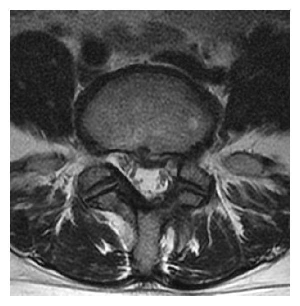
An axial-view T2-weighted magnetic resonance image (MRI) of the lumbar spine at the L4-L5 level. The image demonstrates a herniated nucleus pulposus (HNP) situated in the left lateral recess. The herniation presents as a soft-tissue protrusion extending from the intervertebral disc into the spinal canal, causing significant focal compression and displacement of the left L5 nerve root. Visible anatomical landmarks include the anterior vertebral body, the central spinal canal, the neural foramina, and the posterior elements including the lamina and spinous process. High-intensity signal suggests the acute or recurrent nature of the disc material. This diagnostic image illustrates common pathophysiology associated with lumbar radiculopathy and sciatica, specifically highlighting how localized disc displacement results in neurovascular compromise within the lateral recess.
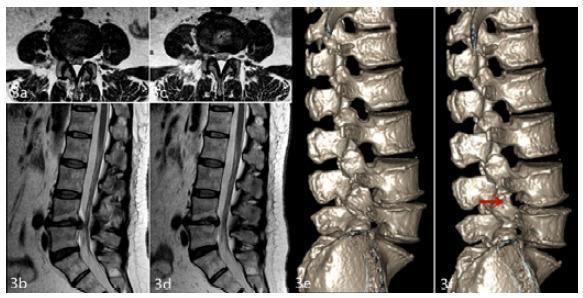
This composite diagnostic image illustrates a clinical case of lumbar disc herniation at the L4-L5 level in a female patient, showcasing pre-operative and post-operative status through multiple imaging modalities. The pre-operative axial and sagittal T2-weighted MRI scans (a, b) demonstrate a right paracentral disc herniation causing significant compression of the L5 nerve root and thecal sac. The 12-month post-operative MRI scans (c, d) show complete resolution of the herniated nucleus pulposus and restoration of the spinal canal space, effectively relieving neural compression. Panels (e) and (f) provide three-dimensional CT reconstructions of the lumbar spine, offering a lateral view of the vertebral architecture. The post-operative reconstruction (f) features a red arrow indicating the enlargement of the L4-L5 intervertebral foramen, a direct result of surgical foraminoplasty. These images collectively provide a comprehensive comparison of disc morphology, canal patency, and foraminal decompression, serving as an educational resource for spinal surgery outcomes and radiological assessment of degenerative disc disease.
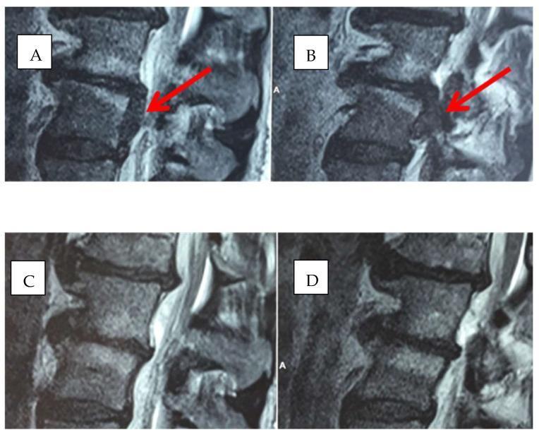
Diagnostic sagittal and axial magnetic resonance imaging (MRI) of the lumbar spine, specifically focusing on the L4-L5 segment. Images A and B represent the baseline state, demonstrating a focal posterior disc herniation (indicated by red arrows) that causes significant encroachment on the neural foramen and potential compression of the traversing nerve root. The intervertebral disc at L4-L5 shows a distinct protrusion beyond the vertebral margins. Images C and D are follow-up scans of the same anatomical region after oxygen-ozone and nutraceutical treatment (ALA, PEA, Myrrh). These post-treatment images show a significant reduction in the size and protrusion of the herniated disc material. The neural foramen appears more patent, with restored epidural fat space and improved alignment of the posterior disc margin. This comparison illustrates the radiological resolution of lumbar disc herniation and the decompression of associated spinal neural structures over time.
spinal canal stenosis thecal sac compression diagram anatomy
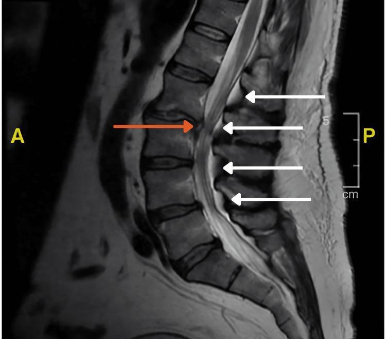
This sagittal T2-weighted MRI scan of the lumbosacral spine demonstrates multilevel thecal sac compression. Key diagnostic features include spinal epidural lipomatosis, characterized by an abnormal accumulation of hyperintense fat in the dorsal epidural space (indicated by white arrows), which narrows the spinal canal and displaces the thecal sac anteriorly. Additionally, a focal L2-L3 intervertebral disc herniation is visible (indicated by the orange arrow), further contributing to canal stenosis by protruding posteriorly. The vertebral bodies show mild degenerative changes, and the overall image illustrates the combined mechanical impact of epidural fat deposition and disc pathology on the neural structures within the lumbar spinal canal. This imaging is characteristic of conditions causing radiculopathy or neurogenic claudication, and serves as an educational example of extrinsic compression of the thecal sac.
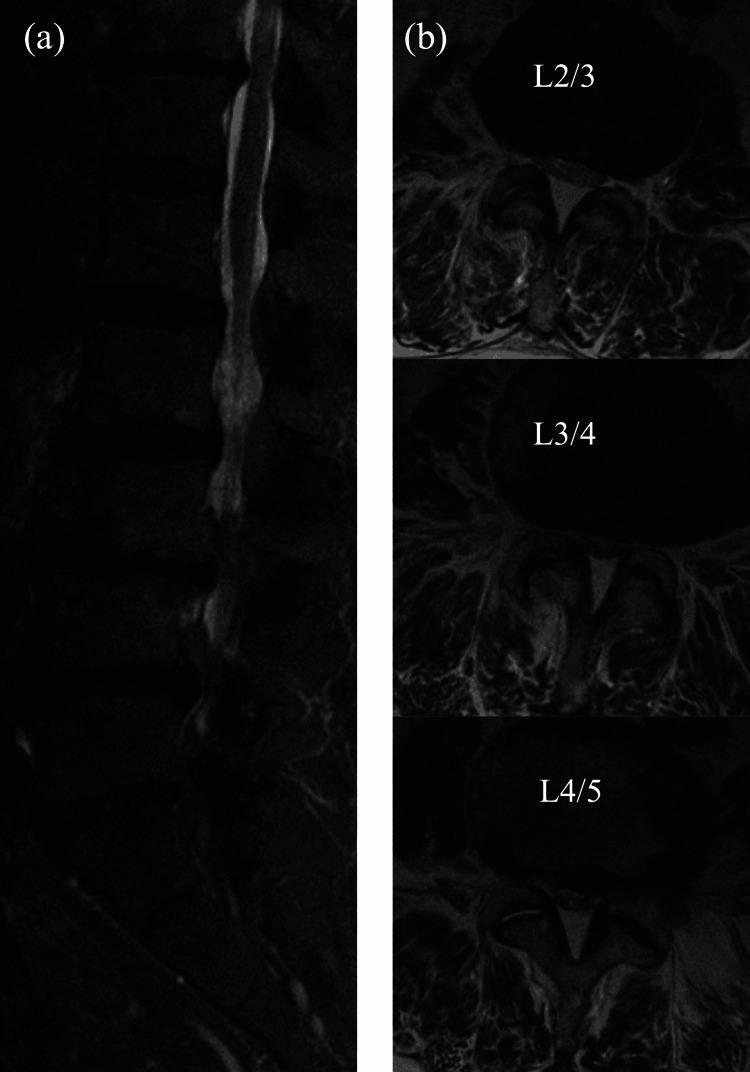
Diagnostic Image: Magnetic Resonance Imaging (MRI) of the lumbar spine. (a) Sagittal plane MRI showing the longitudinal thecal sac. There is evidence of multi-level spinal canal narrowing, particularly notable for areas of relative expansion and hyperintensity in the CSF space between segments of narrowing, indicating multilevel stenosis. (b) Axial plane MRI slices at L2/3, L3/4, and L4/5 levels. At L2/3 and L3/4, the spinal canal appears constricted with heterogeneous signal intensity, suggesting compression of the cauda equina. At the L4/5 level, a distinct 'Y-shaped' hyperintense configuration is visible within the canal, characteristic of dural foldings or anatomical variations associated with high-grade stenosis or post-operative changes. This imaging is clinically relevant for evaluating lumbar spinal stenosis and the risk factors for postoperative spinal epidural hematoma (POSEH). It demonstrates structural changes in the dural sac morphology across different vertebral levels, serving as an educational resource for orthopedic surgeons and radiologists in assessing spinal canal patency.
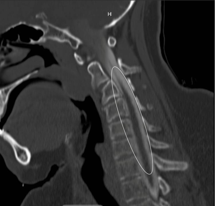
This diagnostic image is a CT cervical myelogram in the sagittal view, demonstrating the anatomy and pathology of the cervical spine from C1 through the upper thoracic vertebrae. The thecal sac is opacified with hyperdense contrast material (appearing bright white), allowing for clear visualization of the spinal cord (appearing relatively hypodense) and the surrounding subarachnoid space. In the highlighted area from C3 to C7, multiple mild disc bulges are visible as focal indentations on the anterior aspect of the contrast-filled thecal sac. The vertebral bodies maintain their rectangular morphology, and the alignment of the anterior and posterior longitudinal ligaments appears intact. The spinal cord itself shows no significant displacement or focal signal changes in this view, and the neural foramina are not significantly stenosed. This imaging modality is essential for evaluating disc-related compression, canal stenosis, and the relationship between the intervertebral discs and the central nervous system structures in patients with neck pain or radiculopathy.
lumbar spinal stenosis treatment physiotherapy exercises
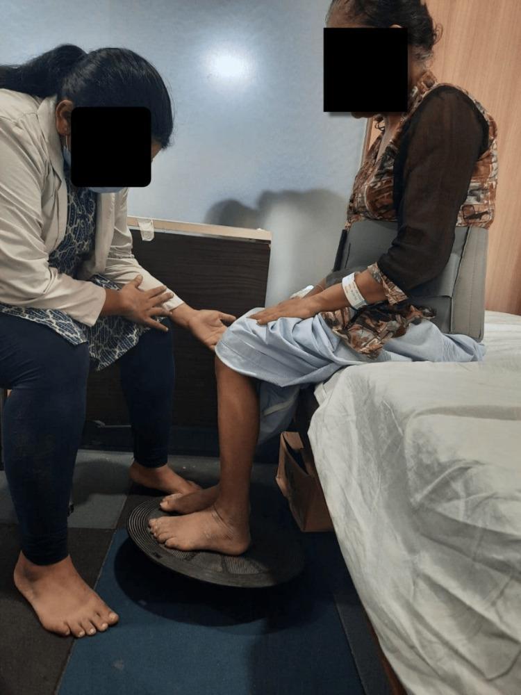
A clinical photograph capturing a postoperative physiotherapy session for a patient following lumbar spine surgery. The patient is seated at the edge of an examination bed, wearing a rigid lumbosacral orthosis (back brace) for spinal stability. The patient's bare feet are positioned on a black circular wobble board placed on a blue non-slip floor mat. A physiotherapist is positioned in a semi-kneeling stance to the patient's side, providing manual guidance and tactile cues at the patient's knee and arm to assist with balance. This setup illustrates a seated proprioceptive and neuromuscular retraining exercise, designed to improve core stability and lower extremity motor control while minimizing spinal load during early mobilization. The context suggests rehabilitation following L3-S1 decompression and fusion for spinal stenosis.
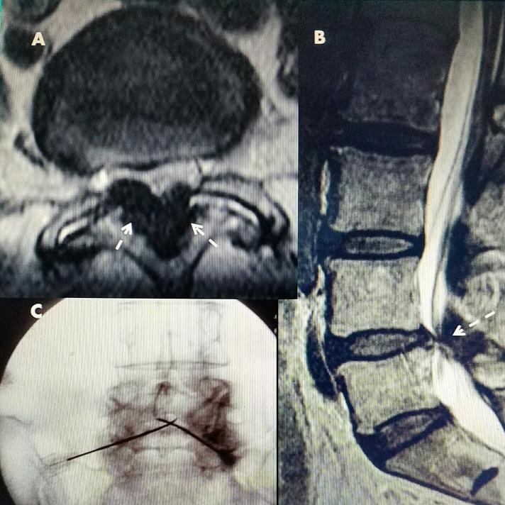
This composite figure illustrates L4-L5 lumbar spinal stenosis and a corresponding radiofrequency (RF) treatment procedure. Panel A is an axial CT image showing marked bilateral ligamentous hypertrophy (white arrows), appearing hypointense and significantly narrowing the spinal canal from the posterior aspect. Panel B is a sagittal T2-weighted MRI demonstrating posterior compression of the thecal sac at the L4-L5 level caused by ligamentous stenosis. Panel C is an anteroposterior (AP) fluoroscopic image showing the percutaneous placement of two RF electrodes. The electrodes are positioned with an inferior-lateral to superior-medial trajectory, meeting within the posterior canal targeting the hypertrophied ligamentum flavum. This instructional sequence highlights the diagnostic radiological findings of soft tissue spinal stenosis and the subsequent targeted interventional approach for decompression using RF thermal ablation.
cervical spine straightening loss of lordosis osteophytes
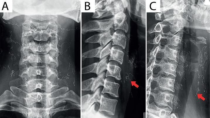
This composite diagnostic image presents three radiographic views of the cervical spine: (A) Anteroposterior (AP), (B) Lateral, and (C) Oblique. The lateral view demonstrates a significant loss of the normal cervical lordosis, indicative of straightening of the spine. Degenerative changes are visible, including reduced intervertebral disc space height at the C4-C5 level and marginal osteophytes at the C5-C7 vertebrae. Most notably, all three views reveal multiple, thin, radiopaque string-like foreign materials (indicated by red arrows in B and C) distributed in a mesh-like pattern over the anterior and bilateral soft tissues of the neck. These findings are characteristic of cosmetic gold thread implantation (gold thread lift). The image is relevant for radiology and musculoskeletal educational indexing, illustrating both degenerative spondylosis and the radiographic appearance of subcutaneous cosmetic implants.
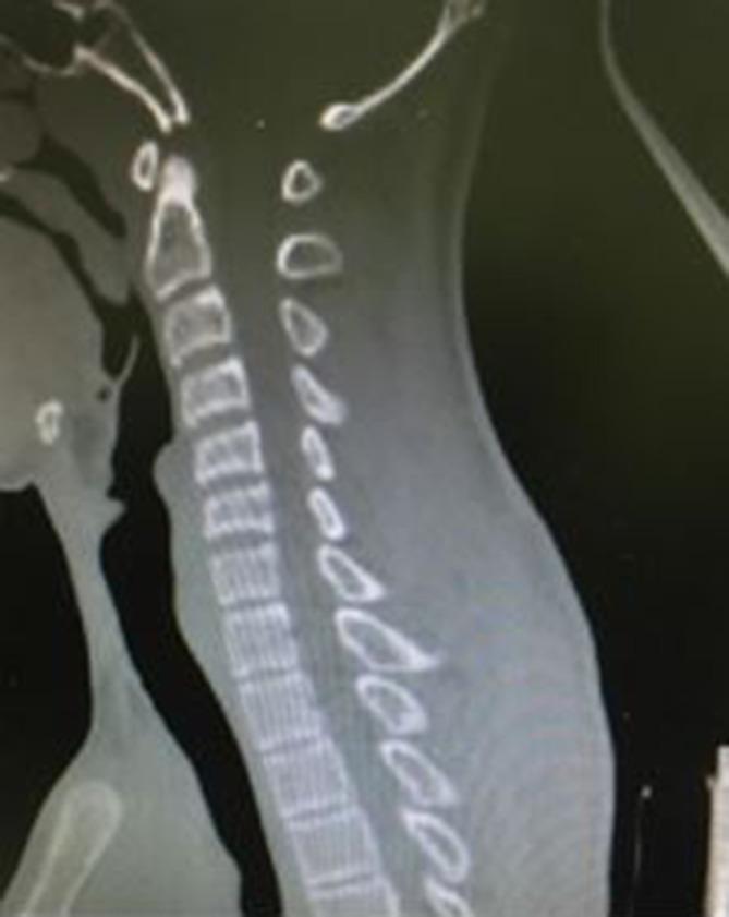
This diagnostic image is a sagittal reformatted Computed Tomography (CT) scan of the cervical spine. The primary finding is a notable straightening of the cervical spine, representing a loss of the normal physiological cervical lordosis. The vertebral bodies and spinous processes appear intact, with no evidence of gross fracture, subluxation, or dislocation visible in this specific plane. The alignment of the vertebral bodies is linear rather than curved. In a clinical context, such straightening can be associated with acute muscle spasms, ligamentous injury, or post-traumatic guarding. This image serves as a baseline assessment for cervical spinal trauma and alignment, highlighting the importance of evaluating the curvature of the spine alongside bony integrity in radiological examinations.
MRI Report Explained - Mr. Mangat Ram (64 yrs, Male)
🔬 What is a "Whole Spine MRI"?
📋 FINDINGS - Region by Region
1. CERVICAL SPINE (Neck - C1 to C7)
- Straightening of cervical spine
- Marginal osteophytes and disc-osteophyte complexes at multiple levels
- C3-C4 and C4-C5: Disc-osteophyte complex + disc bulge causing indentation on the thecal sac
2. DORSAL (THORACIC) SPINE (Mid-Back - T1 to T12)
- Marginal osteophytes and Modic Type II changes at multiple levels
- Normal spinal cord signal - no damage to the cord
- Vertebral heights maintained (no collapsed bones)
3. LUMBAR SPINE (Lower Back - L1 to S1) ⚠️ MOST IMPORTANT AREA
⚠️ L4-L5 (Most Critical Finding)
- Central-paracentral disc protrusion
- Facet joint arthropathy (arthritis)
- Ligamentum flavum thickening
- Together causing compression of the thecal sac and right traversing nerve root compression
- Lower back pain
- Right leg pain, tingling, or numbness (sciatica)
- Possible weakness in the right leg
⚠️ L5-S1 (Second Important Level)
- Diffuse disc bulge compressing the thecal sac
- Bilateral lateral recess narrowing and neural foramina narrowing
4. OTHER NORMAL / REASSURING FINDINGS
| Finding | What it means |
|---|---|
| Cord ends at L1 (normal) | Spinal cord is the right length, no tethering |
| No intraspinal mass | No tumor inside the spinal canal |
| No pre/paravertebral collection | No abscess or fluid collection near the spine |
| Facet joints & ligamentum flavum normal at L5-S1 | The rear joints at this level are not arthritic |
🧠 SUMMARY OF IMPRESSIONS (Doctor's Final Conclusion)
| # | Finding | Severity |
|---|---|---|
| 1 | L4-L5: Disc protrusion + facet arthropathy + ligamentum flavum thickening → right nerve root compression | High (most symptomatic) |
| 2 | L5-S1: Diffuse disc bulge → bilateral foraminal narrowing | Moderate |
| 3 | C3-C4, C4-C5: Disc-osteophyte complex → thecal sac indentation | Mild-Moderate |
| 4 | Whole spine: Marginal osteophytes + Modic II changes (degenerative changes) | Mild (age-related) |
💊 TREATMENT OPTIONS
Important Disclaimer: This is an educational explanation only. Treatment decisions must be made by your treating doctor (Dr. Rahul Khinchi) who referred this MRI, based on your clinical symptoms and physical examination. Do NOT start or stop any treatment based on this alone.
Step 1 - Conservative (Non-Surgical) Treatment - Try This First
A. Medications
| Medicine Type | Purpose |
|---|---|
| NSAIDs (e.g., ibuprofen, diclofenac, etoricoxib) | Reduce disc inflammation and pain |
| Neuropathic agents (e.g., pregabalin, gabapentin) | Reduce nerve pain (tingling/numbness down the leg) |
| Muscle relaxants (e.g., thiocolchicoside, baclofen) | Relieve back muscle spasms |
| Oral steroids (short course, if acute flare) | Reduce swelling around compressed nerve |
| Calcium + Vitamin D + Vitamin B12 | Support bone and nerve health |
B. Physiotherapy (Very Important)
- McKenzie exercises - gentle extension-based exercises for lumbar disc herniation
- Core strengthening - planks, bird-dog exercises to stabilize the spine
- Cervical traction - gentle stretching to relieve neck disc pressure at C3-C4, C4-C5
- Hot/cold packs - for pain relief
- Postural correction - very important; avoid prolonged sitting, use lumbar support
C. Lifestyle Modifications
- Lose weight if overweight - every kilogram lost reduces spinal load significantly
- Avoid prolonged sitting, bending forward, lifting heavy objects
- Sleep on a firm mattress with a pillow under knees (supine) or between knees (side-lying)
- Use a lumbar support belt during activities
Step 2 - Interventional (Minimally Invasive) - If Conservative Fails After 6-8 Weeks
- Epidural steroid injection - a corticosteroid injection directly into the epidural space around L4-L5 or L5-S1 to reduce nerve inflammation. Provides relief in 60-80% of patients
- Facet joint injections / medial branch blocks - for the facet arthropathy component at L4-L5
- Radiofrequency ablation of ligamentum flavum - a newer technique to shrink the thickened ligament
Step 3 - Surgery - Only if Conservative + Interventional Treatment Fails
- Progressive weakness in the leg (foot drop)
- Loss of bladder or bowel control (cauda equina syndrome - emergency)
- Severe unremitting pain not responding to 3+ months of treatment
- Significant functional disability
| Surgery | For Which Level |
|---|---|
| Microdiscectomy (keyhole disc removal) | L4-L5 disc protrusion - right side |
| Laminectomy / Decompression | L4-L5 and/or L5-S1 canal narrowing |
| Anterior Cervical Discectomy and Fusion (ACDF) | C3-C4, C4-C5 if neck symptoms worsen |
| Spinal fusion (if instability) | If vertebral instability is found on dynamic X-rays |
⚠️ Warning Signs - Go to Hospital Immediately if You Have:
- Sudden weakness or paralysis of both legs
- Loss of control over urine or stools
- Numbness in the inner thighs/saddle area
- Rapidly worsening neurological symptoms
📅 Recommended Next Steps
- See your referring doctor (Dr. Rahul Khinchi) immediately to correlate these findings with your symptoms
- Ask for referral to a spine specialist (neurosurgeon or orthopedic spine surgeon)
- Start physiotherapy under a qualified physiotherapist
- Bring the MRI films (actual images) to the specialist, not just this report