
Sx → dx → pseudo obstruction. Tx ✓, still Sx + diarrhea cx of diarrhea? OP! → What to do next ⓐ Ba ⓑ CT ⓒ Culture ⓓ Parasite test
Clinical Scenario Breakdown
Why (c) Culture / C. difficile Testing is Correct
- Pseudo-obstruction treatment involves neostigmine and/or antibiotics for preparation/prevention
- Any antibiotic exposure disrupts normal enteric flora and can trigger C. difficile overgrowth
- The patient now has post-treatment diarrhea - the most classic complication
"Clostridium difficile colitis is extremely common and is the leading cause of nosocomially acquired diarrhea. Colitis is thought to result from overgrowth of this organism after depletion of the normal commensal flora of the gut with the use of antibiotics... even a single dose of an antibiotic may cause the disease."
"The greatest concern in postoperative patients is pseudomembranous colitis... Pseudomembranous colitis is due to the toxin produced by Clostridium difficile. The current recommendation for symptomatic patients is to use a nucleic acid amplification test (NAAT/PCR)."
Why the Other Options Are Wrong
| Option | Why Wrong |
|---|---|
| (a) Barium enema | Contraindicated in active/suspected colitis - risk of perforation, and diagnosis is not the priority here |
| (b) CT scan | Useful if you suspect perforation or severe colitis, but not the first step in an OP (outpatient) setting with simple diarrhea post-treatment |
| (d) Parasite test | No travel history or immunocompromise context given; C. diff is far more likely post-antibiotic treatment |
Next Steps After Confirming C. difficile
- Stop the offending antibiotic (if still on one)
- Mild disease (outpatient): Oral metronidazole x 10 days
- Severe disease: Oral vancomycin
- NAAT/PCR is the preferred diagnostic test (more sensitive/specific than enzyme immunoassay)
- Schwartz's Principles of Surgery, 11th Ed., Infectious Colitis section
- Tintinalli's Emergency Medicine, Complications of Drug Therapy section
Girl – Mexico. Sx Abd pain 2 days ago is relieved → came back, admitted to emergency department Cx?
Diagnosis: Perforated Appendicitis
Why "Mexico" + "Girl" matters
The Classic 3-Phase Pain Pattern of Perforation
| Phase | What happens | Time |
|---|---|---|
| Phase 1 | Visceral pain - periumbilical crampy pain from luminal obstruction of the appendix | Hours 1-12 |
| Phase 2 - "RELIEF" | Perforation occurs → intraluminal pressure is suddenly released → pain temporarily disappears - this is the dangerous "false relief" that delays presentation | ~Day 1-2 |
| Phase 3 - "RETURNS" | Fecal/purulent content spills into peritoneum → generalized peritonitis → severe diffuse pain returns, patient comes to ED | Day 2+ |
"The release of intraluminal obstruction with perforation often results in sudden alleviation of pain; consider appendiceal perforation if the patient's pain has suddenly improved."
"Girl - Mexico" - Additional Differentials to Rule Out
| Differential | Why consider | Why less likely here |
|---|---|---|
| Ruptured ectopic pregnancy | Young female, RLQ pain | No mention of missed period / hCG |
| Amoebic abscess (E. histolytica) | Mexico = endemic area | Subacute course, no dysentery described |
| Ovarian torsion / ruptured cyst | Young female | Does not follow the 2-day pain→relief→return pattern |
| Mesenteric adenitis | Common in girls | Self-limiting, not this severe course |
What to do in the ED
- NPO + IV fluids + antibiotics (gram-negative + anaerobic coverage: pip/tazo or cefoxitin + metronidazole)
- CT abdomen/pelvis with contrast - confirms free air, periappendiceal fluid, abscess
- Urgent surgical consult for emergency appendectomy (or percutaneous drainage if contained abscess)
- Beta-hCG to rule out ectopic pregnancy in any female of reproductive age
- Tintinalli's Emergency Medicine, Appendicitis - Clinical Features
Radiologic tests are useful in all of the suspected conditions EXCEPT: A. Perforated duodenal ulcer B. Acute cholelithiasis C. Renal colic D. Ruptured aortic aneurysm E. Torsion of an ovarian cys
Answer: A. Perforated Duodenal Ulcer
Why Perforated Duodenal Ulcer is the Exception
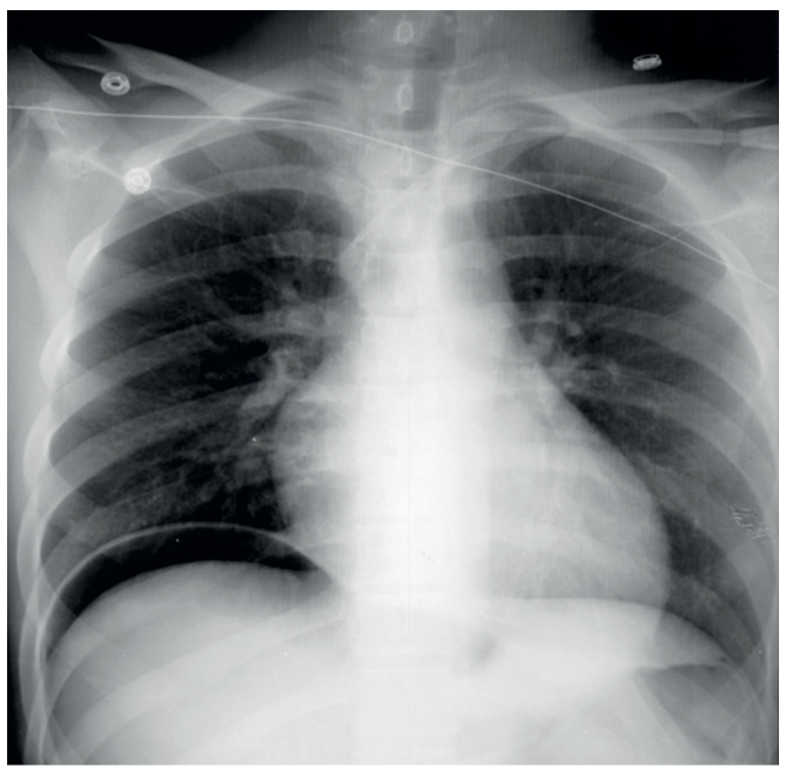
Why Radiology IS Useful in All Other Options
| Condition | Imaging modality | What it shows |
|---|---|---|
| B. Acute cholelithiasis | Ultrasound (first-line) | Gallstones, gallbladder wall thickening, pericholecystic fluid, Murphy's sign on US |
| C. Renal colic | CT KUB (non-contrast) | Stone location, size, degree of obstruction, hydronephrosis |
| D. Ruptured aortic aneurysm | CT angiography (if stable) / Bedside US (if unstable) | Aortic diameter, retroperitoneal hematoma, extravasation |
| E. Torsion of ovarian cyst | Transvaginal Doppler US (first-line) | Enlarged ovary, twisted pedicle, absent/reduced blood flow |
"The diagnosis of ovarian torsion is challenging as clinical symptoms are neither sensitive nor specific and there are no definitive criteria with imaging. Surgical diagnosis (laparoscopy or laparotomy) remains the diagnostic and therapeutic method of choice if there is suspicion of ovarian torsion."
Key Takeaway
- Diagnosis is primarily clinical (sudden rigid abdomen)
- Free air on CXR is absent in ~30-50% of cases - so negative imaging cannot rule it out
- A positive clinical picture = emergent surgery regardless of imaging result
- Radiology confirms but does not diagnose - making it the least "useful" among the options
- Bailey and Love's Short Practice of Surgery, 28th Ed., Fig. 67.22
- Berek & Novak's Gynecology, Ovarian Torsion - Diagnosis
A 24-year-old female presents to the emergency room with a 24 hour history of pain and nausea. The pain originally started in the periumbilical area and has now localized to the right lower quadrant. Her temperature is 38.50C, blood pressure is 120/50, pulse is 110. On examination, her abdomen is slightly distended, and bowel sounds are hypoactive. She is markedly tender in the right lower quadrant with voluntary guarding. Her white blood cell count was 7,000 with 76% neutrophils. Which of the following facts from the patient would be helpful in formulating a differential diagnosis? A. date of her last menstrual period B. whether she is sexually active C. history of sexually transmitted diseases in the past D. history of prior abdominal surgeries E. History of similar episodes similar to this one F. All of the above G. A,C and D H. B,C and E
Answer: F. All of the above
The Clinical Picture First
| All patients | Female-specific additions |
|---|---|
| Appendicitis | Ectopic pregnancy |
| Gastroenteritis | Ovarian torsion |
| IBD | Pelvic inflammatory disease (PID) |
| Bowel obstruction | Ovarian cyst |
| Nephrolithiasis |
Why Each Historical Fact Matters
- Rules in/out ectopic pregnancy - the single most dangerous mimic; a missed period + RLQ pain = ectopic until proven otherwise
- If LMP is late → serum beta-hCG immediately
- Also helps evaluate mittelschmerz (mid-cycle ovulatory pain) or corpus luteum cyst rupture
- Unlocks the question of ectopic pregnancy (requires sexual activity)
- Also raises the prior probability of PID/salpingitis (which can perfectly mimic appendicitis with RLQ pain, fever, leukocytosis)
- If NOT sexually active, ectopic and PID become far less likely, pushing appendicitis to the top
- Prior chlamydia or gonorrhea = major risk factor for PID and tubo-ovarian abscess (TOA)
- Prior STDs also cause tubal scarring, raising risk of ectopic pregnancy significantly
- Fitz-Hugh-Curtis syndrome (perihepatitis from PID) can also cause RUQ or referred RLQ pain
- Prior surgery → adhesions → risk of small bowel obstruction (explains pain, distension, hypoactive bowel sounds in this patient)
- Prior appendectomy would obviously eliminate appendicitis from the differential entirely
- Prior gynecologic surgery raises the risk of adhesion-related complications
- Recurrent similar episodes argue against acute appendicitis (which is progressive and does not resolve spontaneously in classic form) and for:
- Recurrent ovarian cysts with intermittent rupture or torsion
- IBD (Crohn's disease commonly causes RLQ pain with remission/relapse)
- Chronic PID with acute exacerbation
- Irritable bowel syndrome or mesenteric adenitis
Why the WBC Is a Red Herring Here
Bottom Line
- Rosen's Emergency Medicine, Table 79.3 - Differential Considerations in Appendicitis Evaluation
Current recommendations from the American Cancer A. B. Society for screening of colon cancer are: Digital rectal exam and occult blood testing beginning at age 55 Flexible sigmoidoscopy at the age of 50 for anyone at increased risk C. Five consecutive negative sigmoidoscopies, followed by endoscopy every three years D. Yearly physical examinations by a physician including blood work E. Screening CEA levels over the age of 50 F. All of the above G. A,B and C H. B and D
American Cancer Society colon cancer screening recommendations 2024 guidelines age 45
Answer: A - with important caveats (this is an outdated question)
Evaluating Each Option Against Current ACS Guidelines
| Option | Statement | Verdict |
|---|---|---|
| A | Digital rectal exam + occult blood testing beginning at age 55 | WRONG age - should be 45 (updated 2018); DRE alone is not a recommended screening strategy |
| B | Flexible sigmoidoscopy at age 50 for increased risk | WRONG - increased risk patients often need to start earlier than 50, not at 50; also age is now 45 for average risk |
| C | Five consecutive negative sigmoidoscopies → endoscopy every 3 years | WRONG - not an actual guideline; colonoscopy is every 10 years, not every 3 years after sigs |
| D | Yearly physical exam including blood work | WRONG - not a CRC screening strategy |
| E | Screening CEA levels over age 50 | WRONG - CEA is a tumor marker for monitoring, NOT for screening |
What the ACS Actually Recommends (Current 2018-updated guidelines)
- Colonoscopy every 10 years (gold standard)
- Annual high-sensitivity guaiac FOBT (gFOBT) or FIT
- Multi-target stool DNA (Cologuard) every 1-3 years
- CT colonography every 5 years
- Flexible sigmoidoscopy every 5 years
- Flexible sigmoidoscopy every 10 years + annual FIT
Why Each Distractor Fails
- Monitor response to treatment in known CRC
- Detect recurrence post-resection
- Baseline before surgery CEA has poor sensitivity (~30-40%) and specificity for early CRC - making it useless for population screening.
The Intended Answer in an Exam Context
- Harrison's Principles of Internal Medicine 22nd Ed., Table 333-3
- ACS Colorectal Cancer Screening Guidelines (current: start at age 45)
The colon has all of the following anatomic characteristics, EXCEPT A. B. C. D. The left colic flexure is usually higher, has more acute angulation, and is less flexible than the right The descending colon usually lacks a mesocolon The rectosigmoid junction begins at the level of the pelvic brim The lower layer of the transverse mesocolon covers the pancreas and the lower part of duodenum E. The blood supply to the rectosigmoid region is by way of the superior rectal artery and sigmoid arteries
Answer: C. The rectosigmoid junction begins at the level of the pelvic brim
Verifying Each Statement
- Higher in position than the hepatic (right) flexure
- More acutely angled (nearly 90°)
- More fixed/less mobile - held by the phrenocolic ligament to the diaphragm
- Clinically important: makes colonoscopy passage more difficult here, and is the watershed zone for ischemic colitis (Griffiths' point)
- It defines the boundary between sigmoid colon (mobile, mesentery present) and rectum (fixed, no mesentery)
- Important for surgical planning in rectal cancer vs. sigmoid cancer
- Head of the pancreas
- Second part of the duodenum
- Anterior surface of the pancreatic body
- Sigmoid arteries (2-6 branches) arise from the inferior mesenteric artery (IMA) and supply the sigmoid colon
- Superior rectal artery is the terminal branch of the IMA, supplying the upper rectum and rectosigmoid
- The junction between superior rectal and middle rectal (from internal iliac) artery territories creates the critical watershed zone - a region of relative ischemia important in anterior resection anastomotic leak risk
Summary Table
| Option | Statement | True/False |
|---|---|---|
| A | Left flexure higher, more acute, less flexible | TRUE |
| B | Descending colon lacks mesocolon | TRUE |
| C | Rectosigmoid junction at pelvic brim | FALSE - it's at the sacral promontory |
| D | Transverse mesocolon covers pancreas/duodenum | TRUE |
| E | Rectosigmoid supplied by superior rectal + sigmoid arteries | TRUE |
Hepatitis B chronic hepatitis differs from autoimmune hepatitis is that it A. B. C. D. E. typically affects females over 30 years of age often produces acute hepatic failure is characterised by florid physical signs typically progresses slowly without exacerbations is less likely to be complicated by hepatoma
Answer: E. Is less likely to be complicated by hepatoma
Head-to-Head: Chronic HBV vs. Autoimmune Hepatitis
| Feature | Chronic HBV | Autoimmune Hepatitis | Verdict for each option |
|---|---|---|---|
| Demographics | Any age, male = female or male predominance; endemic populations | Predominantly young females (bimodal: 10-30 and 40-60 yrs); 70-80% female | A is FALSE for HBV - this describes AIH |
| Acute hepatic failure | Rare in chronic phase; acute HBV can cause ALF | AIH can present as acute liver failure in 10-40% | B is FALSE for chronic HBV - AIH more often presents acutely |
| Florid physical signs | Relatively few specific signs | AIH: cushingoid features, acne, striae, amenorrhea, arthralgia, rashes, other autoimmune features | C is FALSE for HBV - florid signs are more typical of AIH |
| Slowly progressive without exacerbations | HBV has phases: immune tolerant → immune active → inactive carrier → reactivation flares; NOT typically slow and steady | AIH tends to be more relapsing-remitting with flares too, but can have quiet periods | D is FALSE for HBV - HBV characteristically has reactivation episodes |
| Hepatoma risk | HBV = one of the strongest risk factors for HCC worldwide; can cause HCC even WITHOUT cirrhosis (direct viral oncogenesis via HBx protein integration) | AIH rarely causes HCC; risk is much lower even with cirrhosis | E is the DISTINGUISHING TRUE feature - HBV is MORE likely to cause hepatoma, not less |
Why E is the Answer
- The HBx protein integrates into the host genome and has direct oncogenic potential
- HCC can develop in chronic HBV patients even without cirrhosis (unique among hepatitis causes)
- HBV accounts for ~50% of HCC worldwide
- Autoimmune hepatitis-associated HCC requires established cirrhosis and is far less common
Summary
- Robbins, Cotran & Kumar Pathologic Basis of Disease - Autoimmune Hepatitis, Clinical Features
20-уеаr-old woman presents with abdominal pain, increasing jaundice and arthralgia. She is noted to have hepatosplenomegaly. She recently donated blood. She is found to have an increase in both conjugated and unconjugated bilirubin. Select the most likely diagnosis for this patient. A. Primary biliary cirrhosis B. Chronic active hepatitis C. Hepatitis В D. Alcoholic hepatitis E. Leptospirosis
Answer: C. Hepatitis B
Decoding the Clues in This Case
| Clue | What it points to |
|---|---|
| 20-year-old woman | Acute infectious hepatitis - common in young adults |
| Abdominal pain + increasing jaundice | Hepatocellular disease with biliary involvement |
| Arthralgia | Immune complex-mediated extrahepatic manifestation - classic for HBV |
| Hepatosplenomegaly | Active viral hepatitis with systemic involvement |
| Recently donated blood | Key clue - blood donation = HBsAg screening detected OR recent blood exposure; HBV is the hepatitis classically associated with blood/parenteral transmission |
| Both conjugated AND unconjugated bilirubin elevated | Hepatocellular disease (impaired conjugation + impaired excretion of conjugated bili) |
"Immune-mediated symptoms, including rash, hives, arthralgias, angioneurotic edema, and fever, are observed in 10 to 20% of patients during the preicteric phase."
"Physical examination reveals jaundice and hepatic tenderness. Hepatomegaly and splenomegaly may be present. On laboratory testing, acute viral hepatitis is characterized by elevated total and direct serum bilirubin levels and aminotransferase levels."
Why "Recently Donated Blood" is the Pivot Clue
- She was found to be HBsAg-positive on routine donor screening - the donation triggered the diagnosis
- She received blood products previously (though this reading is less likely given phrasing)
Eliminating the Other Options
- Affects middle-aged women (40-60 yrs), not 20-year-olds
- Presents with pruritus, fatigue, cholestatic picture (predominantly conjugated bilirubin)
- No arthralgia as a feature; anti-mitochondrial antibody (AMA) positive
- No blood donation connection
- This is a chronic condition - the question describes an acute presentation
- Chronic hepatitis does NOT typically cause the acute arthralgia + jaundice combination in this dramatic fashion
- Also, chronic hepatitis B could be a later diagnosis from this acute presentation
- 20-year-old - possible but unlikely as primary diagnosis
- Alcoholic hepatitis does NOT cause arthralgia
- AST:ALT ratio typically >2:1, but no history of alcohol use given
- No blood donation link
- Hepatosplenomegaly in a 20-year-old with no alcohol history doesn't fit
- Does cause jaundice + hepatosplenomegaly (Weil's disease)
- But requires exposure to contaminated water/animals (not blood donation)
- Presents with fever, myalgia, conjunctival suffusion - NOT arthralgia predominantly
- Renal failure is a key feature (not mentioned here)
The Bilirubin Pattern Explained
- Hepatocyte damage impairs conjugation → unconjugated bilirubin rises
- Intrahepatic cholestasis from swollen hepatocytes blocking bile canaliculi → conjugated bilirubin accumulates and regurgitates into blood
- Result: mixed hyperbilirubinemia - the hallmark of hepatocellular disease
- Pure conjugated elevation → obstructive/cholestatic (stones, PBC, PSC)
- Pure unconjugated elevation → hemolysis or Gilbert's syndrome
Answer: C. Hepatitis B
- Goldman-Cecil Medicine, Acute Viral Hepatitis - Clinical Features