
Serological markers of hepatitis b
Reading File
Finding Sources
Finding Sources
Reading File
Reading File
Finding Sources
The Sleisenger GI/Liver textbook has excellent content. Let me read the full diagnosis section and search for images.
Reading File
Finding Sources
Searching Images
hepatitis B serological markers timeline HBsAg HBeAg antibodies
Excellent content retrieved. Let me read more of the Sleisenger diagnosis section and fetch the Harrison's diagrams.
Reading File
Loading Image
Loading Image
Now let me get the detailed HBeAg section and a summary interpretation table from the textbook:
Reading File
Finding Sources
I now have comprehensive information. Let me compile the full answer.
Serological Markers of Hepatitis B
Hepatitis B virus (HBV) infection generates a characteristic sequence of antigens and antibodies in serum. Interpreting these markers allows you to determine the phase of infection, infectivity, immune status, and response to therapy.
The Key Markers at a Glance
| Marker | What it is | Clinical meaning |
|---|---|---|
| HBsAg | Surface antigen (envelope protein) | Active infection (acute or chronic) |
| Anti-HBs | Antibody to surface antigen | Immunity (resolved infection or vaccination) |
| HBcAg | Core antigen | Not detected in serum (intrahepatic only) |
| IgM Anti-HBc | IgM antibody to core antigen | Acute infection / window period marker |
| IgG Anti-HBc (Total Anti-HBc) | IgG antibody to core antigen | Past or ongoing infection |
| HBeAg | e antigen (precore protein) | Active viral replication, high infectivity |
| Anti-HBe | Antibody to e antigen | Reduced replication, seroconversion |
| HBV DNA | Viral genomic DNA (PCR) | Quantifies viral load; guides therapy |
Acute Hepatitis B — Serological Sequence
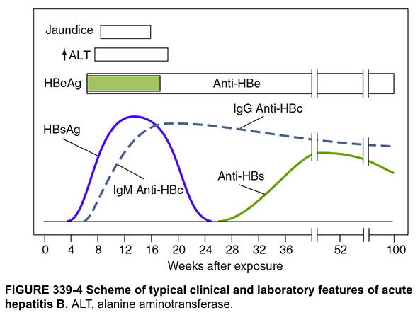
1. HBsAg appears in serum 2–10 weeks after exposure, before symptoms or ALT elevation. In self-limited infection it clears by 4–6 months. Persistence beyond 6 months = chronic infection. — Sleisenger & Fordtran's GI and Liver Disease, p. 1479
2. HBeAg appears early alongside HBsAg, indicating active replication and high infectivity. It disappears at or just after peak ALT. Persistence ≥3 months suggests progression to chronic infection.
3. IgM Anti-HBc rises with HBsAg, peaks around weeks 12–16, and persists for 4–6 months (rarely up to 2 years). This is the key marker of acute infection and the only marker detectable during the window period (when HBsAg has cleared but anti-HBs has not yet appeared). It can also re-emerge during exacerbations of chronic hepatitis B.
4. Anti-HBe appears as HBeAg clears — marking seroconversion to lower-replication state.
5. Anti-HBs appears weeks to months after HBsAg disappears. It indicates immunity and recovery. Persists for life in most patients. Also the marker produced by vaccination (anti-HBs positive, anti-HBc negative = vaccinated).
6. IgG Anti-HBc (Total Anti-HBc) replaces IgM anti-HBc and persists lifelong, indicating prior or ongoing infection.
The "Window Period"
The window period occurs between disappearance of HBsAg and appearance of anti-HBs. During this gap:
- HBsAg: negative
- Anti-HBs: negative
- IgM Anti-HBc: POSITIVE ← diagnostic key
Without testing for IgM anti-HBc, this phase can be missed entirely.
Chronic Hepatitis B — Serological Course
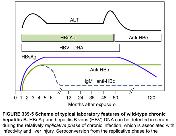
In chronic infection (HBsAg positive >6 months), two main phases are distinguished by HBeAg status:
| Phase | HBsAg | HBeAg | Anti-HBe | HBV DNA | ALT |
|---|---|---|---|---|---|
| Immune tolerant | + | + | − | Very high (up to 10¹²–¹³ IU/mL) | Normal |
| Immune active (HBeAg+) | + | + | − | High (≥20,000 IU/mL) | Elevated |
| Inactive carrier | + | − | + | Low/undetectable | Normal |
| HBeAg-negative chronic hepatitis | + | − | + | Detectable (fluctuating) | Elevated/fluctuating |
| Resolved infection | − | − | ± | Undetectable | Normal |
HBeAg seroconversion (HBeAg → Anti-HBe) signals a shift from high to low replication, but does not mean eradication — HBsAg remains positive.
Isolated Anti-HBc — Interpretations
When only anti-HBc is positive (HBsAg and anti-HBs both negative), possible interpretations include:
| Interpretation | Key Features |
|---|---|
| Resolved/remote infection | Common in endemic areas; HBV DNA undetectable |
| Window period of acute HBV | IgM anti-HBc positive; HBV DNA also detectable |
| False-positive | Weakly reactive; repeat in 3–6 months |
| Occult HBV infection | Rare; HBV DNA detectable at low levels |
— Sleisenger & Fordtran's GI and Liver Disease, Table 79.3, p. 1479
HBV DNA
Quantitative HBV DNA (real-time PCR; sensitivity 5–10 IU/mL) is essential for:
- Establishing indication for antiviral therapy
- Monitoring treatment response
- Detecting drug resistance (HBeAg-negative patients with rising DNA on therapy)
HBsAg quantification (licensed in many countries): HBsAg < 1000 IU/mL in anti-HBe-positive patients distinguishes true inactive carriage from relapsing disease. — Sleisenger & Fordtran's, p. 1480
Summary: Common Patterns
| HBsAg | Anti-HBs | IgM Anti-HBc | Total Anti-HBc | HBeAg | Interpretation |
|---|---|---|---|---|---|
| + | − | + | + | + | Acute HBV infection |
| − | − | + | + | − | Window period |
| − | + | − | + | − | Resolved past infection |
| − | + | − | − | − | Vaccination (immune) |
| + | − | − | + | + | Chronic HBV, replicative |
| + | − | − | + | − | Chronic HBV, low replication / inactive carrier |
| − | − | − | + | − | Isolated anti-HBc (see table above) |
Sources:
- Sleisenger & Fordtran's Gastrointestinal and Liver Disease, pp. 1479–1480
- Harrison's Principles of Internal Medicine, 21st ed., Figs. 339-4 & 339-5
- Medical Microbiology, 9th ed., Chapter 55
Ascites
Your credits are exhausted. Your daily credits reset tomorrow, or you can purchase a top-up for more.
This is a shared conversation. Sign in to Orris to start your own chat.