
Inflammatory breat cancer
"inflammatory breast cancer"
inflammatory breast cancer diagnosis treatment NCI
https://www.cancer.gov/types/breast/breast-cancer-types/infl…
inflammatory breast cancer clinical photograph redness swelling peau d orange
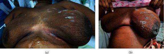
This clinical photograph consists of two panels illustrating gross pathological findings in the breasts. Panel (a) provides a comparative view of both breasts, demonstrating significant asymmetry; the left breast is markedly enlarged and swollen relative to the right. Panel (b) offers a close-up perspective of the left breast, highlighting diffuse erythema (redness) and skin changes consistent with inflammation. A distinct discharging sinus is visible in the medial quadrant, located just medial to the left nipple-areolar complex. The surrounding skin exhibits a thickened, edematous texture, occasionally referred to as peau d'orange appearance in such clinical contexts. These visual signs—swelling, redness, and a draining sinus tract—are characteristic markers of a severe inflammatory process, such as inflammatory breast cancer, complicated mastitis, or a ruptured mammary abscess. The image serves as an educational tool for recognizing clinical signs of advanced breast pathology and associated cutaneous manifestations.
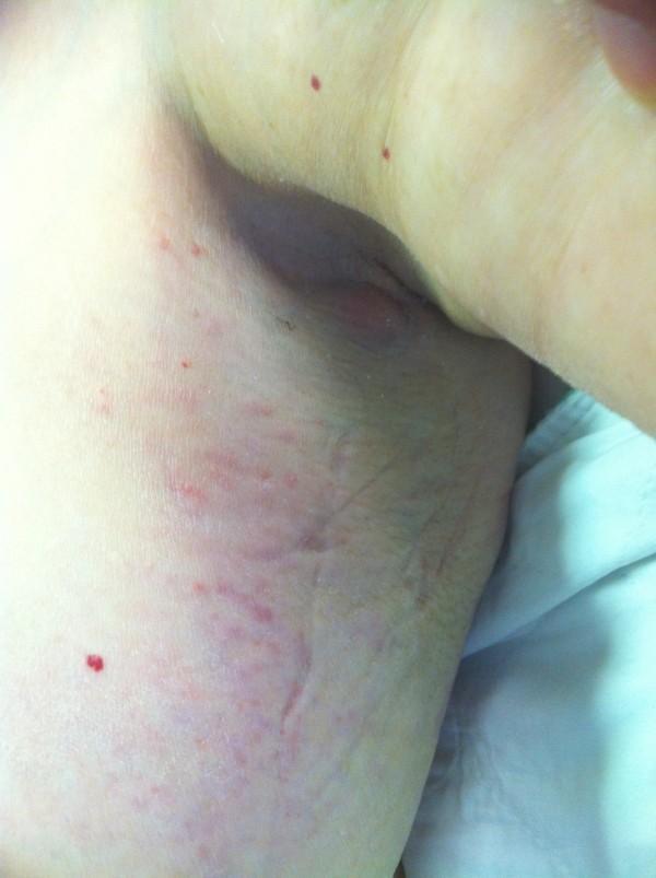
This clinical photograph displays a close-up view of the left breast upper outer quadrant and the left axillary region. The image demonstrates significant dermatological findings including diffuse erythema (redness) and pronounced soft tissue edema (swelling), which distorts the normal anatomical contours of the axillary fold. The skin surface exhibits an uneven texture with areas of thickening and subtle wrinkling, consistent with peau d'orange or inflammatory infiltration. Scattered across the erythematous region are several pinpoint petechiae or small angiomatous red spots. These findings are clinically significant as they can represent an inflammatory presentation of breast malignancies, such as inflammatory breast cancer or, as in this specific clinical context, primary breast lymphoma. The image serves as an educational example of how systemic or malignant processes can manifest with localized skin changes mimicking cellulitis or mastitis.
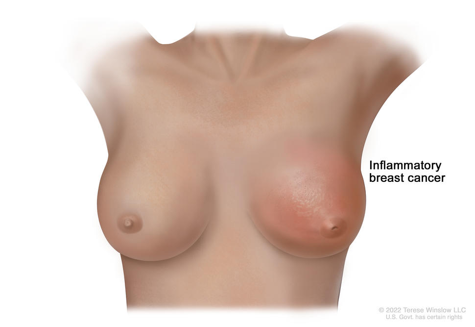
Warning signs
- Red, pink, purple, or bruised-looking skin over a substantial part of one breast
- Rapid breast swelling, warmth, heaviness, tenderness, or enlargement
- Skin thickening or dimpling, called peau d’orange because it resembles orange peel
- New nipple inversion, flattening, or discharge
- Swollen lymph nodes in the armpit or above the collarbone
Diagnosis
- Breast examination plus diagnostic mammogram and ultrasound, often breast MRI.
- Core biopsy of the breast or involved lymph node to confirm cancer.
- Skin punch biopsy if skin is involved.
- Testing for ER, PR, and HER2 biomarkers.
- CT, PET/CT, and/or bone imaging to determine whether it has spread.
Treatment
- Chemotherapy first (neoadjuvant systemic therapy), often with HER2-targeted treatment if HER2-positive.
- Modified radical mastectomy with axillary lymph-node dissection.
- Radiotherapy to the chest wall and regional lymph-node areas.
- Additional treatment based on tumor biology, such as HER2-targeted therapy, endocrine therapy for hormone-receptor-positive cancer, or immunotherapy in selected cases.