
How to do a mechanical thrombectomy
mechanical thrombectomy stroke procedure stent retriever
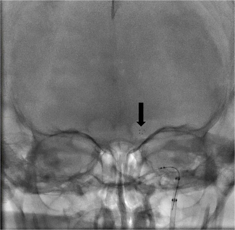
A native fluoroscopic image (X-ray) in an anteroposterior (AP) view showing a mechanical thrombectomy procedure for an ischemic stroke. The image demonstrates a radio-opaque stent retriever deployed within the intracranial vasculature. An arrow points to the distal radio-opaque markers of the device, which is positioned at the level of the left carotid terminus (the bifurcation of the internal carotid artery into the middle cerebral artery and anterior cerebral artery). Below this, the proximal delivery catheter and markers are visible as they ascend through the cervical and petrous segments of the internal carotid artery. The surrounding cranial anatomy, including the calvarium, orbits, and petrous ridges, provides anatomical context for the endovascular positioning. This clinical image illustrates a key step in interventional neuroradiology where the device is used to engage and extract a thrombus to restore cerebral perfusion.
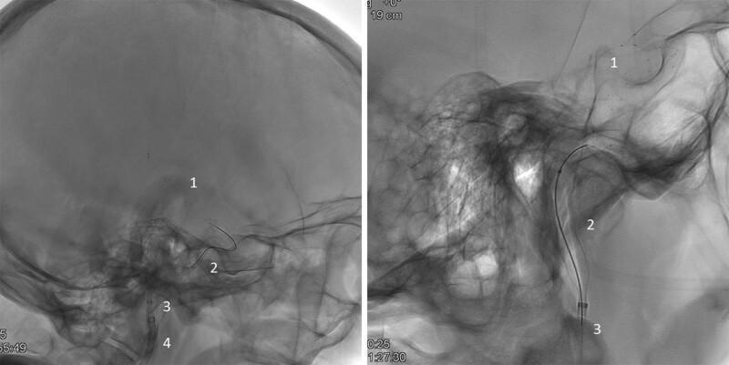
Two lateral fluoroscopic/angiographic views of the skull demonstrating an endovascular mechanical thrombectomy procedure in the cerebral vasculature. The images illustrate the triaxial spatial arrangement of interventional devices. A Solitaire stent retriever (1) is deployed distally within a cerebral artery, likely the internal carotid or middle cerebral artery, to engage a thrombus. Proximally, the Advanced Neurovascular Access (ANA) device is positioned, featuring an expanded, radiopaque braided funnel (2) that apposes the vessel walls to provide local flow restriction and enhanced aspiration. The ANA delivery catheter (3) is seen coaxially aligned with the funnel system. A larger guide catheter (4) is visible in the more proximal vasculature, providing the primary access route. The clinical significance of this setup is to show the combined use of a stent retriever with a specialized aspiration funnel to optimize clot retrieval and minimize distal embolization during acute ischemic stroke intervention. This material is suitable for intermediate to advanced neurointerventional training.
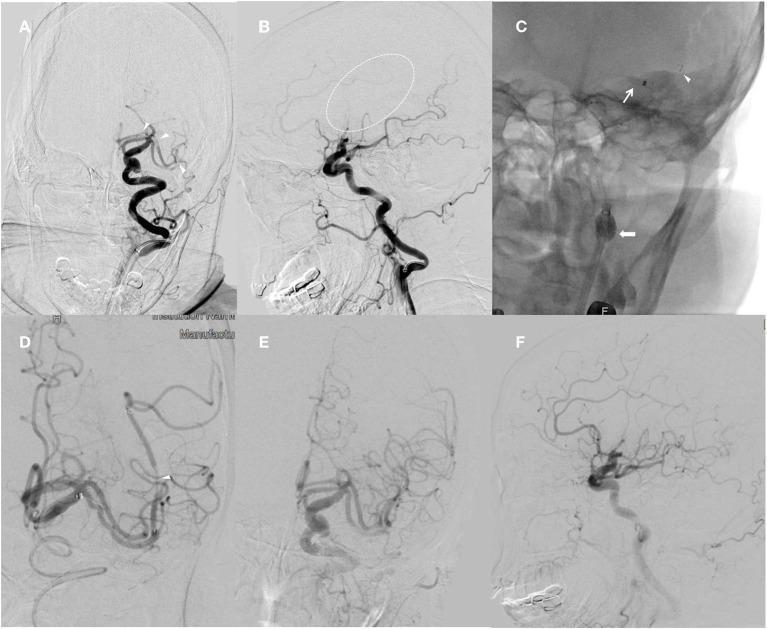
This composite of six digital subtraction angiography (DSA) and fluoroscopic images illustrates a mechanical thrombectomy procedure for acute ischemic stroke. Panels A (anteroposterior) and B (lateral) show the pre-intervention state with proximal occlusions in the left A1 segment of the anterior cerebral artery (ACA), the superior M1 proximal branch, and the inferior Sylvian M2 branch of the middle cerebral artery (MCA), characterized by an abrupt cutoff of contrast flow. Panels C and D demonstrate the intra-procedural combined technique using a balloon guide catheter (BGC), a distal access catheter (DAC), and a stent retriever. The BGC balloon is visibly inflated in the internal carotid artery (ICA) to arrest antegrade flow, while the stent retriever is deployed across the occluded segments. Panels E and F provide the post-intervention angiographic results, showing successful recanalization of the ACA and MCA territories with restored distal perfusion. The series demonstrates the endovascular management of complex intracranial arterial occlusions using modern aspiration and stent-retriever techniques.
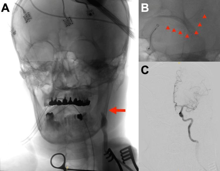
This composite figure illustrates a mechanical thrombectomy procedure for acute ischemic stroke using digital subtraction angiography (DSA) and fluoroscopic guidance. Panel A shows a frontal radiograph of the skull and neck during the initial phase. A red arrow indicates the site of a direct left common carotid artery (CCA) puncture. Visible external monitoring equipment includes multiple EEG electrodes adhered to the scalp and an endotracheal tube. Panel B provides a high-magnification fluoroscopic view of the neurointerventional procedure. Red arrowheads track the deployment of a stent retriever (Solitaire X) extending from the M2 segment of the middle cerebral artery to the terminal internal carotid artery (ICA), alongside a REACT71 aspiration catheter. Panel C presents a post-procedural DSA image demonstrating successful TICI 2b recanalization of the previously occluded left internal carotid artery and its distal branches. The content highlights advanced neurovascular rescue techniques and the specific approach of direct carotid access for mechanical thrombectomy in complex aortic anatomy.
Mechanical Thrombectomy
Part 1 - Intracranial / Stroke Thrombectomy
Patient Selection & Timing
- No significant prestroke disability (mRS ≤ 1)
- Causative occlusion of the ICA or M1 segment of MCA
- NIHSS ≥ 6
- ASPECTS ≥ 6 on imaging
- Bradley and Daroff's Neurology in Clinical Practice, p. 1132
- ROSEN's Emergency Medicine, p. 1439
Step-by-Step Procedure
1. Setup & Access
- Performed in an angiography suite under fluoroscopic guidance
- Femoral artery access is standard (radial or direct carotid access used in complex anatomy)
- A guide catheter (often a balloon guide catheter, BGC) is placed in the cervical ICA or vertebral artery
- The BGC allows balloon inflation to arrest antegrade flow during clot retrieval, reducing distal embolization
2. Diagnostic Angiography
- Digital subtraction angiography (DSA) confirms the vessel occlusion site (ICA terminus, M1, M2, basilar artery)
- Roadmap imaging is acquired to navigate distally
3. Microcatheter Navigation
- A microwire (e.g. Synchro 14) is used to cross the occlusion, followed by a microcatheter
- A distal access catheter (DAC) is advanced coaxially as close to the clot as possible
4. Device Deployment - Two Main Techniques
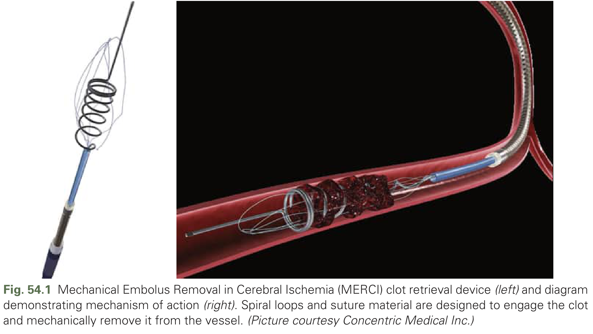
- The stent is deployed and expands within the clot, incorporating it into the mesh
- After 3-5 minutes of dwell time (to allow thrombus integration), the BGC balloon is inflated to stop flow
- The stent retriever is slowly withdrawn with continuous aspiration applied through the DAC or guide catheter
- The ASTER and COMPASS trials showed direct aspiration first-pass (ADAPT) was non-inferior to stent retriever as first-line therapy
- A large-bore aspiration catheter (e.g. Penumbra/Sofia/React 71) is advanced directly to the clot face without a stent retriever
- A vacuum is applied, engaging the thrombus at the catheter tip
- The catheter is withdrawn with continuous suction, removing the clot en bloc
- If the clot does not come free, a stent retriever can be added (combined technique)
5. Post-Retrieval Angiography
- Perform runs to confirm reperfusion grade using TICI scoring:
- TICI 2b/3 = successful reperfusion (goal)
- TICI 0/1 = failed reperfusion
- If residual stenosis is identified (e.g. due to underlying intracranial atherosclerotic disease), angioplasty +/- stenting may be considered
6. Repeat Passes
- Multiple passes may be needed (typically up to 3 attempts)
- Each pass increases risk of vessel injury and hemorrhagic transformation
Part 2 - Peripheral Arterial Thrombectomy
Preoperative Planning
- Mechanical thrombectomy has reduced bleeding risk versus catheter-directed thrombolysis (CDT) when tPA is NOT used
- Suitable for patients who have contraindications to tPA (recent surgery, intracranial hemorrhage)
- Arterial access obtained, vascular sheath placed, diagnostic angiography performed
- Current Surgical Therapy 14e, p. 1637
Devices
- End-hole catheter with side holes that expel saline jets while simultaneously aspirating
- The rheolytic action mechanically disrupts and extracts thrombus
- Technique: wire traverses the occlusion → AngioJet catheter is advanced → device activated → catheter slowly advanced and retracted with intermittent rotation to cover all clot
- "Power Pulse" mode infuses tPA with the expelled fluid for a pharmacomechanical hybrid approach, followed by a conventional pass after a dwell period
- Complications: hemolysis, hemoglobinuria, hyperkalemia, acute kidney injury (limit device run time); microembolization (consider distal protection devices)
- Flexible "CAT" catheter connected to a continuous vacuum engine
- Catheter advanced to thrombus, wire removed, vacuum applied via flow switch
- Catheter retracted until blood seen in canister, then suction rapidly disengaged
- Monitor canister carefully - rapid exsanguination can occur
- Newer models include automated blood-flow cessation systems
Post-Procedure Care
- Serial neurovascular examinations of the limb
- Monitor for reperfusion compartment syndrome - prophylactic 4-compartment fasciotomies recommended if ischemia >6 hours or if limb edema develops post-reperfusion
- Watch for rhabdomyolysis: target urine output >100 cc/hour, urine alkalinization with bicarbonate if CPK is rising
- After successful revascularization, patients require long-term anticoagulation to prevent re-thrombosis
Key Evidence Summary
| Trial | Window | Finding |
|---|---|---|
| MR CLEAN, ESCAPE, EXTEND-IA, SWIFT PRIME, REVASCAT | 0-6 h | Stent retriever thrombectomy superior to standard care for anterior LVO |
| DAWN | 6-24 h | Benefit in selected patients with clinical-imaging mismatch |
| DEFUSE 3 | 6-16 h | Benefit with perfusion mismatch imaging selection |
| ASTER / COMPASS | 0-6 h | Aspiration (ADAPT) non-inferior to stent retriever as first-line |
| ARISE II | - | EmboTrap: 80% TICI ≥2b, 67% mRS 0-2 at 90 days |
- Bradley and Daroff's Neurology in Clinical Practice, p. 1132-1134
- ROSEN's Emergency Medicine, pp. 1439-1441
- Current Surgical Therapy 14e, pp. 1637-1638