


Identify this breast tumor
solid papillary carcinoma breast histology neuroendocrine nested organoid pattern
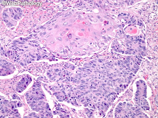
Histology using light microscopy on formalin-fixed paraffin-embedded colonic tissue stained with Hematoxylin and Eosin (H&E). The subject is a high-grade neuroendocrine carcinoma of the large bowel, large cell type, showing an organoid/nested growth pattern with solid sheets and occasional rosettes. Tumor cells are large with abundant cytoplasm and nuclei with vesicular or finely granular chromatin and conspicuous nucleoli; mitotic figures are frequent and necrosis may be present. In some foci there are glands or squamous differentiation, reflecting heterogeneous differentiation within the neoplasm. The architecture is compact, with high cellularity and minimal mucin. The background mucosa may show peritumoral inflammatory infiltrate. The histologic features are consistent with neuroendocrine carcinoma of the colon, large cell variant, which typically demonstrates high-grade cytology and aggressive behavior, and may co-express neuroendocrine markers on immunohistochemistry (not shown). The image exemplifies organoid arrangement reminiscent of pulmonary LCNEC, but arising in the colon with gastrointestinal clinical implications. Diagnostic significance includes distinguishing LCNEC from colorectal adenocarcinoma with neuroendocrine differentiation, small cell carcinoma, or MiNEN. This histology is relevant for prognosis, staging, and therapeutic decision-making, often guiding platinum-based chemotherapy regimens and comprehensive oncologic management. Correlation with imaging and biomarker profiling is recommended for accurate staging and management in practice.
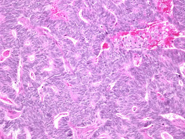
Light microscopy of a hematoxylin and eosin (H&E) stained breast tissue section demonstrates a solid architectural variant of papillary carcinoma with spindle-cell morphology. The primary subject is a malignant epithelial neoplasm of the mammary gland, imaged at microscopic level. The specimen type is a histology slide from breast tissue, and the image represents a high-power field with cellular detail. In the tumor, there are solid sheets and nests of cells with spindle to oval shapes and scant cytoplasm. Nuclei are elongated with mild to moderate nuclear atypia and relatively inconspicuous nucleoli. Mitotic figures are infrequent in this field. Focal papillary structures may be present but the dominant pattern here is solid, with delicate fibrovascular cores observed where visible. The stroma is fibrous, with limited inflammatory infiltrate and no conspicuous necrosis. Overall morphology supports a papillary carcinoma that can mimic metaplastic spindle-cell lesions, underscoring the need for thorough histologic assessment and adjunctive studies. Clinically, solid variant papillary carcinoma of the breast is typically low-to-intermediate grade with favorable prognosis, though occasional aggressive behavior has been reported. Immunohistochemical profiling and radiologic-pathologic correlation aid accurate diagnosis and guide surgical management and adjuvant therapy decisions.
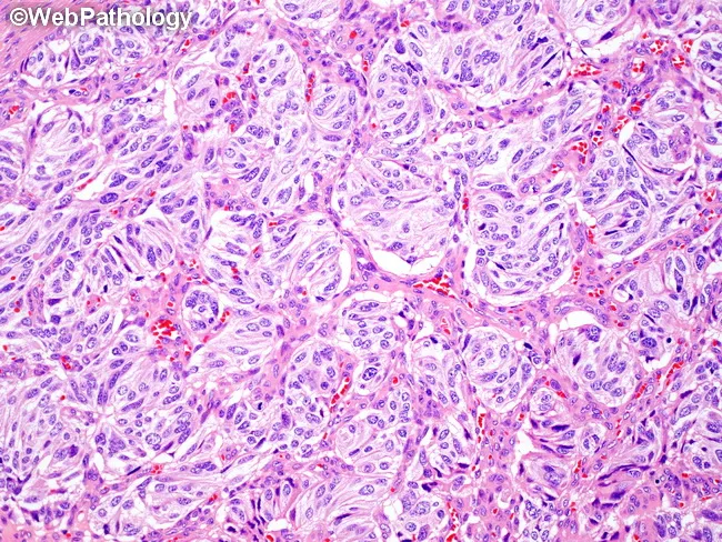
A histopathologic image of pancreatic tissue demonstrates a well-differentiated neuroendocrine tumor (pNET) with solid nests arranged in a paraganglioma-like, zellballen pattern. The material is stained with Hematoxylin and Eosin and viewed under light microscopy. The pancreas is the organ of origin, within the digestive/endocrine system. Tumor cells form cohesive nests and cords, separated by delicate vascular stroma, producing an organoid architecture. Within nests, cells are uniform, polygonal to round, with moderate eosinophilic cytoplasm and round to oval nuclei showing characteristic salt-and-pepper chromatin. Nuclear size is relatively uniform; mitotic figures are infrequent in a low to mid-grade lesion. Background pancreatic parenchyma may show minimal desmoplastic reaction. The overall pattern mimics paraganglioma due to nested arrangement, but clinical context and immunophenotype (not shown) would support pancreatic neuroendocrine differentiation with chromogranin A and synaptophysin positivity and a low to intermediate Ki-67 index. This histology has diagnostic significance for grading, prognosis, and surgical planning, and it informs differential considerations against paraganglioma, solid pseudopapillary neoplasm, and ductal adenocarcinoma. Clinically relevant uses include guiding endocrine evaluation, localization prior to resection, and inclusion in educational resources on pancreatic neuroendocrine neoplasms and nest-like patterns in gastroenterology and pathology curricula.
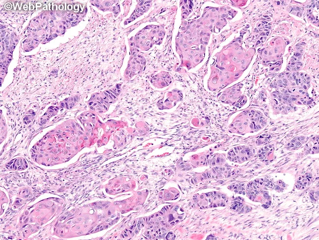
This histopathology image depicts a high-grade neuroendocrine carcinoma of the large bowel, represented by a predominantly large cell neuroendocrine carcinoma morphology with organoid (rosette-like) nests and solid sheets. The tissue shown is from the colorectal mucosa/submucosa. The stain is Hematoxylin and Eosin (H&E) on a formalin-fixed paraffin-embedded section, viewed under light microscopy. Notable features include nests of polygonal tumor cells with vesicular nuclei, salt-and-pepper chromatin, conspicuous mitotic activity, and geographic necrosis. There are areas demonstrating small cell morphology with scant cytoplasm and high nuclear-to-cytoplasmic ratio, reminiscent of SCNEC; also, foci showing glandular and squamous differentiation (adeno- and squamo-differentiated components) within the same lesion, indicating mixed histology. The architectural pattern is organoid in places, with nests separated by delicate fibrovascular stroma. The lesion invades adjacent stroma and shows considerable pleomorphism. Diagnostic significance: high-grade colorectal neuroendocrine carcinoma portends aggressive behavior, often with early metastasis; immunohistochemical confirmation (chromogranin A, synaptophysin, CD56, INSM1) is typically positive, and Ki-67 index is high. Differential diagnoses include colorectal adenocarcinoma with neuroendocrine differentiation, pure small cell colorectal carcinoma, and mixed adeno-neuroendocrine carcinoma. Clinically relevant: influences therapeutic planning toward platinum-based chemotherapy, potential surgical resection, and staging workup; important for prognosis, follow-up strategies, and research into colorectal NEC.
Histopathological Diagnosis: Solid Papillary Carcinoma (SPC) of the Breast with Neuroendocrine Differentiation
Key Histological Features Identified
| Feature | Finding |
|---|---|
| Growth pattern | Organoid/nested arrangement - cohesive solid nests, lobules, and anastomosing cords separated by delicate fibrovascular septa |
| Cell morphology | Monomorphic, round to polygonal cells with moderate, finely granular cytoplasm |
| Nuclear chromatin | Classic "salt-and-pepper" (finely stippled, evenly distributed) - hallmark of neuroendocrine differentiation |
| Stroma | Delicate sinusoidal-type capillaries wrapping nests; no desmoplasia |
| True glandular lumina | Absent in the dominant areas |
| Nuclear pleomorphism | Low-grade |
| Mitoses | Scarce |
Diagnostic Interpretation
- Solid expansile nests with delicate fibrovascular cores (the "papillary" component may not be obvious without reticulin staining)
- Consistent neuroendocrine-type morphology (this is so common that SPC and primary breast neuroendocrine tumor significantly overlap)
- Typically ER/PR strongly positive, HER2 negative
- Frequent intracellular or extracellular mucin production (may not be visible at this magnification)
- Low-grade biological behavior in the in-situ/encapsulated form, though invasive SPC does occur
Recommended Confirmatory IHC Panel
| Marker | Expected Result | Purpose |
|---|---|---|
| Synaptophysin | Strongly positive (diffuse) | Neuroendocrine confirmation |
| Chromogranin A | Positive | Neuroendocrine confirmation |
| INSM1 | Nuclear positive | Sensitive NE marker |
| ER / PR | Strongly positive | Typical in SPC/breast NET |
| HER2 | Negative | Typical profile |
| E-cadherin | Positive (preserved) | Rules out lobular carcinoma |
| Ki-67 | Low (<20%) | Confirms low-grade |
| p63 / CK5 | Negative | Rules out high-grade basal-type |
Key Differentials to Exclude
- Invasive Lobular Carcinoma (solid variant) - similar cell uniformity, but cells are discohesive, E-cadherin is lost, and the organoid vascularity is absent
- High-grade neuroendocrine carcinoma - would show significant pleomorphism, high mitotic rate, necrosis; excluded here by low-grade morphology
- Metastatic neuroendocrine tumor (carcinoid from GI/lung) - must correlate with clinical/radiologic history; TTF-1 (lung), CDX2 (GI) IHC can distinguish
Clinical Pearls
- SPC accounts for roughly 1-2% of breast carcinomas; it predominantly affects older women
- When in situ (encapsulated), prognosis is excellent; invasive SPC is treated as standard invasive carcinoma
- These tumors are virtually always hormone receptor positive and respond well to endocrine therapy
- Mucin production (colloid/mucinous features) is a common associated finding