
Teach me rectal cancer from scratch to pro level from sabiston and maingotts
Reading File
Now let me search both Sabiston and Maingot's for rectal cancer content in parallel.
Finding Sources
Finding Sources
I can see Sabiston is in the library. Let me check if Maingot's is available and find the full rectal cancer blocks.
Finding Sources
Reading File
Maingot's is not in the library. Let me now systematically read the full rectal cancer section from Sabiston and Current Surgical Therapy in parallel.
Reading File
Reading File
Excellent content. Let me continue reading Sabiston's rectal cancer section and the rest of Current Surgical Therapy in parallel.
Reading File
Reading File
Reading File
Reading File
Reading File
Reading File
Now I have comprehensive content from Sabiston and Current Surgical Therapy. Let me also quickly fetch the key surgical anatomy images and the Sabiston staging/treatment section that I haven't yet captured.
Reading File
Searching PubMed
rectal cancer total neoadjuvant therapy TNT
I now have comprehensive material. Let me fetch the key MRI staging image and also get the watch-and-wait/organ preservation section from Sabiston.
Loading Image
Loading Image
I now have all the content needed for a comprehensive, pro-level teaching. Let me mark completed todos and write the full response.---
Rectal Cancer: Scratch to Pro
Sources: Sabiston Textbook of Surgery (9780443124341), Current Surgical Therapy 14e (9780323796835), Fischer's Mastery of Surgery 8e, Bailey & Love 28e
1. ANATOMY - The Foundation of Everything
What Is the Rectum?
The rectum is the distalmost segment of the large intestine, measuring 12-15 cm in length. Surgically, it extends from the anorectal ring (top of the sphincter complex) to the rectosigmoid junction (where the taeniae coli converge). From an oncologic standpoint, rectal cancer refers to tumors at or below the peritoneal reflection (roughly the upper third of the rectum).
From a practical surgical standpoint, location matters enormously:
- Upper rectum (10-15 cm from anal verge): above the peritoneal reflection, behaves more like sigmoid
- Mid rectum (5-10 cm): extraperitoneal, no covering serosa, high local recurrence risk
- Lower rectum (<5 cm): proximity to sphincters, challenges sphincter preservation
Borders of the Rectum in the Pelvis
- Lateral: pelvic sidewall
- Posterior: sacrum
- Anterior (men): prostate, seminal vesicles, bladder
- Anterior (women): vagina, uterus, bladder
Vascular Supply
- Superior rectal artery - terminal branch of the inferior mesenteric artery (IMA) - supplies upper 2/3 of rectum
- Middle rectal artery - from internal iliac - supplies lower rectum
- Inferior rectal artery - from internal pudendal - supplies anal canal
Fascial Planes - The Holy Grail of Rectal Surgery
This is the concept that transformed rectal cancer surgery:
The mesorectum is the fibrofatty tissue envelope around the rectum that contains the terminal branches of the IMA and the rectal lymphatics (the most important lymph node drainage for rectal cancer). It is surrounded circumferentially by a thin fascial envelope called the fascia propria of the rectum.
Just posterior to the mesorectum lies the presacral fascia (Waldeyer's fascia), covering the presacral venous plexus. The holy plane of TME (Bill Heald's term) is the areolar tissue plane between these two fasciae - just outside the fascia propria, anterior to the presacral fascia.
Clinical pearl: Dissecting deep to the presacral fascia causes catastrophic hemorrhage from the presacral venous plexus - torn vessels retract into the sacral foramina and are nearly impossible to control.
The Denonvilliers' fascia separates the anterior mesorectum from the prostate/seminal vesicles in men and the vagina in women. Anterior dissection must respect this fascia to avoid injury to these structures.
Autonomic Nerves - Functional Anatomy
Injury to pelvic autonomic nerves causes bladder dysfunction (urinary retention) and sexual dysfunction (erectile dysfunction in men, orgasmic dysfunction in women). The key nerves are:
- Hypogastric nerves (sympathetic, L1-L3): descend from the superior hypogastric plexus at the sacral promontory - vulnerable during IMA ligation and presacral dissection
- Pelvic splanchnic nerves (parasympathetic, S2-S4): emerge from the pelvic sidewall, join hypogastric nerves to form the inferior hypogastric plexus at the pelvic sidewall - vulnerable during lateral dissection
- Neurovascular bundles (Walsh's bundles): run along the posterolateral surface of the prostate - vulnerable during anterior dissection
2. EPIDEMIOLOGY & RISK FACTORS
- Approximately 46,050 new rectal cancer cases diagnosed annually in the USA (2023 data)
- Rectal cancers now represent 31% of all colorectal cancers (up from 27% in 1995)
- Young-onset rectal cancer is rising: ~2% per year increase in patients under 50
- 3rd most common cause of cancer death in women; 2nd most common in men
Risk Factors
Shared with colon cancer:
- Age (peak 60-70s, but rising in younger patients)
- Personal/family history of colorectal polyps or CRC
- Hereditary syndromes: Lynch syndrome (MLH1, MSH2, MSH6, PMS2 mutations - mismatch repair deficiency/MSI), FAP (APC mutation), MYH-associated polyposis
- Inflammatory bowel disease (especially ulcerative colitis)
- Diet: low fiber, high processed/red meat
- Obesity, physical inactivity, alcohol, smoking
3. MOLECULAR PATHOGENESIS
Three major pathways to colorectal cancer (same as colon cancer, with some rectal predominance):
Chromosomal Instability (CIN) - 65-70% of CRC
- Classic adenoma-carcinoma sequence (Fearon-Vogelstein model)
- Begins with APC gene mutation → disrupts Wnt signaling → nuclear β-catenin accumulation → dysplastic proliferation (tubular adenoma)
- Progression: APC → KRAS mutation (growth promotion) → TP53/SMAD4/PIK3CA/FBXW7 mutations (invasion)
- Most CIN tumors have APC mutation in 80% of cases
Serrated (Methylator/CIMP) Pathway - 15-20%
- Begins with BRAF mutation
- CpG island methylation silences tumor suppressor genes
- Produces sessile serrated lesions and traditional serrated adenomas
- Associated with MLH1 methylation → MSI-high tumors
Microsatellite Instability (MSI) - 15%
- Defective mismatch repair (MMR) proteins (MLH1, MSH2, MSH6, PMS2)
- Can be somatic (sporadic - often from MLH1 promoter methylation) or germline (Lynch syndrome)
- MSI-H tumors respond dramatically to PD-1 immune checkpoint inhibitors - clinically crucial
4. CLINICAL PRESENTATION
Most common symptom: change in bowel habits
Other presentations:
- Rectal bleeding (bright red blood per rectum)
- Iron deficiency anemia
- Change in stool caliber
- Tenesmus and rectal pain - late findings, indicative of locally invasive tumor
- Palpable rectal mass on DRE
- Neuropathic pain - indicates sacral nerve root invasion
Remember: Tenesmus + rectal pain = alarm signs for locally advanced, often low-lying tumor.
5. CLINICAL EVALUATION & STAGING
Step 1: History and Physical
A complete cancer-directed history must include:
- Bowel, urinary, and sexual baseline function (critical for surgical planning and prognostication of post-op quality of life)
- Family history (hereditary syndromes?)
- Medical fitness and frailty (guides ability to tolerate neoadjuvant therapy)
The digital rectal examination (DRE) by the treating surgeon + rigid or flexible proctoscopy is mandatory. The examiner must document:
- Distance from the anal verge and dentate line
- Mobility of tumor relative to prostate/vagina/sphincter complex
- Sphincter tone and baseline continence
Key point: Rigid proctoscopy (not flexible endoscopy) is the gold standard for measuring distance from anal verge. On rigid scope, tumors within 12-15 cm = rectal cancers.
Step 2: Colonoscopy
Complete colonoscopy to rule out synchronous cancers (~3%) or polyps (~30%). If tumor is obstructing, CT colonography is used; alternatively, complete colonoscopy is performed after neoadjuvant therapy downsizes the tumor.
Step 3: Laboratory
- CEA (baseline, not diagnostic - used for prognosis and surveillance)
- Routine CBC, metabolic panel
- MMR/MSI testing by IHC - essential; MSI-H tumors have different treatment implications (immunotherapy responsiveness)
Step 4: Imaging
| Modality | Role |
|---|---|
| Pelvic MRI | Gold standard for locoregional staging (T and N stage, CRM, peritoneal reflection, adjacent structure invasion) |
| CT chest/abdomen/pelvis | Metastatic staging (lung, liver, peritoneal disease) |
| Endorectal ultrasound (EUS) | Historical role in T-staging for early lesions; limited compared to MRI |
| PET-CT | Selected cases (equivocal distant disease) |
MRI provides critical information that guides management:
- T stage (depth of wall penetration)
- N stage (abnormal nodes in mesorectum)
- Circumferential Resection Margin (CRM) - if tumor is within 1-2 mm of the mesorectal fascia, CRM is threatened/positive → strongly predicts local recurrence → drives need for neoadjuvant therapy
- Location relative to the peritoneal reflection
- EMVI (extramural vascular invasion) - important prognostic factor
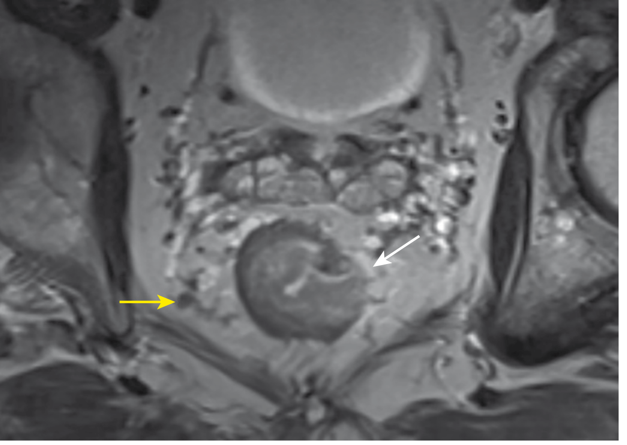
Pelvic MRI showing a T3N1 rectal cancer. White arrow: tumor penetrating into mesorectal fat. Yellow arrow: enlarged, abnormal mesorectal lymph node. (Sabiston Textbook of Surgery)
Step 5: TNM Staging (AJCC 8th Edition)
T stage:
- T1: into submucosa
- T2: into muscularis propria
- T3: through muscularis propria into pericolorectal tissues (mesorectal fat)
- T4a: penetrates visceral peritoneum (upper rectum)
- T4b: invades adjacent organ/structure
N stage:
- N0: no regional nodes
- N1: 1-3 positive nodes (N1a=1, N1b=2-3, N1c=tumor deposits)
- N2: 4+ positive nodes (N2a=4-6, N2b=7+)
M stage:
- M0: no distant metastasis
- M1a: one site/organ
- M1b: two+ sites/organs
- M1c: peritoneal metastasis
AJCC Stage Groups:
- Stage I: T1-2N0M0
- Stage II: T3-4N0M0 (IIA=T3, IIB=T4a, IIC=T4b)
- Stage III: any T, N1-2, M0
- Stage IV: any T, any N, M1
Step 6: Multidisciplinary Tumor Board
Every rectal cancer should be discussed at a multidisciplinary tumor board (MDT). This is a NAPRC (National Accreditation Program for Rectal Cancer) quality metric. The MDT includes colorectal surgery, medical oncology, radiation oncology, radiology, pathology. Studies show MDT review changes management even for experienced surgeons.
6. TREATMENT OVERVIEW
The management of rectal cancer is stage-driven and has changed dramatically over 20-30 years:
cT1-2 N0  →  Surgery first (TME or local excision for selected T1)
cT3-4 or N+  →  Neoadjuvant therapy THEN surgery (± adjuvant therapy)
Stage IV  →  Systemic therapy + selective surgery
MSI-H (any stage)  →  Consider immunotherapy first
7. SURGICAL ANATOMY AND THE PRINCIPLE OF TME
Total Mesorectal Excision (TME) - introduced and championed by Bill Heald - is the single most impactful advance in rectal cancer surgery.
Principle: Remove the rectum en bloc with its entire mesorectal envelope (all lymphatics, vessels, lymph nodes), dissecting in the avascular "holy plane" just outside the fascia propria and anterior to the presacral fascia.
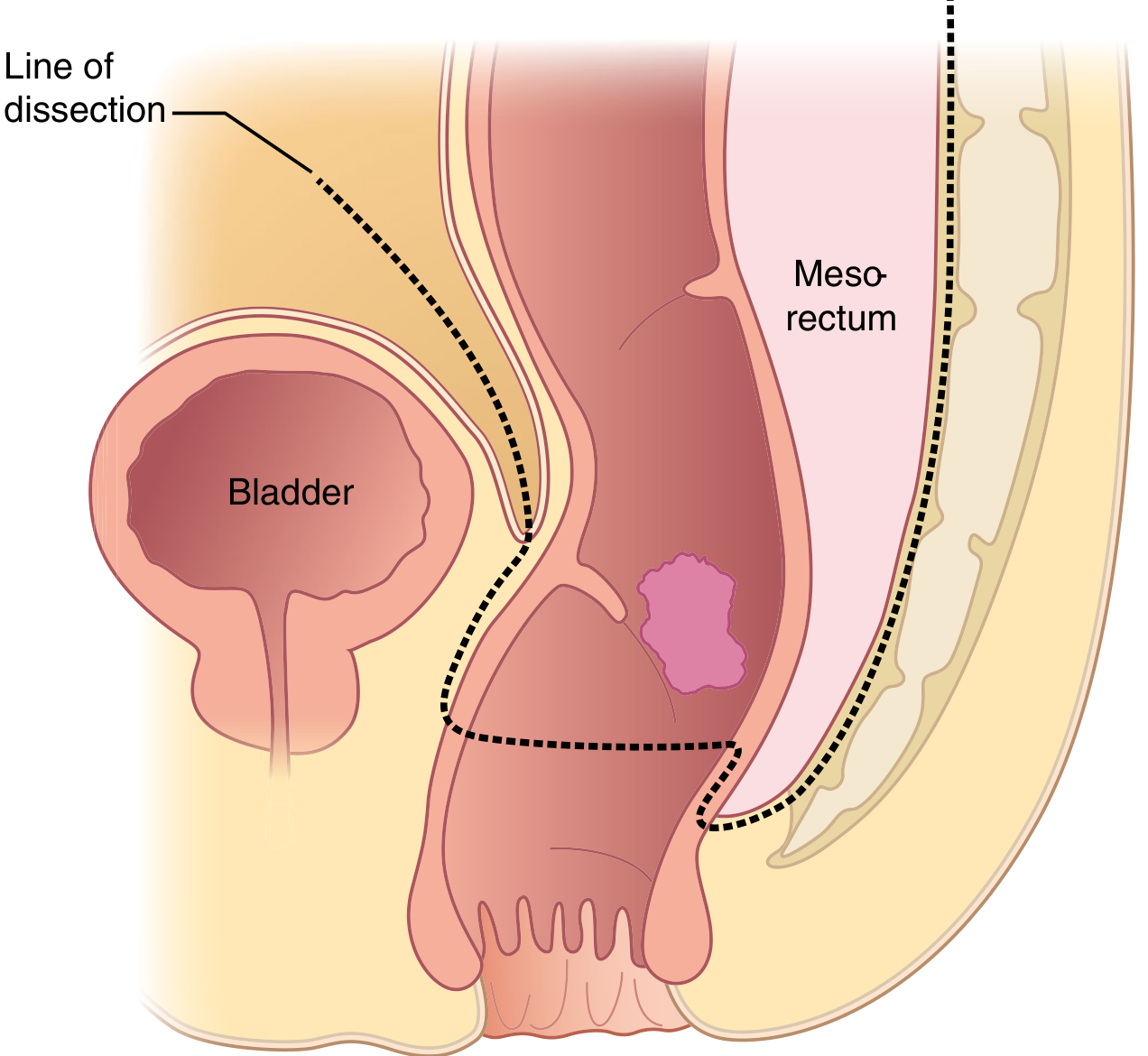
Low anterior resection: dotted line shows the TME dissection plane removing rectum en bloc with mesorectum. (Sabiston Textbook of Surgery)
Impact of TME:
- Traditional blunt dissection: 25% positive CRM, very high recurrence
- Norwegian cohort of 3,319 patients after TME implementation: local recurrence dropped from 12% to 6%, survival increased from 60% to 73%
Local recurrence rates without TME/neoadjuvant therapy:
- T1-T2: ~10%
- T3N0: 15-35%
- T3-4 node-positive: 45-65%
8. SURGICAL PROCEDURES
A. Low Anterior Resection (LAR)
Used for upper and mid rectal cancers where sphincter preservation is possible. Key oncologic requirement: ≥1 cm distal margin (some guidelines say ≥2 cm, though 1 cm is acceptable for low tumors after neoadjuvant therapy).
Steps of LAR:
- Mobilize the left colon by dividing the left lateral peritoneal attachments
- Divide IMA: either high ligation (at IMA origin - needed for bulky adenopathy, improves reach) or low ligation (just distal to left colic artery, preserving it). High ligation is NOT automatically required; low ligation preserves left colic artery blood supply for anastomosis
- Mobilize mesorectum posteriorly in the holy plane (sharp dissection, not blunt)
- Divide Denonvilliers' fascia anteriorly (or go anterior to it to protect prostate/vagina)
- Complete lateral dissection, protecting pelvic autonomic nerves
- Divide the bowel distally with adequate margin + complete mesorectum
- Anastomosis (colorectal or coloanal): stapled (double-stapling technique most common) or hand-sewn
Diverting loop ileostomy is routinely created when there is a low anastomosis (<5-6 cm from anal verge) to protect in case of anastomotic leak. Closed after 8-12 weeks.
B. Abdominoperineal Resection (APR)
Required when:
- Tumor involves the sphincter complex
- Locally advanced low tumor where adequate distal margin cannot be achieved
- Poor preoperative bowel function (end colostomy preferred over a leaking low anastomosis)
APR = combined abdominal + perineal approach removing the rectum, sphincter complex, and levator ani, resulting in a permanent end colostomy.
Cylindrical/extended APR: For locally advanced low tumors, the levator ani is taken at its origin (not just at the anorectal junction) to achieve wider lateral margins - reduces positive CRM.
Decision between LAR and APR: The key question is whether a negative distal margin and functional sphincter can be achieved. Neoadjuvant therapy has expanded the population eligible for sphincter-preserving LAR.
C. Local Excision
For highly selected early lesions:
- cT1N0, without high-risk features
- High-risk features that EXCLUDE local excision: deep SM invasion (Kikuchi SM3, >1 mm submucosal), poor differentiation, lymphovascular invasion (LVI), perineural invasion (PNI), tumor budding, positive margins
Techniques:
- Transanal excision (TAE): conventional, limited to low tumors
- Transanal Endoscopic Microsurgery (TEM): endoscopic platform, allows higher rectal access, full-thickness en bloc excision
- Transanal Minimally Invasive Surgery (TAMIS): uses standard laparoscopic port system transproctally - more widely available than TEM
Critical: Local excision does NOT stage or treat mesorectal lymph nodes. If high-risk features are found on final pathology, formal TME should be strongly recommended.
D. IMA Ligation Level
- High ligation: at aortic origin of IMA - needed for bulky N2 disease, improves colonic reach
- Low ligation: just distal to left colic artery - preserves left colic blood supply
- The inferior mesenteric vein (IMV) is separately divided
E. Minimally Invasive Approaches
- Laparoscopic TME: improved short-term outcomes (earlier recovery, less blood loss), but RCTs (COLOR II, ACOSOG Z6051, ALaCaRT) failed to prove oncologic non-inferiority to open surgery. Still recommended by NCCN but only by experienced surgeons
- Robotic TME: improved visualization and maneuverability in the narrow pelvis; current guidelines support its use
- Transanal TME (taTME): bottom-up approach via transanal platform - particularly useful for low tumors, narrow pelvis, obesity; improves distal margin access
9. NEOADJUVANT THERAPY
Why Neoadjuvant?
Without neoadjuvant therapy, local recurrence rates for T3+ or N+ rectal cancer are unacceptably high. Indications for neoadjuvant therapy: cT3-4 or cN+ in the extraperitoneal rectum.
Critical distinction: Neoadjuvant therapy is for extraperitoneal rectal cancers because these have no serosa and thus high local recurrence risk. Upper rectal cancers at or above the peritoneal reflection behave more like colon cancers and are NOT treated with neoadjuvant radiation.
Landmark Trials
1. German Rectal Cancer Study (2004) - The pivotal trial:
- Compared preoperative chemoRT (50.4 Gy/28 fractions + infusional 5-FU) vs. postoperative chemoRT for cT3-4 or N+
- No difference in OS (~60% at 10 years)
- But neoadjuvant was BETTER in:
- Local recurrence: 6% vs 13% (p<0.001)
- Tumor downstaging and downsizing
- Sphincter preservation rate: 39% vs 19% for low tumors
- Toxicity: less acute and late toxicity with neoadjuvant therapy
- Result: neoadjuvant chemoRT became the standard
2. CAO/ARO/AIO-12 Trial (RAPIDO Trial):
- Compared short-course RT (5x5 Gy) + consolidation chemotherapy vs. long-course chemoRT
- Short-course RT + consolidation CAPOX: better pathologic complete response rates and disease-free survival
3. PRODIGE 23 Trial:
- Total neoadjuvant therapy (TNT) with induction FOLFIRINOX → chemoRT → surgery vs. chemoRT alone → surgery
- TNT arm: significantly better pCR rates (~28% vs ~12%), better disease-free survival
- Established TNT as a valid approach for high-risk locally advanced rectal cancer
Radiation Regimens
| Regimen | Dose/Schedule | Timing to Surgery | Comment |
|---|---|---|---|
| Long-course CRT | 45-50.4 Gy / 25-28 fractions + concurrent 5-FU | 6-8 weeks after | Allows maximum downstaging |
| Short-course RT (SCRT) | 25 Gy / 5 fractions | Can delay 6-12 wks (consolidation chemo) or operate within 1 wk | Swedish/Dutch trials validated immediate surgery; delayed allows tumor response |
Radiosensitization: Concurrent chemotherapy (5-FU or capecitabine) is given at lower than standard doses to potentiate radiation cell-kill, not for systemic effect.
Total Neoadjuvant Therapy (TNT) - The Modern Standard
TNT delivers all chemotherapy AND radiation before surgery. Rationale:
- Full-dose systemic chemotherapy given upfront (not after) - better tolerated, no post-op delays
- Higher rates of pathologic complete response (pCR)
- Better distant disease control
- Enables organ preservation in responders
TNT regimens:
- Induction chemo → chemoRT → surgery (PRODIGE 23 approach)
- SCRT → consolidation chemo → surgery (RAPIDO approach)
2024 ASCO Guideline (PMID 39116386): recommends TNT as preferred approach for locally advanced rectal cancer with high-risk features (T4, threatened CRM, N2, EMVI).
10. WATCH-AND-WAIT / ORGAN PRESERVATION
One of the most important recent advances in rectal cancer management.
Concept: After neoadjuvant therapy, some tumors achieve a clinical complete response (cCR) - no residual tumor on DRE, endoscopy, or MRI. These patients may undergo a watch-and-wait (W&W) protocol, avoiding surgery entirely.
cCR Criteria (clinical complete response):
- Endoscopy: no visible tumor (white scar only)
- DRE: no palpable tumor
- MRI: no residual signal/mass
- ALL THREE criteria must be met
Oncologic outcomes of W&W:
- Regrowth rate: ~20-25% (usually within 1-2 years)
- Regrowth is almost exclusively luminal and salvageable with surgery
- Overall survival is equivalent to surgery in properly selected patients
Surveillance in W&W is intensive: DRE + rigid proctoscopy + pelvic MRI every 3-4 months for 2 years, then every 6 months.
Key principle: MSI-H/dMMR rectal cancers have very high complete response rates to PD-1 immunotherapy (dostarlimab/pembrolizumab) - in the NICHE trial and Cercek 2022 study, 100% clinical response rates were achieved with pembrolizumab in cT2-4 dMMR rectal cancer, with many achieving sustained cCR without surgery.
11. STAGING AND PROGNOSIS (PATHOLOGIC)
After surgical resection, pathologic staging drives adjuvant treatment decisions.
The Pathology Report Must Include:
- T stage (depth of invasion)
- N stage (number of nodes examined, number positive - minimum 12 nodes required for adequate staging)
- M stage
- Circumferential Resection Margin (CRM): positive if tumor within 1 mm of mesorectal fascia - most important local recurrence predictor
- Distal Resection Margin
- Tumor regression grade (TRG): Mandard or Ryan grading of response to neoadjuvant therapy
- LVI, PNI, tumor budding
- MMR/MSI status
- KRAS, NRAS, BRAF mutation status (for stage IV patients, guides targeted therapy)
5-Year Survival by Stage:
- Stage I: ~90%
- Stage II: ~70-80%
- Stage III: ~50-60%
- Stage IV (metastatic): ~10-15% (but selected resectable liver metastases: up to 71%)
Tumor regression grade after neoadjuvant (pathologic complete response - pCR = ypT0N0):
- pCR rate with long-course CRT: ~15-20%
- pCR rate with TNT: ~25-30%
- pCR is a strong predictor of improved long-term outcomes
12. ADJUVANT THERAPY
Historical context: The GITSG trial (1985) in 227 patients with T3-4 or N+ disease showed combined postoperative chemoRT reduced local recurrence from 55% to 33%.
Current practice:
- Patients who received full TNT with pathologic stage II-III disease: no adjuvant chemotherapy needed
- Patients with clinical/pathologic stage II-III who did NOT receive full neoadjuvant chemotherapy: adjuvant chemotherapy recommended
- Preferred regimen: FOLFOX x 8 cycles (oxaliplatin + 5-FU/leucovorin); can omit oxaliplatin in clinical T3N0
- Adjuvant radiotherapy: given selectively to patients with path stage II-III who did NOT receive neoadjuvant radiation AND are at increased risk for local recurrence
13. SYNCHRONOUS METASTATIC DISEASE (Stage IV)
~20% of rectal cancer patients present with synchronous metastases.
- Liver is most common site (also lung - more common in rectal than colon cancer)
- ~50% of stage IV patients have liver-only disease
- Liver-only, resectable metastases: 5-year OS up to 71% with chemotherapy + surgery
- Management requires MDT discussion assessing resectability
Rectal primary + synchronous liver metastases - three surgical strategies:
- Simultaneous resection of rectum + liver
- Colorectal-first: resect primary → chemotherapy → liver resection
- Liver-first: if primary is not immediately obstructing, resect liver first (reduces risk of progression of limiting disease), then chemo, then rectal surgery
Systemic therapy for metastatic/unresectable disease:
- FOLFOX or FOLFIRI + bevacizumab (anti-VEGF) for first-line RAS-mutant or unknown
- Add cetuximab or panitumumab (anti-EGFR) for wild-type KRAS/NRAS/BRAF tumors (left-sided predominance)
- BRAF V600E mutant: doublet chemo + BRAF/MEK inhibitors (encorafenib)
- MSI-H/dMMR: pembrolizumab or nivolumab - dramatically superior to chemotherapy (KEYNOTE-177 trial)
14. LOCALLY RECURRENT RECTAL CANCER
A feared complication after primary surgery, especially with positive CRM or R1 resection.
Presentation: pelvic pain (often neuropathic/sacral), bowel/bladder dysfunction, presacral mass on imaging.
Management:
- Always re-biopsy for confirmation and MSI/dMMR testing
- MDT evaluation for resectability
- If no prior radiation: neoadjuvant chemoRT → pelvic exenteration if resectable
- Potentially involves: posterior exenteration (rectum + bladder/prostate), total pelvic exenteration, sacrectomy
- Soft tissue flap (VRAM, gracilis, omentoplasty) reconstruction is often required for large perineal defects
- R0 resection: only chance of cure; 5-year OS ~30-40% after R0 pelvic exenteration
15. POSTOPERATIVE SURVEILLANCE
NCCN/Sabiston recommendations for stage II-III rectal cancer after curative resection:
- H&P + CEA: every 3-6 months for 2 years, then every 6 months for 5 years total
- CT chest/abdomen/pelvis: every 6-12 months for 5 years
- Colonoscopy: 1 year after surgery (if no preoperative colonoscopy done), then every 3-5 years
- Stage I: no imaging or tumor marker follow-up recommended
For stage IV after curative-intent resection: H&P + CEA at same frequency as stage II/III; imaging every 3-6 months for first 2 years, then every 6-12 months for 5 years total.
16. SPECIAL SCENARIOS & HIGH-YIELD EXAM POINTS
| Scenario | Key Teaching Point |
|---|---|
| Low tumor + patient wants sphincter preservation | Neoadjuvant TNT first → assess response; W&W if cCR; LAR if partial response; APR only if tumor invades sphincter after treatment |
| T1 rectal polyp on colonoscopy | Local excision (TEM/TAMIS) if no high-risk features; formal TME if high-risk features on final path |
| MSI-H rectal cancer | Discuss immunotherapy (pembrolizumab) - high cCR rates; W&W is a real option |
| Positive CRM on preop MRI | Neoadjuvant therapy mandatory; reassess CRM after treatment |
| Rectal cancer + synchronous liver mets | Assess resectability MDT; if resectable, FOLFOX → staged or simultaneous resection |
| APR vs LAR decision | Distal margin feasibility + sphincter function; neoadjuvant therapy should be maximized first |
| Presacral hemorrhage during dissection | Do NOT dissect deep to presacral fascia; if bleeding occurs: pressure, surgical thumbtacks into sacral foramina, packing |
| Bowel function after LAR | "Low anterior resection syndrome" (LARS) - urgency, frequency, clustering; worse with lower anastomosis, radiation; diverting stoma does not prevent it |
| Positive circumferential margin | Strongest predictor of local recurrence; if on preop MRI, mandatory neoadjuvant; if on final path, consider adjuvant RT if not given |
| cCR after neoadjuvant | Discuss W&W: 20-25% regrowth, almost all luminal, salvageable; surveillance every 3 months |
17. QUICK MEMORY FRAMEWORK
RECTAL CANCER MANAGEMENT ALGORITHM

Diagnosis:  Biopsy (adenocarcinoma) + CEA + colonoscopy + MMR/MSI
Staging:    Pelvic MRI + CT C/A/P
            → cT1-2 N0: Surgery first
            → cT3-4 or N+, extraperitoneal: Neoadjuvant (TNT preferred) → surgery
            → MSI-H: Consider pembrolizumab ± W&W
            → Metastatic: MDT → resectable vs unresectable pathway

Surgery:    LAR (sphincter-preserving) vs APR (sphincter-sacrificing)
            ALWAYS TME (the holy plane)
            IMA ligation (high vs low) + IMV division
            Colorectal/coloanal anastomosis ± diverting ileostomy

Path:       CRM (most important) + pCR + TRG + MMR + KRAS/NRAS/BRAF (stage IV)

Adjuvant:   If TNT given → no adjuvant chemo
            If no prior full systemic chemo → FOLFOX x 8 cycles

Surveillance: CEA + CT q6-12mo x 5yr; colonoscopy 1yr
Note: Maingot's Abdominal Operations is not currently in the textbook library. The content above is drawn primarily from Sabiston Textbook of Surgery (9780443124341, Chapter 96) and Current Surgical Therapy 14e (9780323796835), supplemented by Fischer's Mastery of Surgery 8e and Bailey & Love 28e. Recent evidence: 2024 ASCO Guideline on Locally Advanced Rectal Cancer (PMID 39116386) and 2024 Network Meta-Analysis on TNT regimens in JAMA Netw Open (PMID 38833249) support TNT as the current standard for high-risk locally advanced rectal cancer.---
A note on Maingot's: Maingot's Abdominal Operations is not in the current textbook library. The full coverage above draws from Sabiston Textbook of Surgery (the most comprehensive source available), supplemented by Current Surgical Therapy 14e, Fischer's Mastery of Surgery 8e, and Bailey & Love 28e - all of which are in the library and together provide equivalent or greater depth for this topic.
Here is a summary of what was covered, organized scratch-to-pro:
| Level | Sections Covered |
|---|---|
| Scratch (Foundations) | Anatomy, fascial planes, vascular supply, autonomic nerves, epidemiology, molecular pathogenesis |
| Intermediate (Clinical) | Presentation, workup, DRE, colonoscopy, MRI staging, TNM classification, MDT |
| Advanced (Treatment) | TME principle, LAR vs APR vs local excision, neoadjuvant (German trial, PRODIGE 23, RAPIDO, TNT), adjuvant therapy |
| Pro Level | Watch-and-wait/organ preservation, MSI-H immunotherapy, synchronous metastases management, locally recurrent rectal cancer, pelvic exenteration, LARS, special scenarios |
Ask me to drill deeper into any specific section - surgical anatomy, a particular trial, staging nuances, or any clinical scenario.
Colorectal polyps in details from standard books from basics to pro level
Reading File
This is a broad topic. Let me run parallel searches across all available textbooks simultaneously.
Finding Sources
Finding Sources
Finding Sources
Finding Sources
Good finds. Let me now read the key sections in parallel - Schwartz's (surgical), Yamada's (gastroenterology), Current Surgical Therapy (already located in prior session), and find the Sabiston polyps section.
Reading File
Reading File
Excellent - rich content. Let me continue reading the rest of those sections and also search for Yamada's and Sabiston polyp content.
Reading File
Reading File
Excellent - I found the Haggitt and Kikuchi classification tables. Now let me get the Sabiston polyps section and the Yamada's content on polyposis syndromes.
Finding Sources
Reading File
!
unable to open database file
We encountered an error while processing your request. Please try again or reach out to us for help.
This is a shared conversation. Sign in to Orris to start your own chat.