
What MIC in drug sensitivity report
MIC stands for Minimum Inhibitory Concentration - it's the lowest concentration of an antibiotic that visibly inhibits growth of a microorganism in vitro. In a drug sensitivity (culture & sensitivity) report, MIC tells you the exact potency needed to suppress the pathogen.
Searching sources...
Reading File
Searching the Web
MIC minimum inhibitory concentration drug sensitivity report interpretation
Finding Sources
Reading File
Reading File
Here is a thorough explanation of MIC in a drug sensitivity (culture & sensitivity) report:
MIC - Minimum Inhibitory Concentration
Definition
The MIC is the lowest concentration of an antimicrobial agent (expressed in µg/mL) that completely prevents visible bacterial growth after 24 hours of incubation in a standardized lab test.
- Lippincott Illustrated Reviews: Pharmacology, p. 914: "The minimum inhibitory concentration (MIC) is the lowest antimicrobial concentration that prevents the visible growth of a microorganism after 24 hours of incubation."
Why It Matters Clinically
The MIC tells you how much drug is needed to suppress the bug. When comparing the MIC value to established breakpoints (set by CLSI, FDA, or EUCAST), the lab interprets the organism as:
| Interpretation | What It Means |
|---|---|
| S - Susceptible | Drug at standard dose achieves tissue concentrations well above the MIC - infection likely to respond |
| I - Intermediate / Susceptible-Dose Dependent (SDD) | Drug works, but requires higher or optimized dosing to achieve adequate concentrations at the infection site |
| R - Resistant | Normal achievable drug concentrations cannot reach or exceed the MIC - drug unlikely to work |
Important: A lower MIC number is better (less drug needed to inhibit growth), but you should not pick an antibiotic just because it has the lowest MIC. Breakpoints differ between antibiotics, so comparisons across drugs using raw MIC values are misleading.
How It Is Measured in the Lab
Two main methods are used:
-
Broth Dilution (most common today) - The organism is inoculated into tubes/wells containing serial dilutions of antibiotic. The lowest clear (no visible growth) concentration = MIC. Computer-automated systems (e.g., VITEK, MicroScan) run this rapidly and are the standard in most clinical labs.
-
Disk Diffusion (Kirby-Bauer) - Antibiotic-impregnated paper disks are placed on an agar plate seeded with the organism. The zone of inhibition around the disk is inversely proportional to MIC: larger zone = lower MIC = more susceptible (Sherris & Ryan's Medical Microbiology, p. 938).
-
E-test (Epsilometer test) - A plastic strip with a continuous antibiotic gradient is placed on agar; where bacterial growth meets the strip, you can read off the MIC directly. Useful for fastidious and anaerobic bacteria.
MIC in PK/PD (Pharmacokinetic/Pharmacodynamic) Dosing
MIC is used in modern dose optimization. Key relationships:
| PK/PD Parameter | Drug Examples | Goal |
|---|---|---|
| Time > MIC | Beta-lactams, carbapenems | Drug concentration stays above MIC for 40-70% of dosing interval |
| AUC/MIC ratio | Vancomycin, fluoroquinolones | AUC:MIC ratio target (e.g., vancomycin AUC/MIC of 400-600 mg·h/L) |
| Cmax/MIC ratio | Aminoglycosides | Peak concentration well above MIC (8-10x) |
For example, The Washington Manual of Medical Therapeutics states that for vancomycin, a target AUC:MIC of 400-600 mg·h/L (assuming MIC of 1 mg/L) is recommended.
MIC vs. MBC
| MIC | MBC (Minimum Bactericidal Concentration) | |
|---|---|---|
| Definition | Lowest concentration that inhibits visible growth | Lowest concentration that kills ≥99.9% of bacteria |
| Clinical use | Routine - used every day | Rarely used (time-consuming, labor-intensive) |
| Relationship | MBC ≥ MIC always | MBC can be 2-4x MIC for bactericidal drugs |
A drug is called bactericidal when MBC ≈ MIC (within 4-fold), and bacteriostatic when MBC is much higher than MIC.
Key Takeaway
When you read a sensitivity report, look at the S/I/R interpretation first - not the raw MIC number. The MIC becomes especially useful when:
- Infection is in a difficult-to-reach site (e.g., CNS, prostate)
- You need to optimize the dose (PK/PD dosing for serious infections)
- Monitoring for emerging resistance over time (rising MIC = "MIC creep")
Site of lesion for decorticate and decerebrate posturing
Decorticate = lesion above the red nucleus (cortex/internal capsule/cerebral hemispheres). Decerebrate = lesion at or below the red nucleus (midbrain/upper pons). Decerebrate generally indicates a worse prognosis.
Searching sources...
Finding Sources
Searching the Web
decorticate decerebrate posturing site of lesion neuroanatomy
Reading File
Reading File
Reading File
Now let me fetch the key diagram from Ganong's that illustrates both postures:
Loading Image
Loading Image
Loading Image
Here is a complete breakdown from the authoritative textbooks:
Decorticate vs. Decerebrate Posturing - Site of Lesion
The Key Anatomical Dividing Line: The Red Nucleus
The red nucleus (in the midbrain tegmentum, at the level of the superior colliculus) is the critical landmark that separates these two postures. Specifically, the dividing line is the intercollicular level of the brainstem.
1. Decorticate Posturing (Flexor Posturing)
Site of lesion: ABOVE the red nucleus
- Cerebral cortex, internal capsule, cerebral hemispheres, or upper midbrain (rostral to/above the red nucleus)
Clinical appearance:
- Upper limbs: flexed at elbows, wrists, and fingers - arms pulled up toward the chest ("toward the cortex" - mnemonic)
- Lower limbs: extended, internally rotated, plantar-flexed
- Head may be extended
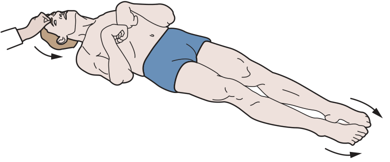
Mechanism: The red nucleus (which drives upper limb flexion via the rubrospinal tract) is intact and unopposed because the inhibitory cortical input above it is lost. The rubrospinal tract dominates, causing arm flexion.
2. Decerebrate Posturing (Extensor Posturing)
Site of lesion: AT or BELOW the red nucleus
- Lower midbrain and upper pons (caudal to the red nucleus)
- Equivalent to Sherrington's intercollicular transection in animal experiments
Clinical appearance:
- Upper limbs: extended, arms pronated, wrists flexed, fingers extended
- Lower limbs: extended, internally rotated, plantar-flexed
- Neck and head: extended (opisthotonus)
- Jaw: clenched
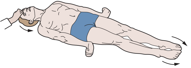
Mechanism: Both the red nucleus (rubrospinal - flexor) AND the cortex are disconnected. Only the vestibulospinal and reticulospinal tracts (extensor drive) remain active, producing unopposed extensor tone in all four limbs.
Summary Table
| Feature | Decorticate | Decerebrate |
|---|---|---|
| Site of lesion | Above red nucleus | At/below red nucleus |
| Anatomical level | Cortex / internal capsule / upper midbrain | Lower midbrain / upper pons |
| Upper limbs | Flexed (arms curl up) | Extended + pronated |
| Lower limbs | Extended | Extended |
| Pathway intact | Rubrospinal (flexor) | Vestibulospinal/reticulospinal (extensor) only |
| Pathway lost | Corticospinal | Corticospinal + rubrospinal |
| Prognosis | Serious | More severe / worse prognosis |
Mnemonic
DeCORticate = arms toward the CORe (chest) DEcerebrate = arms Down and Extended
Important Clinical Caveat
As emphasized in Neuroanatomy through Clinical Cases (3rd ed.) and Adams & Victor's Principles of Neurology (12th ed.): in humans, posturing does not always localize as precisely as in animal experiments. The same extensor posturing can occur with:
- Midbrain compression from a supratentorial (hemispheric) mass
- Cerebellar/posterior fossa lesions
- Metabolic encephalopathies (anoxia, hypoglycemia)
- Uncal herniation
Therefore, many neurologists now prefer the terms "flexor posturing" and "extensor posturing" to avoid implying a precise anatomical level, while still noting which limbs are involved.
Types of cerebral edema and its management
Cerebral edema has 3 classic types: vasogenic (BBB breakdown), cytotoxic (cell swelling), and interstitial (transependymal CSF). Management centers on osmotherapy, steroids (for vasogenic), and ICP reduction measures.
Searching sources...
Finding Sources
Searching the Web
types of cerebral edema vasogenic cytotoxic management 2024
Reading File
Reading File
Reading File
Reading File
Finding Sources
Reading File
Reading File
Reading File
Loading Image
Here is a complete, textbook-based breakdown:
Types of Cerebral Edema and Management
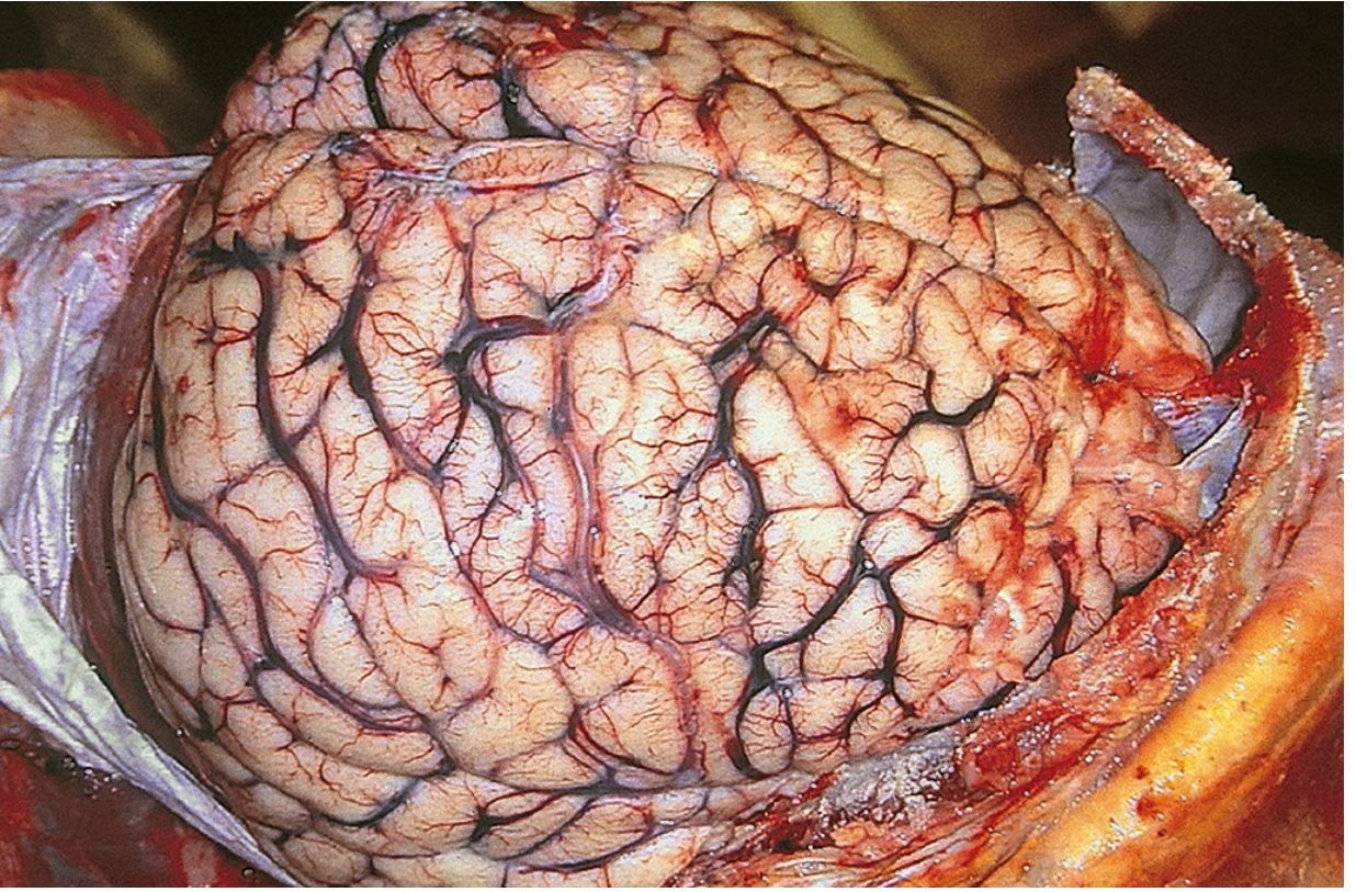
Classification - 4 Types
Bradley & Daroff's Neurology classifies cerebral edema into four types:
| Type | BBB Status | Fluid Location | Primary Mechanism |
|---|---|---|---|
| Vasogenic | Disrupted | Extracellular | Vascular leakage |
| Cytotoxic | Intact | Intracellular | Cell membrane/pump failure |
| Ionic | Intact | Extracellular | Na+ accumulation without cell death |
| Interstitial | Intact | Interstitial/periventricular | Transependymal CSF seepage |
In practice, most conditions produce a combination of types - particularly vasogenic + cytotoxic together.
1. Vasogenic Edema
Mechanism: BBB disruption increases vascular permeability, allowing plasma proteins and fluid to leak into the extracellular space. Spreads more in white matter (loose fiber tracts) than gray matter.
Causes:
- Brain tumors (most classic)
- Brain abscess
- Contusions / trauma
- Inflammatory lesions (MS plaques, encephalitis)
- Hypertensive encephalopathy
- High-altitude cerebral edema (HACE)
Key feature: Reversible if the underlying vessel injury resolves. Responds to steroids.
2. Cytotoxic Edema
Mechanism: Energy failure (ischemia, hypoxia, trauma) disrupts membrane Na+/K+-ATPase. Na+ accumulates intracellularly, creating osmotic gradient that draws water INTO cells. The extracellular space shrinks (unlike vasogenic).
Causes:
- Cerebral ischemia / stroke
- Hypoxia
- Metabolic encephalopathy
- Severe hypoglycemia
- Toxic injury
Key feature: Direct cell injury - generally NOT reversible. Does NOT respond to steroids. Worse prognosis than vasogenic.
3. Ionic (Early Cytotoxic / Pre-cytotoxic) Edema
Mechanism: Glutamate release from ischemic/traumatic injury opens Ca2+ channels. 1 Ca2+ exchanged for 3 Na+ ions causes Na+ buildup and water influx, but cell membrane is still intact at this stage. Considered a reversible precursor to full cytotoxic edema.
Causes: Early phase of ischemia, trauma - the same as cytotoxic but caught earlier.
4. Interstitial Edema
Mechanism: Obstructive hydrocephalus raises intraventricular pressure, forcing CSF to seep transependymally into the periventricular white matter. Also occurs with acute hyponatremia (osmotic movement of water into brain cells).
Causes:
- Obstructive hydrocephalus
- Acute hyponatremia (water intoxication)
Key feature: Can resolve with CSF drainage (VP shunt or EVD).
Management of Cerebral Edema
Management is ideally targeted at the underlying cause while simultaneously reducing ICP. The stepwise approach:
Step 1 - General / Supportive Measures
- Head elevation to 30 degrees - promotes venous drainage, reduces ICP
- Airway management and normoventilation (PaCO2 35-45 mmHg target normally)
- Avoid hypotension - maintain adequate cerebral perfusion pressure (CPP = MAP - ICP; target CPP > 60 mmHg)
- Avoid hyperthermia (increases metabolic demand)
- Treat underlying cause (antibiotics for abscess, anti-epileptics for seizures)
Step 2 - Sedation and Analgesia (for intubated/ICU patients)
- Propofol is preferred - short half-life, reduces ICP, allows rapid neurological assessment on discontinuation
- Fentanyl for analgesia (preferred in TBI over morphine/hydromorphone due to hemodynamic stability)
- Avoid routine neuromuscular blockade (risk of critical illness myopathy/neuropathy)
Step 3 - Osmotherapy (for raised ICP)
The cornerstone of medical ICP management. Works by creating an osmotic gradient across the intact BBB, drawing water out of brain tissue.
Mannitol
- Dose: 0.25-1 g/kg IV, can repeat every 4-6 hours
- Target serum osmolality: 300-315 mOsm/L
- Mechanism: osmotic diuresis + free radical scavenging
- Caution: transient rise in intravascular volume (avoid in heart failure, renal failure); can cause rebound edema; avoid in intracranial hemorrhage before craniotomy (hematoma expansion risk)
Hypertonic Saline (3% NaCl)
- Dose: 5-10 mL/kg over 30 min, OR continuous infusion titrated to Na+ 145-155 mEq/L
- Advantages over mannitol: less hypovolemia/hypotension, may be preferred in hemodynamically unstable patients
- Caution: use central line (extravasation risk); can cause hyperchloremic metabolic acidosis; avoid rapid correction in hyponatremic patients (risk of osmotic demyelination syndrome / central pontine myelinolysis)
- No clear evidence that one agent is superior over the other
Step 4 - Corticosteroids (for Vasogenic Edema ONLY)
- Dexamethasone is highly effective for tumor-associated, abscess-related, and radiation-induced vasogenic edema
- Mechanism: upregulates tight junction proteins, closes the disrupted BBB
- Also used in acute MS exacerbations: methylprednisolone 1g/day for 3-5 days
- NOT effective in cytotoxic edema (stroke, TBI) - corticosteroids are explicitly contraindicated in stroke edema
- NOT effective in DKA-related cerebral edema
Step 5 - Hyperventilation
- Target PaCO2 30-33 mmHg (moderate hyperventilation)
- Mechanism: hypocapnia causes cerebral vasoconstriction, reducing CBF and CBV rapidly
- Short-term / bridge therapy only - prolonged use causes cerebral ischemia from excessive vasoconstriction
- Used acutely when herniation is imminent while other measures are being set up
Step 6 - CSF Drainage
- External ventricular drain (EVD) / ventriculostomy: first-line ICP intervention in many ICUs after sedation
- Continuous drainage preferred over intermittent (BTF guidelines) but watch for ventricular collapse
- VP shunt: for chronic interstitial edema from hydrocephalus
Step 7 - Decompressive Craniectomy
- Reserved for refractory ICP > 25 mmHg not responding to medical therapy
- Evidence: reduces mortality in severe TBI with sustained raised ICP
- Also used in malignant MCA infarction (hemicraniectomy with duroplasty)
Step 8 - Barbiturate Coma (Last Resort)
- High-dose barbiturates (pentobarbital/thiopental) for refractory ICP
- Mechanism: suppress cerebral metabolism, reduce CBF and ICP
- Requires continuous EEG monitoring; significant side effects (hypotension, immunosuppression)
Quick Reference Summary Table
| Measure | Vasogenic | Cytotoxic | Interstitial |
|---|---|---|---|
| Steroids (Dexamethasone) | YES | No | No |
| Mannitol / Hypertonic saline | Yes | Yes | Yes |
| Hyperventilation (acute) | Yes | Yes | Yes |
| EVD / CSF drainage | - | - | YES |
| Decompressive craniectomy | Selected | Selected (malignant stroke, TBI) | - |
| Treat underlying cause | Tumor/abscess Rx | Revascularization (stroke) | Shunt (hydrocephalus) |
Sources: Bradley & Daroff's Neurology in Clinical Practice; Morgan & Mikhail's Clinical Anesthesiology 7e; Barash et al. Clinical Anesthesia 9e; Mulholland & Greenfield's Surgery 7e; Robbins & Cotran Pathologic Basis of Disease
Types of brain herniation and their symptoms according to ATLS 11 edition
Brain herniation syndromes include uncal (transtentorial), central, subfalcine, and tonsillar types. ATLS focuses on clinical recognition - uncal herniation classically presents with ipsilateral fixed dilated pupil and contralateral hemiplegia.
Searching sources...
Finding Sources
Searching the Web
ATLS 11th edition brain herniation types symptoms management traumatic brain injury
Reading File
Reading File
Reading File
Reading File
Reading File
Loading Image
Loading Image
Note on ATLS 11th edition: ATLS 11 was released in 2025. The library's most current emergency medicine texts (Tintinalli, Rosen's) align closely with ATLS principles. The web search confirmed ATLS 11 updated xABCDE sequencing but the herniation classification and clinical findings remain consistent with prior editions. The breakdown below reflects current ATLS/emergency medicine standard teaching.
Types of Brain Herniation and Their Symptoms
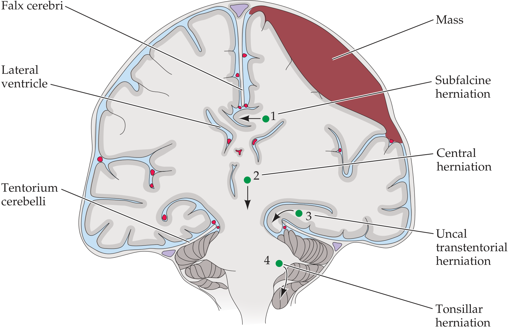
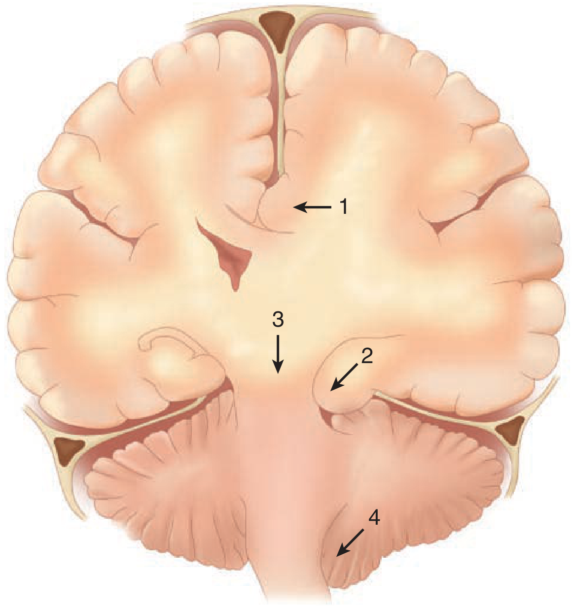
There are 4 major herniation syndromes:
1. Uncal Transtentorial Herniation (Most Common)
Mechanism: The uncus (medial temporal lobe) herniates inferiorly through the medial edge of the tentorium cerebelli. Most commonly caused by an expanding lateral temporal/middle fossa hematoma (e.g., epidural or subdural).
Symptoms - Progression:
| Stage | Finding | Mechanism |
|---|---|---|
| Early | Anisocoria, ptosis, sluggish pupil (ipsilateral) | CN III compression - parasympathetic fibers on outer surface compressed first |
| Progressive | Ipsilateral fixed dilated pupil ("blown pupil") | Complete CN III palsy - unopposed sympathetic tone |
| Early motor | Contralateral Babinski sign | Ipsilateral cerebral peduncle compressed |
| Progressive motor | Contralateral hemiparesis/hemiplegia | Ipsilateral corticospinal tract in midbrain compressed |
| LOC | Agitation → confusion → lethargy → coma | Midbrain reticular activating system compression |
| Respiratory | Normal → sustained hyperventilation → ataxic breathing | Progressive brainstem compression |
| Late | Bilateral decerebrate posturing | Bilateral brainstem involvement |
| Terminal | Cardiovascular collapse, brain death | Brainstem failure |
Important variant - Kernohan's Notch Phenomenon:
The midbrain is pushed so far that the contralateral cerebral peduncle is compressed against the opposite tentorial edge. This causes ipsilateral hemiparesis (same side as the mass and the dilated pupil) - a false-localizing sign.
2. Central Transtentorial Herniation
Mechanism: Bilateral central downward displacement of the diencephalon and midbrain through the tentorial incisura. Caused by midline/vertex/frontal/occipital lesions or diffuse cerebral edema - pressure is exerted from above bilaterally.
Symptoms - Progression (rostrocaudal deterioration):
| Stage | Finding |
|---|---|
| Early | Subtle change in mental status / decreased LOC, bilateral motor weakness |
| Pupils | Bilateral pinpoint pupils (2 mm) - light reflex still present but difficult to detect |
| Muscle tone | Increased bilaterally; bilateral Babinski signs |
| Progressive | Both pupils become midpoint and unreactive |
| Respiratory | Yawns and sighs → sustained hyperventilation → slow irregular breathing |
| Posturing | Decorticate posturing (to noxious stimuli) → bilateral decorticate → spontaneous decerebrate |
| Late | Fixed pupils, absent brainstem reflexes, death |
3. Subfalcine Herniation
Mechanism: The cingulate gyrus is displaced horizontally under the falx cerebri across the midline. Caused by unilateral hemispheric mass lesions.
Symptoms:
- Often clinically silent in early stages (most frequently detected on CT/MRI)
- Contralateral leg weakness (anterior cerebral artery territory - paracentral lobule involvement)
- Can compress the anterior cerebral artery (ACA) causing ischemia
- Frequently coexists with uncal herniation as the mass enlarges
4. Cerebellar Tonsillar Herniation (Foramen Magnum Herniation)
Mechanism: Cerebellar tonsils herniate downward through the foramen magnum, compressing the medulla and upper cervical cord. Caused by posterior fossa masses, elevated supratentorial ICP propagated downward, or lumbar puncture in setting of raised ICP.
Symptoms:
- Pinpoint pupils
- Flaccid paralysis (medullary compression disrupts upper motor neuron and reticulospinal tracts)
- Neck stiffness / head tilt
- Loss of protective reflexes (gag, cough)
- Cardiovascular instability (Cushing's response: hypertension + bradycardia + irregular respirations)
- Sudden respiratory arrest and death - most feared outcome
5. Upward Posterior Fossa (Upward Transtentorial) Herniation
Mechanism: A posterior fossa mass pushes cerebellar structures upward through the tentorial notch (opposite direction to the usual downward transtentorial herniation).
Symptoms:
- Conjugate downward gaze ("setting sun" sign)
- Absence of vertical eye movements
- Pinpoint pupils
- Loss of consciousness
Summary Comparison Table
| Type | Cause | Pupils | Motor | LOC | Key Feature |
|---|---|---|---|---|---|
| Uncal (most common) | Lateral temporal mass (EDH, SDH) | Ipsilateral fixed dilated (blown) | Contralateral hemiparesis; Kernohan's = ipsilateral | Progressive coma | Ipsilateral CN III palsy first |
| Central | Midline/diffuse/vertex lesion | Bilateral pinpoint → midpoint → fixed | Bilateral Babinski; decorticate → decerebrate | Gradual bilateral deterioration | Rostrocaudal deterioration |
| Subfalcine | Unilateral hemisphere mass | Normal (early) | Contralateral leg weakness (ACA) | Usually intact early | Often silent; CT finding |
| Tonsillar | Posterior fossa mass or raised ICP | Pinpoint | Flaccid paralysis | Rapid deterioration | Sudden respiratory arrest |
| Upward PF | Posterior fossa mass | Pinpoint | Variable | Coma | Downgaze, no vertical movement |
ATLS Key Clinical Points for Herniation Recognition
- Any change in pupil reactivity or size in a TBI patient must be treated as herniation until proven otherwise
- GCS motor score is the strongest predictor of outcome
- The "talk and die" patient - brief lucid interval followed by rapid deterioration = classic epidural hematoma causing uncal herniation
- Cushing's triad (hypertension + bradycardia + irregular respirations) = late sign of severe raised ICP/brainstem compression - requires immediate intervention
- Temporizing measures while preparing for neurosurgery: head elevation, osmotherapy (mannitol 1g/kg IV or hypertonic saline), controlled hyperventilation (PaCO2 30-35 mmHg - bridge only)
Sources: Tintinalli's Emergency Medicine; Rosen's Emergency Medicine; Schwartz's Principles of Surgery 11e; Neuroanatomy through Clinical Cases 3rd ed.; ATLS 11 principles per JournalFeed 2026 update summary
Testicular torsion management
Testicular torsion is a urological emergency requiring immediate surgical exploration. The key principle: time = testis. Detorsion and bilateral orchiopexy within 6 hours gives ~100% salvage; beyond 24 hours, salvage drops to <10%.
Searching sources...
Finding Sources
Searching the Web
testicular torsion management guidelines 2024 surgical detorsion orchiopexy
Reading File
Reading File
Reading File
Reading File
Reading File
Finding Sources
Reading File
Reading File
Loading Image
Here is a complete, evidence-based breakdown from multiple surgical and emergency medicine textbooks:
Testicular Torsion - Management
Background & Pathophysiology
Testicular torsion is a urological emergency where the testis twists on its spermatic cord, causing venous obstruction followed by arterial ischemia and infarction if not rapidly reversed.
Two anatomical types:
| Type | Age Group | Mechanism | Contralateral Orchiopexy? |
|---|---|---|---|
| Intravaginal (most common ~90%) | Adolescents / young adults | Bell-clapper deformity - tunica vaginalis completely covers the testis, allowing free rotation | YES - bilateral anomaly |
| Extravaginal | Neonates (~8-10%) | Torsion of entire cord outside tunica vaginalis, usually an in utero event | NO - not bilateral anomaly |
Bell-Clapper Deformity: The tunica vaginalis has an abnormally high attachment, the testis has no posterior fixation to the scrotal wall, and hangs freely like a clapper inside a bell - predisposing to rotation.
The degree of twist and duration are the two factors that determine testicular damage:
- Twists of 720° cause more rapid ischemia than 360°
- Duration: the critical window is 6 hours
Salvage Rate vs. Time - The Critical Window
| Time from Onset | Testicular Salvage Rate |
|---|---|
| < 6 hours | ~90-100% |
| 6-12 hours | ~50% |
| 12-24 hours | ~20% |
| > 24 hours | < 10% |
| > 48 hours | Near 0% |
(Bailey & Love's Surgery 28e; Sabiston Textbook of Surgery; Tintinalli's Emergency Medicine)
Clinical Features
- Sudden severe scrotal/groin pain - often waking patient from sleep
- Nausea and vomiting (common - from visceral pain)
- High-riding, transversely lying testis (Brunzel's sign)
- Swollen, tender, edematous scrotum (not initially erythematous)
- Absent cremasteric reflex on affected side (most reliable sign)
- Tender twisted cord palpable above testis
- Prehn's sign: elevation worsens pain (opposite of epididymitis where elevation relieves pain)
Diagnosis
- Clinical diagnosis first - do not delay surgery for imaging if clinical suspicion is high
- Scrotal Doppler ultrasound: decreased or absent intratesticular blood flow; can visualize spermatic cord twisting
- Urine analysis: typically normal (pyuria/leukocytes suggest epididymitis)
- Radioisotope scanning: most specific but rarely practical in emergency
Key rule: If testicular torsion cannot be clinically excluded, proceed to surgical exploration. A negative exploration is preferable to a missed torsion.
Management - Step by Step
Step 1: Immediate Urological Consultation + OR Preparation (Simultaneously)
Call urology immediately. Do not wait for imaging if clinical presentation is classic. Consent for possible orchiectomy.
Step 2: Manual Detorsion (Bedside Temporizing Measure - NOT Definitive)
Attempt while preparing for the operating room - never as a substitute for surgery.
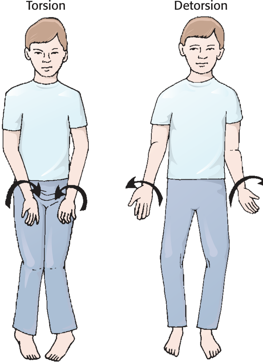
Technique:
- Administer IV analgesia (fentanyl preferred) ± spermatic cord block with 1% lidocaine at the superficial inguinal ring
- "Open the book" maneuver: Rotate the affected testicle medial to lateral (outward toward the thigh) - like opening a book
- Rotate 180° initially; may need 2-3 full rotations total
- If resistance or worsening pain, try the opposite direction (torsion is lateral in ~33% of cases)
- End point: Relief of pain OR return of intratesticular blood flow on Doppler ultrasound
Important caveats:
- Even after clinically successful manual detorsion, proceed to OR immediately - residual torsion has been reported in up to 32% of cases
- Infarction can occur with as little as 180° of remaining torsion
Step 3: Emergency Surgical Exploration (Definitive Management)
Approach: Scrotal midline raphe incision (gives bilateral access via one incision) OR two separate anterolateral hemiscrotal incisions.
Procedure steps:
- Incise skin → dartos fascia → open tunica vaginalis
- Approach affected side first
- Drain any reactive hydrocele
- Deliver testis, inspect viability, assess direction/degrees of torsion
- Detorse the spermatic cord
- Wrap in warm saline sponges for 10 minutes to assess reperfusion
Step 4: Assess Viability - Orchiopexy vs. Orchiectomy
Test for viability:
- Make a short incision in the tunica albuginea through the medulla
- Observe for active bleeding within 10 minutes
- Bleeding present = viable → proceed with orchiopexy
- No bleeding = not viable → proceed with orchiectomy
If Viable - Orchiopexy
- Trim excess tunica vaginalis, achieve hemostasis
- Place 3-point fixation sutures (medially, laterally, inferiorly) - tack tunica albuginea to dartos layer with absorbable sutures (e.g., 4-0 PDS)
- Goal: place testis in extravaginal position to prevent recurrent intravaginal torsion
- Close dartos with absorbable sutures; skin with 4-0/5-0 absorbable subcuticular sutures
If Non-Viable - Orchiectomy
- Divide cord structures between clamps, ligate with 2-0 Vicryl/silk
- In postpubertal males: consider combined orchiectomy + testicular prosthesis placement in the same sitting (avoids second anesthetic; good outcomes reported)
Step 5: Contralateral Orchiopexy (ALWAYS in Intravaginal Torsion)
Mandatory for adolescents/young adults because the bell-clapper deformity is bilateral in most cases - the contralateral testis is at high risk of asynchronous torsion.
- Same 3-point fixation technique as above
- NOT indicated for neonatal (extravaginal) torsion - different mechanism, not bilateral
Special Situations
Neonatal Torsion
- Usually extravaginal, typically in utero
- Presents as firm, darkened, minimally tender hemiscrotum
- Testis usually non-viable - orchiectomy required
- No contralateral orchiopexy needed
Intermittent Testicular Torsion (ITT)
- Testis untwists spontaneously; recurrent acute pain episodes
- Diagnosis often missed; orchiopexy is recommended electively given low surgical morbidity and high risk of eventual infarction
Torsion of Testicular Appendage (Appendix Testis / Hydatid of Morgagni)
- More common in prepubescent boys
- Less intense pain at superior pole of testis
- "Blue dot sign" (visible blue spot through scrotal skin)
- Conservative management (analgesia, NSAIDs); does not threaten fertility
- Surgery only if cannot distinguish from testicular torsion
Summary Algorithm
Acute Scrotal Pain
↓
Clinical assessment (high-riding testis, absent cremasteric reflex, sudden onset)
↓
High suspicion → IMMEDIATE urology consult + OR preparation
↓
Attempt manual detorsion (bedside) simultaneously
↓
Scrotal exploration
↓
Viable?
YES → Orchiopexy (ipsilateral) + Contralateral orchiopexy
NO → Orchiectomy (± prosthesis) + Contralateral orchiopexy
Sources: Bailey & Love's Short Practice of Surgery 28e; Sabiston Textbook of Surgery; Rosen's Emergency Medicine; Roberts & Hedges' Clinical Procedures in Emergency Medicine; Hinman's Atlas of Urologic Surgery; Campbell-Walsh-Wein Urology
This is a shared conversation. Sign in to Orris to start your own chat.