
Syphilis Staph aureus Clostridium Pneumonia Dipteria Micro biology from apurba sharsthry
Treponema pallidum syphilis spirochete dark field microscopy
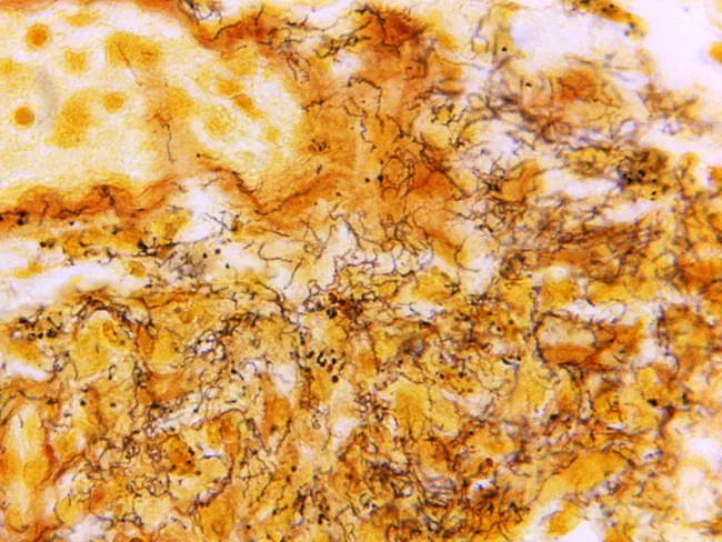
This histology image depicts a lymph node section stained with Steiner silver stain, visualizing Treponema pallidum organisms within the node. Light microscopy reveals numerous dark, corkscrew-shaped spirochetes aligned along walls of capsular vessels and scattered through the interstitial stroma, consistent with syphilitic lymphadenitis. The tissue architecture shows preserved lymphoid elements with areas of mild follicular hyperplasia and perivascular inflammation, overlaid by slender, hyaline backgrounds produced by the stain. Steiner silver staining yields high-contrast, fine filaments that stand out against the pale counterstain, enabling direct visualization of spirochetes in situ. Although histology can be variable in syphilis, the detection of multiple organisms strongly supports infection and guides confirmation with serology. This image exemplifies the complementary roles of histopathology, immunohistochemistry, and molecular methods (PCR on lymph node biopsy or fine-needle aspirate) in diagnosing syphilitic lymphadenitis. Clinically, the diagnosis is most reliably established by serologic tests (treponemal and non-treponemal assays) alongside clinical context and patient history. The image is part of the CDC educational collection (Skip Van Orden), serving as a teaching resource for infectious disease pathology, dermatopathology, and surgical pathology workflows in recognizing T. pallidum. This description emphasizes spirochete visualization as a diagnostic clue.
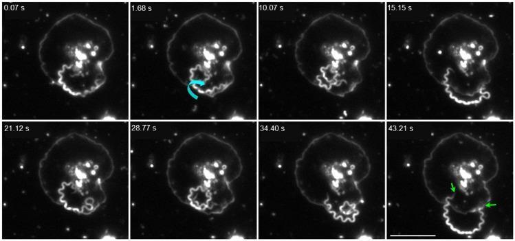
This darkfield video microscopy time series (1000x magnification) illustrates the dynamic interaction between the spirochete Treponema pallidum and a human platelet over a period of 43.21 seconds. The imagery displays a single treponeme, characterized by its distinctive helical morphology, interacting with a fully activated, spread platelet. The sequence captures various behavioral states: at 1.68 s, a cyan curved arrow indicates coiling and vigorous axial rotation against the platelet membrane. In subsequent frames (15.15 s – 34.40 s), the spirochete is seen extending away from or above the platelet surface. The final frame at 43.21 s highlights tip-mediated adhesion, where green arrows point to both distal ends of the treponeme anchored to the platelet membrane. This visual evidence demonstrates the ability of T. pallidum to recognize and physically engage with activated components of the human hemostatic system. A 10 μm scale bar is provided for reference in the final frame.
Staphylococcus aureus gram positive cocci clusters
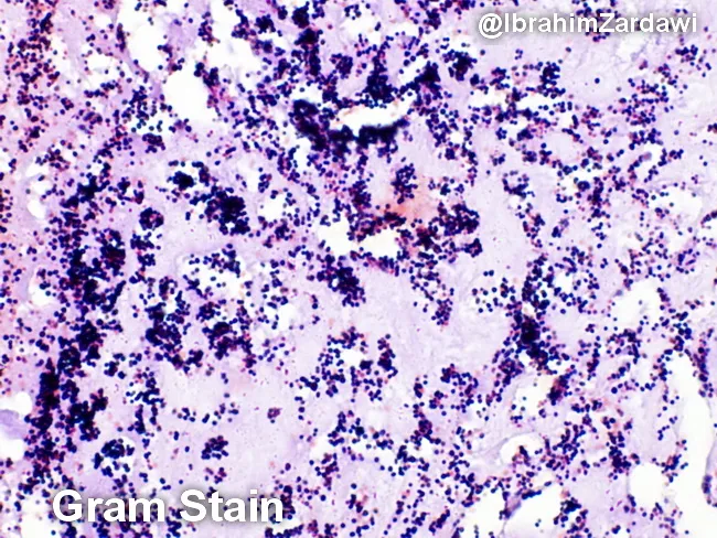
Imaging modality: Light microscopy; Gram staining of a histologic section of a cardiac valve vegetation from suspected infective endocarditis. The specimen shows a dense, friable vegetative mass within valve tissue characterized by a fibrinous, platelet-rich matrix in which numerous Gram-positive cocci are adherent. On the Gram stain, the cocci appear as purple spherical cells arranged in clusters and sometimes in short chains, consistent with Gram-positive organisms. The background demonstrates inflammatory cells and debris typical of endocardial infection. The histologic architecture indicates vegetative lesions with microbial colonization and a biofilm component. These findings correlate with infective endocarditis due to cocci such as Staphylococcus aureus or viridans-group streptococci in clinical practice, though Gram stain alone cannot specify species. The image is a micrograph provided with permission by Dr. Ibrahim Zardawi; image shows classic histopathology of bacterial endocarditis. The diagnostic significance lies in confirming bacterial endocarditis and guiding immediate management including empiric antibiotic therapy targeting Gram-positive cocci while awaiting culture results. This image is valuable for pathology education, microbiology correlation, and endocarditis research. Potential clinical use cases include training in Gram stain interpretation, distinguishing bacterial endocarditis from sterile vegetations, and supporting diagnosis when blood cultures are pending.
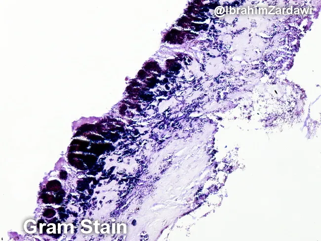
This histology image is a light-micrograph of a Gram-stained valvular vegetation from a case of infective endocarditis. The specimen is cardiac valvular tissue; the vegetation comprises a fibrin-rich, platelet-laden thrombus with embedded bacterial colonies along the endocardial surface. Gram staining highlights Gram-positive cocci as purple to deep purple clusters within the thrombus, indicating organisms such as Streptococcus spp., Staphylococcus spp., or Enterococcus. The bacterial colonies are adherent to the valvar surface within a dense inflammatory milieu, with neutrophilic infiltrate and tissue necrosis limited to microdomains. The histologic architecture demonstrates vegetations typical of endocarditis: a friable, topographically irregular lesion on the valve with microabscess-like foci and bacterial encasement within fibrin. The presence of Gram-positive bacteria on histology supports a bacterial endocarditis diagnosis and guides antimicrobial therapy pending culture confirmation. Clinically, such histologic confirmation is critical for distinguishing infectious from non-infectious valve pathology, for clarifying the etiologic category of pathogens (oral flora-related viridans Streptococcus, Staphylococcus aureus, enterococci), and for correlating with blood culture, echocardiographic findings, and patient risk factors. This image is valuable for educational purposes, illustrating Gram stain morphology in endocardial vegetations and reinforcing the need for targeted antibiotics and possible surgical consultation.
diphtheria pseudomembrane Corynebacterium
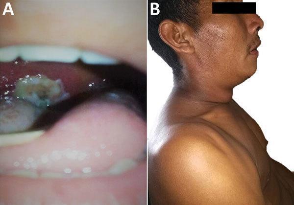
This composite clinical photograph displays the hallmark signs of respiratory diphtheria in a 31-year-old male. Image A is an intraoral view of the oropharynx showing a thick, adherent, gray-white pseudomembrane covering the tonsillar and pharyngeal area. The membrane appears leathery and rough in texture, a characteristic finding caused by the necrotizing exotoxin of Corynebacterium diphtheriae. Image B is a lateral profile of the same patient demonstrating significant soft tissue swelling of the cervical region, commonly referred to as a 'bull-neck' appearance. This manifestation is due to massive cervical lymphadenopathy and associated perilymphatic edema. These images serve as a classic diagnostic reference for medical professionals and students to identify the infectious manifestations of diphtheria, emphasizing the clinical triad of pharyngitis, pseudomembrane formation, and profound cervical swelling.
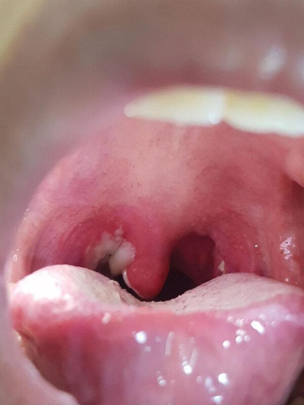
This clinical photograph displays a close-up intraoral view of the oropharynx in a pediatric patient. The primary finding is a classic, thick, grayish-white pseudomembrane that is firmly adherent to the pharyngeal wall and tonsillar region. The surrounding mucosal tissues, including the uvula and soft palate, exhibit significant erythema and inflammation. This visual presentation is characteristic of pharyngeal diphtheria caused by toxigenic Corynebacterium diphtheriae. The pseudomembrane is a hallmark diagnostic sign, formed by a dense collection of necrotic epithelium, fibrin, leukocytes, and bacteria. In the foreground, the tongue and lower teeth are visible, providing anatomical orientation. This image serves as a critical educational example for infectious disease diagnosis, particularly in the context of vaccine-preventable respiratory infections and their clinical manifestations like 'bull neck' lymphadenopathy and systemic toxemia.
Streptococcus pneumoniae lobar pneumonia chest X-ray consolidation
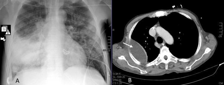
This dual-panel image displays diagnostic thoracic imaging from a patient with necrotizing pneumonia. Panel A is a posterior-anterior (PA) chest X-ray showing significant homogeneous opacity and consolidation in the middle and lower zones of the right lung, characteristic of lobar pneumonia. Normal lung markings are obscured on the right, while the left lung field shows more multifocal, patchy interstitial opacities. Panel B is a transverse (axial) contrast-enhanced CT scan of the thorax. A white arrow identifies a large, well-defined, peripherally located area of low attenuation in the posterior-lateral aspect of the right lung, representing a lung abscess. The lesion is lens-shaped, abuts the pleural surface, and exerts a mass effect on the adjacent pulmonary parenchyma. Central mediastinal structures, including the heart and great vessels, are visible and appear within normal limits. These images illustrate the progression of community-acquired pneumonia to necrotizing disease and abscess formation, a significant complication often associated with Streptococcus pneumoniae and SARS-CoV-2 coinfection.
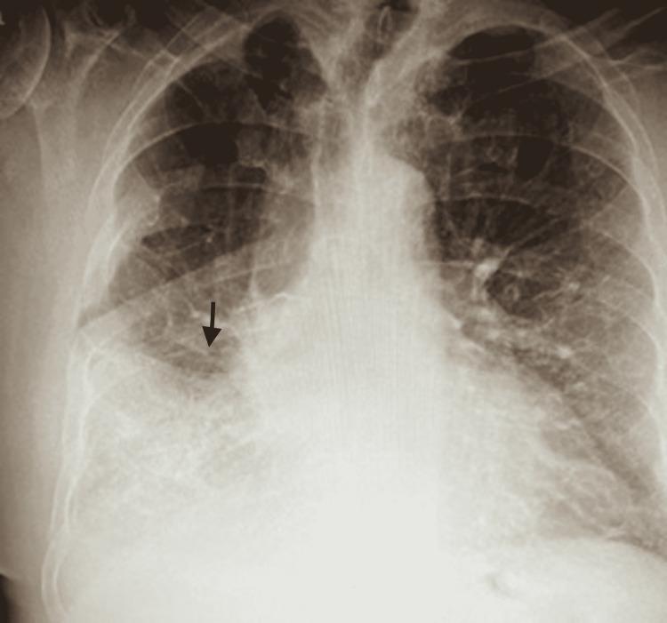
This diagnostic image is an anteroposterior (AP) chest X-ray demonstrating a significant area of increased opacity in the right mid-to-lower lung field. The opacity exhibits a patchy, heterogeneous texture and ill-defined borders, characteristic of an alveolar infiltrate or consolidation. A black arrow points to the superior margin of the infiltrate, which correlates clinically with consolidation in the right lower lobe. Visible air bronchograms within the dense area suggest the replacement of alveolar air with inflammatory fluid or exudate, common in community-acquired pneumonia (CAP). The left lung field remains relatively clear with normal vascular markings. This radiograph serves as a key educational example of lobar consolidation patterns in pulmonary infectious diseases, illustrating the radiographic hallmarks of bacterial pneumonia, such as Legionella or Streptococcus pneumoniae, and their manifestation as focal opacities on chest radiography.
Clostridium perfringens gas gangrene myonecrosis
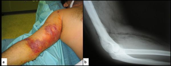
This composite educational material displays clinical and radiological findings of gas gangrene (clostridial myonecrosis) in the upper limb of a patient with a history of intravenous drug use. Figure (a) is a clinical photograph of the right arm showing severe localized edema, circumferential erythema, and large, dusky, hemorrhagic bullae (blistering) across the bicep and antecubital regions. Areas of purple-to-black skin discoloration suggest impending tissue necrosis. Figure (b) is a lateral X-ray of the same limb, demonstrating the pathognomonic sign of gas gangrene: extensive subcutaneous emphysema. This is characterized by radiolucent (dark) gas pockets and streaks dissecting through the soft tissue planes and muscle compartments, indicating gas production by anaerobic organisms such as Clostridium perfringens. The image serves to teach the correlation between clinical signs of rapidly progressing necrotizing soft tissue infection (crepitus, bullae, edema) and the critical radiographic finding of soft tissue gas.
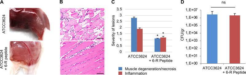
Educational figure illustrating the inhibitory effects of the 6-R peptide on Clostridium perfringens type A-induced gas gangrene in a mouse model. (A) Clinical photographs showing macroscopic pathology: the limb infected with ATCC 3624 alone displays severe dark-red discoloration and edema indicative of hemorrhage and necrosis, while the 6-R peptide-treated limb shows significantly reduced inflammation. (B) Histopathological H&E-stained sections (scale bar: 50 μm) comparing microscopic changes: ATCC 3624 alone causes severe myonecrosis with disorganized, fragmented muscle fibers and widespread cellular damage; in contrast, the 6-R peptide-treated sample maintains organized muscle architecture with minimal inflammatory neutrophil infiltration. (C) Bar graph quantifying histological scores for muscle degeneration/necrosis and inflammation, showing a statistically significant reduction (p < 0.05) in lesion severity with 6-R peptide treatment. (D) Growth yield comparison (CFU/gr) showing no significant difference (ns) in bacterial recovery between the groups, indicating that the peptide reduces virulence rather than inhibiting bacterial growth in vivo. This figure demonstrates the role of quorum-sensing inhibition in mitigating toxin-mediated tissue damage.
Microbiology Notes: 5 High-Yield Topics
1. SYPHILIS
Transmission
- Sexually transmitted (direct contact with infectious lesion)
- Transplacental - congenital syphilis
- Blood transfusion (rare)
Stages of Syphilis
| Stage | Features |
|---|---|
| Primary | Painless indurated ulcer (chancre) at site of inoculation; appears 2-6 weeks after exposure; regional lymphadenopathy; heals spontaneously in 3-8 weeks |
| Secondary | Systemic dissemination; fever, generalized lymphadenopathy, maculopapular rash (palms + soles - hallmark), condylomata lata, mucous patches, hepatitis, nephritis, CNS involvement; highly infectious |
| Latent | Reactive serology, no clinical signs. Early latent (<1 year), late latent (>1 year) |
| Tertiary (Late) | Cardiovascular (aortic aneurysm, aortic regurgitation, coronary stenosis), neurosyphilis (general paresis, tabes dorsalis, Argyll Robertson pupil), gummas |
Congenital Syphilis
- Early: Rhinitis ("snuffles"), skin rash, hepatosplenomegaly, osteochondritis
- Late: Hutchinson's teeth, interstitial keratitis, 8th nerve deafness (Hutchinson's triad), saddle nose, saber shins
Diagnosis
| Test | Use |
|---|---|
| Dark-field microscopy | Primary syphilis (chancre exudate) |
| VDRL / RPR (non-treponemal) | Screening; titers correlate with disease activity; used to monitor treatment response |
| FTA-ABS / MHA-TP / TPHA (treponemal) | Confirmatory; remain positive for life |
| CSF-VDRL | Neurosyphilis |
Secondary syphilis is the stage where serology is most reliable: RPR is 99% sensitive, MHA-TP 100% sensitive.
Treatment
- Primary/Secondary/Early latent: Benzathine penicillin G 2.4 MU IM single dose
- Late/Tertiary: Benzathine penicillin G 2.4 MU IM weekly x 3 doses
- Neurosyphilis: Aqueous penicillin G IV 10-14 days
- Penicillin allergy: Doxycycline or tetracycline (non-pregnant); desensitization in pregnancy
- Jarisch-Herxheimer reaction: Fever, chills, headache 2-8 hours after first treatment dose - due to massive release of treponemal antigens
2. STAPHYLOCOCCUS AUREUS
Key Characteristics
- Catalase positive (distinguishes Staph from Strep)
- Coagulase positive (distinguishes S. aureus from coagulase-negative staph)
- Golden colonies on blood agar (pigment = staphyloxanthin)
- Beta-hemolytic on blood agar
- Ferments mannitol (on mannitol-salt agar - selective medium)
- Protein A on cell wall binds Fc region of IgG - evades opsonization
Virulence Factors
| Factor | Effect |
|---|---|
| Coagulase | Converts fibrinogen to fibrin - "walling off" infection |
| Exfoliative toxin A & B | Splits desmoglein-1 - scalded skin syndrome |
| TSST-1 (Toxic Shock Syndrome Toxin) | Superantigen - massive cytokine release |
| Enterotoxins A-E | Heat-stable; pre-formed in food; cause food poisoning |
| Leukocidin (PVL) | Kills WBCs; associated with necrotizing pneumonia and furunculosis |
| Hyaluronidase | Spreading factor |
| Protein A | Binds IgG Fc - immune evasion |
| Coagulase + clumping factor | Fibrin clot formation |
Diseases
- Scalded Skin Syndrome (SSSS/Ritter disease): Exfoliative toxins; neonates; superficial blister formation; Nikolsky sign positive; no organisms in bullae
- Toxic Shock Syndrome (TSS): TSST-1 (superantigen); fever, hypotension, diffuse erythematous rash, multiorgan failure; associated with tampon use (menstrual TSS) and wound infections
- Food Poisoning: Preformed heat-stable enterotoxin; rapid onset (1-6 hours); vomiting > diarrhea; self-limiting (<24 hours)
- Impetigo, folliculitis, furuncle (boil), carbuncle
- Bacteremia and infective endocarditis (right-sided in IV drug users)
- Pneumonia: Especially post-influenza; necrotizing; cavitation; pneumatoceles in children
- Osteomyelitis (metaphysis of long bones in children)
- Septic arthritis
MRSA (Methicillin-Resistant S. aureus)
- Resistance via mecA gene - encodes altered PBP2a (penicillin-binding protein)
- Hospital-acquired (HA-MRSA): Multi-drug resistant
- Community-acquired (CA-MRSA): Often PVL-positive; causes skin/soft tissue infections and necrotizing pneumonia
Diagnosis
- Gram stain: Gram-positive cocci in clusters
- Coagulase test (tube and slide)
- Mannitol-salt agar (ferments mannitol = yellow colonies)
- DNase test positive
- MRSA: chromogenic agar, NAAT
Treatment
| Infection | Drug |
|---|---|
| MSSA | Flucloxacillin / Nafcillin / Oxacillin |
| MRSA (systemic) | Vancomycin (drug of choice) |
| MRSA (oral) | TMP-SMX, doxycycline, clindamycin, linezolid |
| Alternatives | Daptomycin, tigecycline, linezolid |
| Food poisoning | Symptomatic only |
3. CLOSTRIDIUM
Major Clostridial Species
A. Clostridium difficile (C. diff)
- Toxin A (enterotoxin): Attracts neutrophils, stimulates cytokine release, fluid secretion
- Toxin B (cytotoxin): Increases intestinal permeability; more potent; used for lab diagnosis
- Antibiotic-associated diarrhea (5-10 days after antibiotics)
- Pseudomembranous colitis: Profuse watery diarrhea, abdominal cramps, fever; whitish plaques (pseudomembranes) on colonic mucosa
- Discontinue the offending antibiotic
- Mild-moderate: Metronidazole (oral)
- Severe/recurrent: Vancomycin (oral) or fidaxomicin
- Refractory: Fecal microbiota transplant (FMT)
B. Clostridium perfringens
- Alpha toxin (lecithinase/phospholipase C): Most important; causes myonecrosis, massive hemolysis, platelet destruction; "Nagler reaction" positive
- Enterotoxin (heat-sensitive): Food poisoning - binds small intestinal epithelium causing watery diarrhea
- Type A (most human infections): Gas gangrene, food poisoning, necrotizing enteritis
- Type C: Necrotizing enterocolitis (pigbel disease)
- Contaminated wounds (trauma, surgery)
- Incubation: <24 hours
- Rapidly spreading muscle necrosis; gas in tissue (crepitus); bronze-brown skin discoloration; sweet/foul odor; systemic toxemia
- X-ray: Gas in soft tissues (pathognomonic)
- Contaminated meat (held at 5-60°C)
- Incubation: 8-24 hours
- Watery diarrhea + cramps; no vomiting; self-limiting
- Gas gangrene: Surgical debridement (emergency) + high-dose IV penicillin G + hyperbaric oxygen
- Food poisoning: Symptomatic
C. Clostridium tetani
- Trismus ("lockjaw") - first sign
- Risus sardonicus (sardonic smile)
- Opisthotonos (arched back)
- Reflex spasms; autonomic instability
D. Clostridium botulinum
- Food-borne: Canned/preserved foods (toxin ingested pre-formed); descending paralysis, no fever
- Infant botulism: Honey ingestion; "floppy baby"; most common form in USA
- Wound botulism: Rare; IV drug users
4. PNEUMONIA (Microbiology Focus)
Classification of Pneumonia by Organism
| Type | Key Organisms |
|---|---|
| Community-Acquired (CAP) | S. pneumoniae (most common), H. influenzae, Atypicals (Mycoplasma, Chlamydophila, Legionella), S. aureus (post-viral) |
| Hospital-Acquired (HAP) | Gram-negative rods (Klebsiella, Pseudomonas, E. coli), MRSA |
| Aspiration | Oral anaerobes (Bacteroides, Peptostreptococcus), S. pneumoniae |
| Immunocompromised | Pneumocystis jirovecii (PCP), Aspergillus, CMV, Nocardia |
| Neonatal | Group B Strep, Klebsiella, E. coli |
Key Organisms - High-Yield
- Most common cause of CAP, meningitis, otitis media, sinusitis
- Gram-positive lancet-shaped diplococci; alpha-hemolytic; bile-soluble; optochin sensitive
- Virulence: Polysaccharide capsule (anti-phagocytic) - Quellung reaction
- Diagnosis: Sputum Gram stain; blood culture; urinary antigen test
- Treatment: Amoxicillin / Amoxicillin-clavulanate; ceftriaxone; penicillin-resistant strains: levofloxacin or vancomycin
- Prevention: PCV13 (conjugate) and PPSV23 (polysaccharide) vaccines
- Gram-negative rod; polysaccharide capsule
- "Currant jelly sputum" (bloody, mucoid)
- Risk groups: Alcoholics, diabetics, debilitated patients
- Causes lobar pneumonia with cavitation in upper lobe
- Treatment: Cephalosporins, carbapenems; note: KPC strains (carbapenem-resistant) are emerging
- No cell wall (Gram stain negative); not visible on Gram stain
- "Walking pneumonia" - mild, gradual onset; young adults, school outbreaks
- Cold agglutinins (IgM) - agglutinate RBCs at 4°C
- Chest X-ray: patchy bilateral infiltrates; worse than clinical signs suggest
- Diagnosis: PCR; cold agglutinin titer; serology (complement fixation)
- Treatment: Macrolides (azithromycin) or doxycycline; NOT beta-lactams (no cell wall)
- Gram-negative rod; not seen on Gram stain (stains poorly); silver stain or DFA
- Water-cooled AC systems, cooling towers; inhalation of aerosols
- Legionnaires' disease: Severe pneumonia + hyponatremia + GI symptoms + confusion; "Pontiac fever" = milder flu-like illness
- Diagnosis: Urinary antigen test (detects serogroup 1 - most common); culture on BCYE agar (L-cysteine dependent)
- Treatment: Fluoroquinolones or azithromycin
- Post-influenza (superinfection), IV drug users (hematogenous)
- Cavitation, abscesses, pneumatoceles
- Rapid, severe; necrotizing pneumonia (CA-MRSA with PVL toxin)
- Treatment: Vancomycin (MRSA) or nafcillin (MSSA)
- Fungus (previously classified as protozoa)
- Causes pneumonia in immunocompromised (HIV with CD4 <200, transplant recipients)
- Bilateral interstitial infiltrates; "ground-glass" appearance on CT
- Diagnosis: BAL with Gomori methenamine silver (GMS) stain
- Treatment: TMP-SMX (drug of choice); dapsone or atovaquone as alternatives
- Prophylaxis: TMP-SMX when CD4 <200
Radiological Patterns
| Pattern | Organisms |
|---|---|
| Lobar consolidation | S. pneumoniae, Klebsiella |
| Bronchopneumonia (patchy) | S. aureus, H. influenzae |
| Interstitial (diffuse) | Mycoplasma, viruses, PCP |
| Cavitation | S. aureus, Klebsiella, TB, anaerobes |
5. DIPHTHERIA
- Slender, pleomorphic gram-positive rod with club-shaped ends
- "Chinese letter" or "picket fence" arrangement on microscopy
- Metachromatic granules (Babes-Ernst granules / Volutin granules) - stain reddish-purple with methylene blue (Albert's stain)
Key Features
- Toxigenic strains carry bacteriophage beta (β) encoding the diphtheria toxin
- Only toxigenic strains cause disease
- Toxigenicity tested by: Elek's test (in vitro immunoprecipitation test - gold standard) and NAAT
Diphtheria Toxin - Mechanism
- A-B toxin: B fragment binds host cell; A fragment is enzymatically active
- Fragment A: ADP-ribosylates Elongation Factor-2 (EF-2) → inhibits protein synthesis → cell death
- Even a single toxin molecule can kill a cell by inactivating >1 million EF-2 molecules
Clinical Features - Respiratory Diphtheria
- Sore throat, low-grade fever
- Pseudomembrane - tough, grey-white, firmly adherent membrane on tonsils/pharynx; bleeds when removed; extends to larynx/trachea ("spread upward or downward")
- Bull-neck - massive cervical lymphadenopathy + soft tissue edema (severe cases)
- Laryngeal involvement: hoarseness, barking cough, stridor, potential asphyxia
- Myocarditis (leading cause of death): Fatty change, myofiber necrosis; arrhythmias; cardiac failure; occurs in weeks 1-2
- Neuropathy (Polyneuritis): Demyelination; palatal palsy (nasal voice, nasal regurgitation) earliest; then cranial nerve palsies (oculomotor - squint), peripheral neuropathy
- Renal tubular damage (rare)

Cutaneous Diphtheria
- Chronic, non-healing ulcer with dirty grey membrane
- Does NOT cause systemic toxicity
- Common in tropical regions and homeless populations
Diagnosis
| Method | Details |
|---|---|
| Gram stain | Gram-positive club-shaped rods in "Chinese letter" arrangement |
| Albert's stain / Methylene blue | Metachromatic granules (Babes-Ernst bodies) |
| Culture | Loeffler's serum slope (rapid growth), Tellurite agar (black colonies - selective) |
| Elek's test | Gold standard for toxin detection (immunoprecipitation) |
| PCR | Detects tox gene |
Treatment
- Diphtheria antitoxin (DAT) - most important; given immediately on clinical suspicion (does not wait for culture); neutralizes circulating toxin only (cannot reverse bound toxin)
- Antibiotics (eliminate the organism, prevent further toxin production and transmission):
- Penicillin G (IV/IM) or erythromycin x 14 days
- Patient isolation and contact tracing
Prevention - Immunization
- DTP vaccine (Diphtheria Toxoid + Tetanus Toxoid + Pertussis)
- Toxoid = formalin-inactivated toxin - stimulates antitoxin antibodies
- Schedule: 6 weeks, 10 weeks, 14 weeks (primary), 15-18 months (booster), pre-school
- Schick test (historical): Intradermal injection of diphtheria toxin; positive = susceptible (no antibodies); negative = immune
Quick Summary Table
| Topic | Organism | Key Feature | Treatment |
|---|---|---|---|
| Syphilis | T. pallidum (spirochete) | Painless chancre → rash on palms/soles → gummas/tabes | Benzathine Penicillin G |
| Staph aureus | Gram+ cocci in clusters | Coagulase+, TSST-1, exfoliative toxin, MRSA | Vancomycin (MRSA); Flucloxacillin (MSSA) |
| C. difficile | Gram+ anaerobic rod | Toxin A+B, pseudomembranous colitis | Vancomycin (oral) / Fidaxomicin / FMT |
| C. perfringens | Gram+ anaerobic rod | Alpha toxin, gas gangrene, food poisoning | Surgery + Penicillin G |
| C. tetani | Gram+ anaerobic rod | Tetanospasmin, spastic paralysis | Metronidazole + HTIG |
| C. botulinum | Gram+ anaerobic rod | Botulinum toxin, flaccid paralysis | Antitoxin + supportive |
| Pneumonia | Multiple organisms | S. pneumoniae most common CAP | Amoxicillin / Macrolide (CAP) |
| Diphtheria | C. diphtheriae | Pseudomembrane, EF-2 inhibition, toxin | DAT + Penicillin G/Erythromycin |
Sources: Murray's Medical Microbiology 9e, Robbins Pathologic Basis of Disease, Textbook of Family Medicine 9e - correlating with Apurba Sastry's Microbiology syllabus framework.