
From recent advances point of view, answer the following 1. IHC for CUP 2. IHC for lymphoma 3. IHC for metastatic tumors 4. IHC for breast carcinoma 5. IHC for lung tumors 6. IHC for undifferentiated tumors
Reading File
Now let me search the medical library in parallel for all 6 topics simultaneously.
Finding Sources
Finding Sources
Finding Sources
Finding Sources
Finding Sources
Finding Sources
Let me now read the key Harrison's CUP IHC section and search for more specific topics in parallel.
Reading File
Finding Sources
Finding Sources
Reading File
Reading File
Searching PubMed
immunohistochemistry carcinoma unknown primary CUP diagnosis
Searching the Web
IHC panel immunohistochemistry CUP lymphoma breast lung undifferentiated tumors 2024 2025 updates
Excellent - I have very rich data. Let me now fetch the CK7/CK20 flowchart image and the lung IHC panel image, and also read the lymphoma/undifferentiated section from Henry's Clinical Pathology in parallel.
Loading Image
Reading File
Reading File
Reading File
I now have comprehensive data from multiple authoritative sources. Let me fetch the lung IHC image and the full IHC table for lymphoma from Henry's, then compile the full answer.
Loading Image
Reading File
Reading File
I now have comprehensive, well-sourced information to write a thorough answer on all 6 topics. Let me compile the complete response.
IHC in Pathology: Recent Advances - A Comprehensive Review
1. IHC for Carcinoma of Unknown Primary (CUP)
CUP accounts for ~3-5% of all malignancies. On light microscopy, 60-65% are adenocarcinomas, 5% squamous cell carcinoma, and 30-35% poorly differentiated carcinoma or undifferentiated neoplasm. IHC is the cornerstone of workup, but the emphasis in recent guidelines (SEOM-GECOD 2025, NCCN 2025) has shifted: tissue must now be conserved for molecular profiling (NGS/CGP), and IHC should be used in a tiered, targeted manner rather than exhaustively.
The CK7/CK20 Framework (First Step)
The cytokeratin expression pattern is the initial filter for adenocarcinomas:
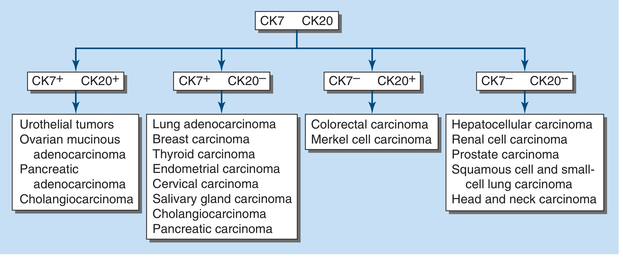
| CK7 | CK20 | Likely Primaries |
|---|---|---|
| + | + | Urothelial, ovarian mucinous, pancreatic, cholangiocarcinoma |
| + | - | Lung adenocarcinoma, breast, thyroid, endometrial, cervical, salivary gland, cholangiocarcinoma |
| - | + | Colorectal carcinoma, Merkel cell carcinoma |
| - | - | Hepatocellular carcinoma, renal cell carcinoma, prostate, SCC and SCLC, head & neck |
The CK7+/CK20- pattern is the most common in CUP but is not site-specific on its own, requiring further second-tier markers.
Expanded IHC Panel by Suspected Primary (Table 97-2, Harrison's 22E, 2025)
| Likely Primary | Key IHC Markers |
|---|---|
| Breast | ER, GCDFP-15, mammaglobin, HER2/neu, GATA3 |
| Ovarian/mullerian | ER, WT1, CK7, PAX8, PAX2 |
| Lung adenocarcinoma | TTF-1 (nuclear), napsin A, SP-A1 |
| Germ cell | beta-hCG, AFP, OCT3/4, CKIT, CD30 (embryonal), SALL4 |
| Prostate | PSA, AMACR/P504S, P501S, PSMA, NKX3-1 |
| Intestinal | CK20, CDX-2, CEA |
| Neuroendocrine | Chromogranin, synaptophysin, CD56 |
| Sarcoma | Desmin, factor VIII, CD31, smooth muscle actin, MyoD1 |
| Renal | RCC antigen, CD10, PAX8 |
| Hepatocellular | Hep Par-1, Arg-1, glypican-3 |
| Melanoma | S100, SOX-10, HMB-45, tyrosinase, melan-A |
| Urothelial | CK7, CK20, thrombomodulin, uroplakin III |
| Mesothelioma | Calretinin, WT1, D2-40, mesothelin |
| Lymphoma | LCA (CD45), CD3, CD4, CD5, CD20 |
| SCC | p63, p40, CK5/6 |
Recent advances:
- GATA3 is a newer, highly sensitive marker for breast and urothelial carcinomas
- NKX3-1 has replaced or supplements PSA for prostate - more specific
- PSMA (prostate-specific membrane antigen) is increasingly used for prostate CUP
- INSM1 is a newer neuroendocrine marker (nuclear transcription factor), more sensitive than chromogranin in poorly preserved specimens
- Molecular profiling (RNA-seq/CGP) is now recommended alongside IHC per the 2025 SEOM-GECOD guidelines; the CUPISCO trial (Lancet Oncol 2024) showed survival benefit when NGS guides site-specific treatment in CUP
- IHC alone is insufficient to guide site-specific treatment - results are "suggestive" not "definitive"
2. IHC for Lymphoma
IHC is essential for lymphoma subclassification according to WHO Classification of Haematolymphoid Tumours (5th edition, 2022). The approach is stepwise.
Step 1: Lineage Determination
| Marker | Lineage |
|---|---|
| LCA (CD45) | All lymphoid tumors |
| CD20, CD19, CD22, CD79a, PAX5 | B-cell |
| CD3, CD2, CD5, CD7 | T-cell |
| CD56, cytoplasmic CD3 | NK-cell |
Step 2: B-Cell Lymphoma Subclassification
| Lymphoma Type | Key IHC Markers |
|---|---|
| Follicular lymphoma | CD20+, CD10+, BCL2+, BCL6+, CD5-, cyclin D1- |
| Diffuse Large B-Cell (DLBCL) | CD20+, CD19+, CD22+, CD79a+; GCB type: CD10+/BCL6+/MUM1-; non-GCB: MUM1+ |
| Mantle Cell Lymphoma (MCL) | CD20+, CD5+, cyclin D1+, BCL2+, SOX11+; CD23-, CD10- |
| CLL/SLL | CD20(dim)+, CD5+, CD23+, CD200+, cyclin D1- |
| Burkitt Lymphoma | CD20+, CD10+, BCL6+, BCL2-, Ki-67 ~100%, c-MYC+ |
| Marginal Zone Lymphoma | CD20+, CD5-, CD10-, BCL2+, cyclin D1- |
| Hairy Cell Leukemia | CD20+, CD103+, CD25+, annexin A1+, BRAF V600E (IHC) |
DLBCL GCB vs. non-GCB (Hans algorithm) using IHC:
- GCB type: CD10+ (>30%) OR CD10-/BCL6+/MUM1- → better prognosis
- Non-GCB: MUM1+ → worse prognosis, responds differently to treatment
Step 3: T-Cell/NK Lymphoma Subclassification
| Lymphoma Type | Key IHC Markers |
|---|---|
| Peripheral T-cell, NOS | CD3+, CD4+>CD8+, loss of CD5/CD7 |
| Anaplastic Large Cell (ALCL) | CD30+, ALK+/-, EMA+, CD3+/- |
| Angioimmunoblastic T-cell | CD3+, CD4+, PD-1+, CXCL13+, ICOS+, BCL6+ |
| NK/T-cell nasal | CD56+, cytoplasmic CD3+, EBER+ (ISH), TIA1+, granzyme B+ |
Recent advances in lymphoma IHC:
- SOX11: Useful marker for cyclin D1-negative MCL; SOX11-negative MCL tends to be more indolent
- BRAF V600E IHC: Now used as a surrogate for mutation testing in hairy cell leukemia and some follicular lymphomas
- PD-L1 IHC: Increasingly important for DLBCL, primary mediastinal B-cell lymphoma, and Hodgkin lymphoma - guides checkpoint inhibitor eligibility
- MYC/BCL2/BCL6 double/triple-hit: IHC for MYC (>40%), BCL2 (>50%) triggers FISH confirmation; "double expressor" lymphomas have poorer prognosis
- EBER in situ hybridization (not strictly IHC but part of the panel) is mandatory for NK/T-cell and EBV-associated lymphomas
- IRF4/MUM1: Distinguishes GCB from non-GCB subtypes of DLBCL
3. IHC for Metastatic Tumors
The challenge is distinguishing primary from metastatic disease, especially when the primary is unknown or when multiple sites are present.
General Principles
- No single marker is 100% sensitive or specific - panels outperform individual markers
- TTF-1 stains ~75% of pulmonary adenocarcinomas but also stains thyroid carcinoma and occasionally ovarian tumors
- Organ-specific markers (PSA for prostate, thyroglobulin for thyroid) are highly specific but these rarely present as CUP
Common Differential Diagnoses and IHC
| Metastatic Site/Scenario | Primary | Key Positive Markers | Key Negative Markers |
|---|---|---|---|
| Liver metastasis | Colorectal | CK20+, CDX-2+, CEA+ | CK7-, TTF-1- |
| Liver metastasis | HCC (primary) | Hep Par-1+, Arg-1+, glypican-3+, AFP+ | CK7-, CK20- |
| Liver metastasis | Cholangiocarcinoma | CK7+, CK19+, K20± | Hep Par-1- |
| Pleural metastasis | Breast | ER+, GATA3+, mammaglobin+ | WT1-, mesothelin- |
| Pleural metastasis | Mesothelioma (vs mets) | Calretinin+, WT1+, D2-40+, mesothelin+ | CEA-, MOC31-, TTF-1- |
| Bone metastasis | Prostate | PSA+, NKX3-1+, PSMA+ | - |
| Brain metastasis | Lung adeno | TTF-1+, napsin A+ | - |
| Brain metastasis | RCC | PAX8+, CD10+, carbonic anhydrase IX (CAIX)+ | - |
| Axillary LN | Breast | ER+, GATA3+, GCDFP-15+ | - |
| Skin metastasis | Lung (non-small cell) | TTF-1+, CK7+, napsin A+ | CK20-, CDX-2- |
| Skin metastasis | Melanoma | S100+, SOX10+, HMB-45+, melan-A+ | Keratins- |
Recent advances:
- PAX8: One of the most useful newer markers - positive in renal, thyroid, ovarian/mullerian, and thymic tumors
- CAIX (carbonic anhydrase IX): Highly specific for clear cell RCC metastases
- SATB2: Highly specific for colorectal and appendiceal origin; supplements CDX-2
- NKX3-1: Now the preferred prostate-specific marker - more sensitive than PSA in poorly differentiated metastatic prostate carcinoma
- INSM1: Better neuroendocrine marker in small crushed biopsies vs chromogranin/synaptophysin
- Claudin-18.2: Emerging marker for gastric/GEJ adenocarcinoma metastases - also a therapeutic target (zolbetuximab)
- TRPS1: A newer, highly sensitive marker for breast origin (more sensitive than GATA3 in some studies)
4. IHC for Breast Carcinoma
IHC in breast carcinoma serves two main purposes: (a) diagnosis/classification, and (b) guiding therapy.
Routine Therapeutic Markers (Mandatory in all invasive breast carcinoma)
| Marker | Significance | Scoring/Threshold |
|---|---|---|
| ER (Estrogen Receptor) | Hormonal therapy eligibility (tamoxifen, aromatase inhibitors) | Allred score; ≥1% nuclear staining = positive (ASCO/CAP 2020 guidelines) |
| PR (Progesterone Receptor) | Complementary prognostic marker | Same threshold as ER |
| HER2/neu (ERBB2) | Trastuzumab, pertuzumab, T-DM1, T-DXd eligibility | IHC 0/1+/2+/3+; 3+ = positive; 2+ requires ISH confirmation |
| Ki-67 | Proliferation index; guides chemotherapy decisions | >20% (or >14% per St. Gallen) = high proliferation |
Molecular Subtype Classification by IHC Surrogates
| Subtype | ER | PR | HER2 | Ki-67 | Prognosis |
|---|---|---|---|---|---|
| Luminal A | + | + | - | Low (<14%) | Best |
| Luminal B (HER2-) | + | +/- | - | High (>14-20%) | Intermediate |
| Luminal B (HER2+) | + | +/- | + | Any | Intermediate |
| HER2-enriched | - | - | + | High | Poor |
| Triple-negative (TNBC) | - | - | - | High | Worst |
Additional Diagnostic/Subclassification Markers
| Marker | Use |
|---|---|
| GATA3 | Breast lineage marker; highly sensitive (~90%) for breast origin |
| GCDFP-15 (BRST-2) | Specific (~95%) but less sensitive (~50%) for breast origin |
| Mammaglobin | Moderate sensitivity/specificity; used with GCDFP-15 |
| E-cadherin | Distinguishes lobular (negative) from ductal (positive) carcinoma |
| p120 catenin | Cytoplasmic in lobular carcinoma; membranous in ductal |
| Androgen Receptor (AR) | Positive in ~70-90% luminal A; therapeutic target in TNBC (LAR subtype) |
| CK5/6, EGFR | Basal-like phenotype within TNBC |
| PD-L1 | Pembrolizumab eligibility in TNBC (CPS score with 22C3 antibody) |
Recent advances (2023-2025):
- HER2-low (IHC 1+ or 2+/ISH-) is now a distinct therapeutic category - eligible for trastuzumab deruxtecan (T-DXd); this has redefined HER2 reporting
- HER2-ultralow (IHC >0 and <1+): Emerging category with potential T-DXd benefit (under investigation)
- TRPS1: Newer breast marker with high sensitivity, even in triple-negative cases
- BIRD3/LRRC15: Emerging markers for stromal biology in TNBC
- PIK3CA IHC surrogates: Being investigated as predictors of alpelisib response
5. IHC for Lung Tumors
The 2021 WHO Classification of Thoracic Tumors has expanded IHC requirements for lung tumor subclassification.
Adenocarcinoma vs. Squamous Cell Carcinoma (Core Panel)
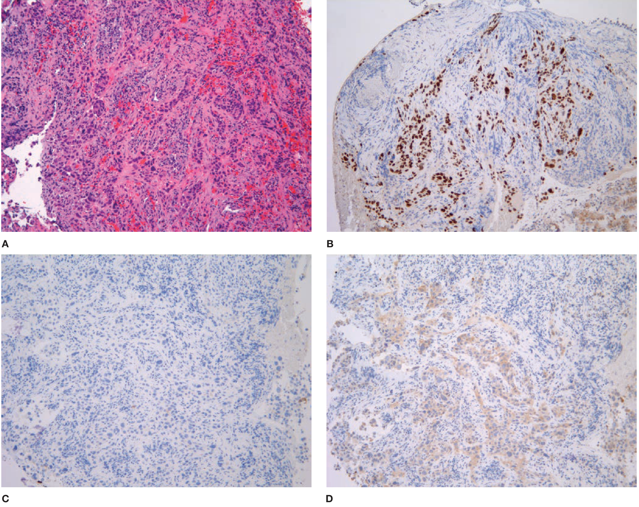
The minimum recommended panel for poorly differentiated NSCLC (Fishman's Pulmonary Diseases, citing Best Practices recommendations):
| Marker | Adenocarcinoma | Squamous Cell Carcinoma |
|---|---|---|
| TTF-1 | + (~75% of cases) | - |
| Napsin A | + (more specific than TTF-1) | - |
| p40 | - | + (most specific for SCC) |
| CK5/6 | - | + |
| p63 | - | + (less specific than p40) |
| CK7 | + | +/- |
Minimum panel: TTF-1 + p40 (±napsin A or CK5/6)
This IHC panel is clinically critical because:
- Adenocarcinoma → molecular testing for EGFR, ALK, ROS1, KRAS, BRAF, MET, RET, NTRK, PD-L1
- SCC → immunotherapy assessment; bevacizumab is contraindicated
Neuroendocrine Tumors (NET) Panel
| Tumor | Synaptophysin | Chromogranin | CD56 | Ki-67 | TTF-1 |
|---|---|---|---|---|---|
| Typical carcinoid | + | + | + | <5% | - |
| Atypical carcinoid | + | + | + | 5-20% | - |
| Large cell NEC | + | +/- | + | >50% | +/- |
| SCLC | + | +/- | + | 80-100% | + (~90%) |
- INSM1 is now recommended as a supplementary neuroendocrine marker; it is nuclear, works well in crushed small biopsies where cytoplasmic markers are lost
- Ki-67 is used as a surrogate for mitotic counting in small biopsies
Other Lung Tumor IHC
| Tumor | Key Markers |
|---|---|
| Pulmonary carcinoid | Synaptophysin+, chromogranin+, CK7+, TTF-1 usually -, low Ki-67 |
| Mesothelioma vs. lung adeno | Calretinin+, WT1+, D2-40+, mesothelin+ (meso) vs TTF-1+, napsin A+, MOC-31+ (adeno) |
| Pulmonary large cell carcinoma | Diagnosis of exclusion - negative for TTF-1, napsin A, p40, CK5/6, neuroendocrine markers |
| NUT carcinoma | NUT IHC (nuclear, speckled pattern) - highly specific; SMARCA4 loss |
| SMARCA4-deficient thoracic sarcoma | SMARCA4 (BRG1) loss by IHC |
Recent advances:
- PD-L1 (22C3 clone): Mandatory for all advanced NSCLC; TPS ≥50% = pembrolizumab monotherapy eligible; TPS ≥1% = combination eligible
- ALK (D5F3 clone): IHC now accepted as a standalone test without mandatory FISH confirmation in many centers
- ROS1 IHC (D4D6 clone): Screening marker - positive cases confirmed by FISH/NGS
- INSM1: Added to 2021 WHO-recommended neuroendocrine panel
- SMARCA4/SMARCB1 loss: IHC markers for rare undifferentiated thoracic malignancies
- Claudin-18: In lung adenocarcinoma subtyping and as a potential therapeutic target
- HER2 IHC: Being validated for lung adenocarcinoma (as in breast) - trastuzumab deruxtecan now approved for HER2-mutant NSCLC
6. IHC for Undifferentiated Tumors
When H&E morphology cannot determine tumor lineage, the goal of IHC is to classify undifferentiated neoplasms into one of five broad categories before applying subtype-specific panels:
Step 1: The "Big Five" Lineage Screen
| Marker | What It Identifies |
|---|---|
| Broad-spectrum keratin (MNF116, AE1/AE3, CAM5.2) | Carcinoma / epithelial origin |
| LCA (CD45) | Lymphoma / hematolymphoid |
| S100 / SOX10 / HMB-45 | Melanoma |
| Vimentin + CD31/CD34/SMA/desmin | Sarcoma (mesenchymal) |
| OCT3/4 / SALL4 | Germ cell tumor |
NCCN 2025 OCC-A panel for undifferentiated neoplasms:
Initial screen → broad keratin (AE1/AE3) + LCA (CD45) + S100 + vimentin
                ↓                    ↓              ↓
           Carcinoma            Lymphoma         Melanoma/sarcoma
             panel                panel              panel
Step 2: After Lineage Confirmation
If carcinoma confirmed: proceed with CK7/CK20 algorithm (see Section 1 above)
If lymphoma suspected: LCA+ → B-cell (CD20/PAX5) vs T-cell (CD3) vs NK-cell panel
If germ cell suspected: SALL4, OCT3/4, AFP, beta-hCG, CD30, PLAP
If sarcoma suspected:
| Sarcoma Type | Key Markers |
|---|---|
| Rhabdomyosarcoma | MyoD1+, myogenin+ |
| Leiomyosarcoma | SMA+, desmin+, caldesmon+ |
| Angiosarcoma | CD31+, CD34+, ERG+, factor VIII+ |
| GIST | CD117 (c-KIT)+, DOG1+, CD34+ |
| Synovial sarcoma | TLE1+, SS18 rearrangement (FISH), CK+/- |
| Ewing sarcoma | CD99+ (membranous), NKX2.2+, FLI1+ |
| Epithelioid sarcoma | CD34+, INI1 (SMARCB1) loss, EMA+ |
| Alveolar soft part sarcoma | TFE3+ (nuclear) |
If melanoma suspected: S100+, SOX10+, HMB-45+, melan-A+, tyrosinase+; keratins-
Recent Advances for Undifferentiated Tumors
- INI1 (SMARCB1) loss: Key marker for malignant rhabdoid tumor, epithelioid sarcoma, poorly differentiated chordoma, some renal medullary carcinoma - nuclear loss is highly specific
- SMARCA4 (BRG1) loss: SMARCA4-deficient undifferentiated carcinoma, thoracic sarcoma; increasingly recognized entity
- NUT IHC: Pathognomonic speckled nuclear pattern for NUT (NUTM1-rearranged) carcinoma - any undifferentiated midline carcinoma in young patients
- INSM1: Superior neuroendocrine marker for crush artifact specimens; better retained in undifferentiated neuroendocrine carcinomas
- TFE3 IHC: For alveolar soft part sarcoma and Xp11 translocation RCC in young patients
- TRPS1: Emerging breast lineage marker
- Claudin-4: Pan-epithelial marker useful when AE1/AE3 is equivocal
- p40 vs p63: p40 (ΔNp63) is now preferred over p63 for squamous identification as it is more specific (p63 is positive in myoepithelial, prostate basal cells)
- SOX10: More specific than S100 for melanoma; also positive in nerve sheath tumors
Summary Table: "Signature" IHC Markers by Context (2025)
| Context | Newer/Preferred Markers (2023-25 updates) |
|---|---|
| CUP - prostate | NKX3-1, PSMA (over PSA alone) |
| CUP - breast | GATA3, TRPS1 (over GCDFP-15 alone) |
| CUP - colorectal | SATB2 + CDX-2 (more specific together) |
| CUP - neuroendocrine | INSM1 (supplements chromogranin/synaptophysin) |
| Lymphoma - MCL | SOX11 (cyclin D1-negative MCL), BRAF V600E IHC |
| Lymphoma - DLBCL | MYC + BCL2 double-expressor; PD-L1 for therapy |
| Breast | HER2-low reporting; TRPS1; PD-L1 (TNBC) |
| Lung adeno vs SCC | p40 (preferred over p63) + TTF-1 minimum panel |
| Lung NET | INSM1; Ki-67 grading |
| Lung targeted therapy | PD-L1 (22C3), ALK (D5F3), ROS1 (D4D6) |
| Undifferentiated | INI1 loss, SMARCA4 loss, NUT IHC, TFE3 |
| CUP molecular era | NGS/CGP now complements or replaces extensive IHC |
Key principle across all 6 categories: IHC results indicate a "suggestive primary profile" - no panel is 100% sensitive or specific. Pattern recognition using coexpression of multiple markers is more reliable than any single marker. Per the 2025 SEOM-GECOD and NCCN guidelines, tissue conservation for NGS/molecular profiling is now a priority alongside IHC, particularly in CUP and undifferentiated tumors.
Sources: Harrison's Principles of Internal Medicine 22E (2025), Fishman's Pulmonary Diseases 5E, Henry's Clinical Diagnosis and Management, Dermatology 2-Volume Set 5E; SEOM-GECOD CUP Guidelines 2025; NCCN Occult Primary v2.2025; Beauchamp et al., Cancer Metastasis Reviews 2023 (PMID: 37394540); Qiao et al., Front Oncol 2024 (PMID: 39659790)
This is a shared conversation. Sign in to Orris to start your own chat.