

image.png
detail answer as muhs mark imp points
Reading File
Finding Sources
Finding Sources
Finding Sources
Finding Sources
Finding Sources
Finding Sources
Finding Sources
Finding Sources
Reading File
Reading File
Reading File
Reading File
Reading File
Reading File
Reading File
Reading File
Reading File
Searching Images
nutmeg liver passive congestion heart failure histology
Searching Images
pulmonary edema alveolar spaces heart failure cells microscopy
Reading File
I now have comprehensive data from Robbins Pathology. Let me compile the full detailed answers.Here is the complete MUHS-format detailed answer covering all SN and LAQ topics from the image:
Derangements of Homeostasis and Haemodynamics - MUHS Important Answers
SHORT NOTES (SN)
SN 1 - INFARCT AND ITS TYPES
Definition:
An infarct is an area of ischemic coagulative necrosis caused by occlusion of the vascular supply to a tissue (arterial or venous occlusion).
Pathogenesis:
Most infarcts result from thrombotic or embolic occlusion of an artery. Arterial occlusion leads to ischemia; if the tissue has no alternative blood supply and ischemia is prolonged, necrosis ensues.
Types of Infarcts:
1. Red (Hemorrhagic) Infarct
- Occurs in:
- Tissues with dual blood supply (lung, small bowel)
- Loose/spongy tissues (lung) where blood can seep in
- Venous occlusion (e.g., ovarian torsion)
- Re-perfused areas (e.g., after thrombolysis in heart/brain)
- Gross: Wedge-shaped, red/hemorrhagic, base towards periphery, apex pointing to hilum
- Example: Pulmonary infarct, intestinal infarct
2. White (Anemic/Pale) Infarct
- Occurs in:
- Solid organs with end-arterial supply (heart, kidney, spleen)
- Where collateral blood supply is absent
- Gross: Wedge-shaped, pale/yellowish, firm, surrounded by a rim of congestion/hemorrhage
- Example: Myocardial infarct, renal infarct, splenic infarct
3. Septic Infarct
- Infected thrombus or embolus lodges in vessel
- Subsequent bacterial infection converts infarct into an abscess
- Example: Infective endocarditis producing septic emboli
Microscopy (General):
- Coagulative necrosis - cell outlines preserved but nuclei lost (pyknosis, karyorrhexis, karyolysis)
- Surrounding zone of acute inflammation
- Later: granulation tissue, fibrosis, and scar formation
Factors Influencing Infarct Development:
- Nature of blood supply (dual vs end-artery)
- Rate of occlusion (gradual = time for collateral formation)
- Tissue vulnerability to hypoxia (neurons most sensitive - 3-4 min; myocardium - 20-30 min)
- Oxygen content of blood (anemia worsens outcome)
- Presence of pre-existing disease (atherosclerosis, stenosis)
(Robbins & Kumar Basic Pathology)
SN 2 - GROSS AND MICROSCOPIC FEATURES OF LIVER AND SPLEEN IN RIGHT-SIDED HEART FAILURE
Pathogenesis of Changes:
Right-sided heart failure causes systemic venous congestion - back pressure transmitted to inferior vena cava, hepatic veins, splenic vein.
LIVER
Gross Features:
- Enlarged, heavy, tense capsule, cyanotic (blue-red discoloration)
- Rounded edges
- Cut surface: variegated mottled red-brown appearance - centrilobular congested areas (dark red) surrounded by pale periportal hepatocytes = "NUTMEG LIVER" (resembles cut surface of a nutmeg)
- If left-sided failure also present: necrotic central areas become depressed
Microscopic Features:
- Acute congestion: Centrilobular sinusoids dilated and engorged with blood; centrilobular hepatocytes compressed and undergoing necrosis
- Chronic passive congestion: Centrilobular hepatocytes atrophied, liver cell plates markedly thin
- Periportal hepatocytes (closer to hepatic arterioles) are relatively spared, may show fatty change
- Combination of retrograde congestion + hypoperfusion = centrilobular hemorrhagic necrosis
- Sustained chronic heart failure: centrilobular fibrosis = "cardiac sclerosis" - may rarely progress to cardiac cirrhosis
The combination of left + right failure acts synergistically causing centrilobular hemorrhagic necrosis. The nutmeg appearance reflects hemorrhage and necrosis in centrilobular regions vs tan periportal hepatocytes.
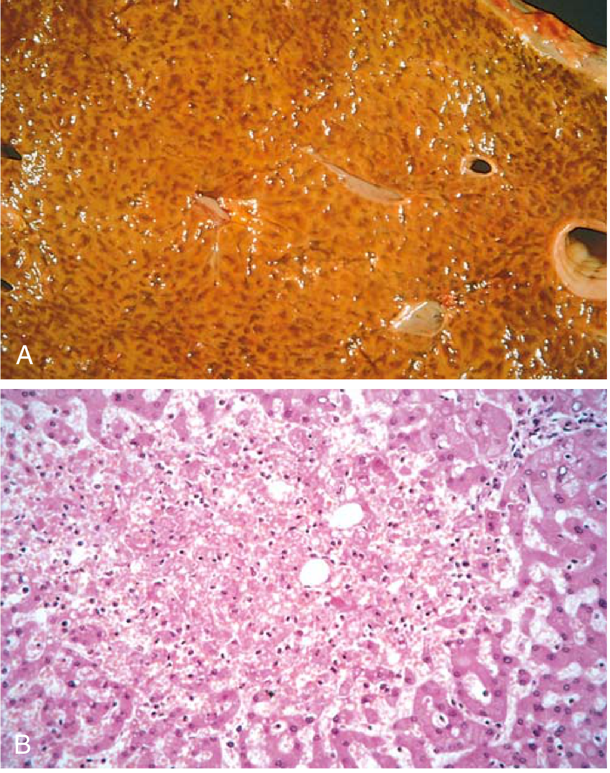
SPLEEN
Gross Features:
- Enlarged (congestive splenomegaly)
- Dark red, firm
- Tense capsule
Microscopic Features:
- Dilated, congested sinusoids
- Thickened sinusoidal walls (fibro-siderotic changes in long-standing congestion)
- Red pulp congestion
- Hemosiderin deposits in macrophages (from red cell breakdown)
- Follicles may be compressed and small
(Robbins & Kumar Basic Pathology)
SN 3 - VIRCHOW'S TRIAD - ROLE IN THROMBUS FORMATION
Definition: Virchow's triad describes the three main factors that predispose to venous thrombus formation, first described by Rudolf Virchow in the mid-19th century.
The triad consists of:
| Factor | Details | Examples |
|---|---|---|
| 1. Endothelial Injury | Most important factor; can initiate thrombosis alone. Exposes subendothelial collagen, vWF, tissue factor | MI, hypertension, atherosclerosis, vasculitis, toxins, cigarette smoke |
| 2. Abnormal Blood Flow (Stasis/Turbulence) | Disrupts laminar flow; brings platelets into contact with endothelium; prevents dilution of activated clotting factors | Aneurysms, atherosclerotic plaques, atrial fibrillation, prolonged bed rest, immobilization, cardiac failure |
| 3. Hypercoagulability of Blood | Increases tendency for clotting; Primary (genetic) or Secondary (acquired) | Factor V Leiden mutation, prothrombin gene mutation, antithrombin III deficiency (primary); malignancy, pregnancy, OCP use, nephrotic syndrome (secondary) |
How Each Component Leads to Thrombosis:
-
Endothelial injury exposes subendothelial matrix - platelets adhere via GpIb-vWF interaction - platelet activation and aggregation - primary plug - then coagulation cascade activation via tissue factor - fibrin clot formation
-
Stasis prevents washout of activated clotting factors, allows platelet contact with endothelium, causes local hypoxia that injures endothelium
-
Turbulence causes endothelial injury and creates countercurrents that allow platelet aggregation
Important: In venous thrombosis, stasis + hypercoagulability dominate. In arterial thrombosis, endothelial injury + turbulence dominate.
(Robbins & Kumar Basic Pathology; Fishman's Pulmonary Diseases)
SN 4 - AIR EMBOLISM
Definition:
Air embolism is a condition where gas (air/nitrogen) bubbles obstruct the vascular lumen, causing ischemic injury.
Types:
1. Nitrogen (Decompression Sickness / Caisson Disease / "The Bends"):
- Occurs in divers, tunnel workers, hyperbaric chamber workers
- On rapid ascent (decompression): nitrogen dissolved in blood/tissues rapidly comes out of solution as bubbles
- Gas bubbles form in skeletal muscle/joints (bends - severe joint pain), lungs (chokes - respiratory distress), brain/spinal cord (neurological deficits)
- Treatment: Slow decompression or hyperbaric O2 chamber (re-dissolves nitrogen)
- Chronic: Aseptic necrosis of femoral heads (avascular necrosis)
2. Iatrogenic/Traumatic Air Embolism:
- Surgical procedures (neurosurgery in sitting position), IV catheter insertion (accidentally introducing air), chest trauma, obstetric procedures
- Air introduced into venous system
- Small amounts (< 100 mL) usually tolerated
- Large amounts (> 150 mL) can cause sudden death - air lock in right ventricle/pulmonary artery
Pathophysiology of Large Air Embolism:
- Air bubbles accumulate in right ventricle - frothy blood cannot be pumped effectively - cardiac output falls - sudden death ("air lock")
- Pulmonary capillary obstruction - V/Q mismatch - hypoxia
Clinical Features:
- Mill-wheel murmur (churning sound over precordium from air in right ventricle)
- Sudden hypotension, cyanosis, dyspnea
- Death in massive embolism
(Robbins & Kumar Basic Pathology)
SN 5 - PULMONARY THROMBOEMBOLISM
Definition:
Pulmonary thromboembolism (PTE) refers to obstruction of pulmonary arteries by emboli, most commonly arising from deep vein thrombosis (DVT) in leg veins.
Source:
-
95% arise from DVT of deep veins of legs (popliteal vein and above)
- Right heart thrombi (rare)
- Pelvic veins (in obstetric patients)
Risk Factors: Virchow's triad factors - surgery (especially hip/knee), trauma, cancer, CHF, OCP, prolonged bed rest, hypercoagulable states, post-partum
Pathophysiology:
Two main consequences:
- Acute rise in pulmonary artery pressure (blockage + vasospasm from thromboxane A2, serotonin release)
- Ischemia of downstream pulmonary parenchyma
Classification and Clinical Effects:
| Type | Description | Effect |
|---|---|---|
| Massive (>60% of vasculature) | Saddle embolus or bilateral large vessel occlusion | Sudden death, acute cor pulmonale, shock |
| Moderate/Multiple | Occludes segmental arteries | Pulmonary infarction, pleuritic chest pain, hemoptysis, dyspnea, fever |
| Small/Microemboli | Small vessels | Clinically silent; repeated emboli → pulmonary hypertension |
Pulmonary Infarction:
- Occurs in only ~10% of PTE (dual blood supply of lung from pulmonary + bronchial arteries is protective)
- Infarction occurs when there is concurrent cardiac failure or poor bronchial circulation
- Gross: Wedge-shaped, hemorrhagic, base toward pleura
- Micro: Coagulative necrosis of alveolar walls with hemorrhage
Hypoxemia Mechanisms:
- V/Q mismatch
- Atelectasis (reduced surfactant)
- Decreased cardiac output (widened A-V O2 difference)
- Right-to-left shunt through patent foramen ovale (30% of people)
Fate:
- Organization and recanalization by fibrinolysis
- Pulmonary hypertension if recurrent
(Robbins & Kumar Basic Pathology)
LONG ANSWER QUESTIONS (LAQ)
LAQ 1 (Med Mayur IMP) - CLINICAL CASE: CHF WITH PULMONARY EDEMA + CIRRHOSIS WITH BILATERAL ANKLE SWELLING
65-year-old chronic heart failure with sudden breathlessness, frothy pink sputum, crackles. Autopsy: heavy, wet lungs with fluid oozing from cut surface.
A. OEDEMA - DEFINITION, TYPES, AND PATHOGENESIS
Definition:
Oedema is the accumulation of excess interstitial fluid in tissues. When it occurs in body cavities, it is called effusion.
- Hydrothorax = pleural cavity
- Hydropericardium = pericardial cavity
- Hydroperitoneum (Ascites) = peritoneal cavity
- Anasarca = severe generalized oedema with widespread subcutaneous and body cavity fluid accumulation
Normal Fluid Balance (Starling's Forces):
Fluid movement is governed by opposing forces:
- Vascular hydrostatic pressure (pushes fluid OUT of capillaries)
- Plasma colloid osmotic pressure (oncotic pressure, pulls fluid IN)
- At arteriolar end: hydrostatic > oncotic → fluid filters out
- At venular end: oncotic > hydrostatic → fluid returns
- Small net outflow is drained by lymphatics
Edema results when this balance is disrupted.
Types and Pathogenesis of Oedema:
| Mechanism | Examples | Type of Fluid |
|---|---|---|
| Increased hydrostatic pressure | CHF (venous backpressure), portal hypertension, venous obstruction | Transudate (low protein, low cells) |
| Decreased plasma oncotic pressure (hypoproteinemia) | Nephrotic syndrome (protein loss), liver cirrhosis (low albumin synthesis), protein malnutrition (kwashiorkor) | Transudate |
| Lymphatic obstruction (Lymphoedema) | Post-mastectomy, filariasis, tumour compression | Protein-rich fluid |
| Sodium and water retention | CHF (RAAS activation), renal failure | Transudate |
| Increased vascular permeability | Inflammation, allergic reactions, burns, sepsis | Exudate (protein-rich, cells present) |
Pitting vs Non-pitting:
- Pitting oedema: CHF, hypoproteinaemia, renal failure (watery transudate)
- Non-pitting oedema: Lymphoedema (protein-rich) or myxoedema (mucopolysaccharide deposits in hypothyroidism)
B. PULMONARY OEDEMA IN PRIMARY HEART FAILURE - PATHOPHYSIOLOGY, GROSS AND MICROSCOPIC CHANGES
Pathophysiologic Mechanism:
Left-sided heart failure
        ↓
Left ventricular pump failure → Reduced cardiac output
        ↓
Elevated left ventricular end-diastolic pressure (LVEDP)
        ↓
Back-pressure → Elevated left atrial pressure
        ↓
Elevated pulmonary venous pressure
        ↓
Transmitted to pulmonary capillaries → Elevated pulmonary capillary pressure
        ↓
Hydrostatic pressure exceeds plasma oncotic pressure
        ↓
Fluid leaks into interstitium (interstitial oedema)
        ↓
Lymphatics overwhelmed → Fluid enters alveolar spaces (alveolar oedema)
        ↓
PULMONARY OEDEMA
Additional Mechanisms:
- RAAS activation (reduced renal perfusion) → Na+ and water retention → expands blood volume → worsens back-pressure
- Sympathetic activation → tachycardia, vasoconstriction → temporarily maintains BP but worsens cardiac work
Gross Changes:
- Lungs are heavy and wet (normal lung weight: ~400g each; in oedema can weigh 1000g or more)
- Frothy, pink-tinged fluid oozes from cut surface (fluid + air + a small amount of red cells from leaky capillaries)
- Lungs do not collapse normally on sectioning
- Congested, dark red appearance
Microscopic Changes:
Acute Pulmonary Oedema:
- Blood-engorged alveolar capillaries (congestion)
- Alveolar septal widening (interstitial transudation)
- Pink homogeneous fluid in alveolar spaces (proteinaceous transudate)
- Variable intraalveolar hemorrhage (red cells in alveoli)
Chronic Pulmonary Oedema (in chronic heart failure):
- Alveolar septa become thickened and fibrotic
- Alveolar spaces contain hemosiderin-laden macrophages = "Heart Failure Cells" (macrophages that have phagocytosed extravasated red cells, the hemoglobin being converted to hemosiderin)
- Prussian blue stain highlights hemosiderin granules in these macrophages
- Perivascular and peribronchial fibrosis
Clinical Correlates from the Case:
| Feature | Explanation |
|---|---|
| Sudden breathlessness | Alveolar oedema → impaired gas exchange |
| Frothy pink sputum | Alveolar fluid + air mixing + small hemorrhage (pink tinge) |
| Crackles (crepitations) at both bases | Fluid in alveolar spaces |
| Heavy, wet lungs at autopsy | Oedema fluid (can be 2-3x normal weight) |
| Fluid oozing from cut surface | Accumulated transudate in alveolar spaces |
C. TRANSUDATE vs EXUDATE
| Feature | Transudate | Exudate |
|---|---|---|
| Pathogenesis | Mechanical (hydrostatic/oncotic imbalance) | Inflammation (increased vascular permeability) |
| Protein content | Low (<3 g/dL) | High (>3 g/dL) |
| Specific gravity | <1.012 | >1.020 |
| LDH | Low | High |
| Cells | Few (mainly mesothelial cells) | Many (neutrophils, lymphocytes, macrophages) |
| Appearance | Clear, straw-yellow | Turbid, cloudy, may be purulent or blood-stained |
| Light's Criteria (for pleural fluid) | Does not meet any criterion | Pleural fluid protein/serum protein >0.5 OR pleural LDH/serum LDH >0.6 OR pleural LDH >2/3 upper limit of normal |
| Causes | CHF, cirrhosis, nephrotic syndrome, hypoalbuminemia | Pneumonia, TB, malignancy, rheumatoid arthritis, PE |
| Fibrin/Clot formation | Does not clot | May clot (high fibrinogen) |
| Glucose | Normal (= serum) | Low (consumed by cells/bacteria) |
| pH | Normal | May be low (empyema, malignancy) |
(Robbins & Kumar Basic Pathology)
LAQ 2 - EMBOLISM - DEFINITION, TYPES, PATHOGENESIS, MORPHOLOGICAL APPEARANCE, FATE
Definition:
An embolus is a solid, liquid, or gaseous mass carried by blood to a site distant from its origin, where it lodges and causes obstruction. Most emboli are dislodged thrombi (thromboembolism).
Types of Embolism:
1. Pulmonary Thromboembolism (most common - see SN5 above)
2. Systemic Thromboembolism
- Source: Left-sided cardiac mural/valvular thrombi, aortic aneurysms, atherosclerotic plaques
- Sites: Lower extremities (75%), brain, intestines, kidneys, spleen
- Effect depends on: Site + alternative blood supply (collaterals)
3. Fat Embolism
- Pathogenesis: After crushing injury/fractures of long bones or severe burns → fat globules released into torn vessels → enter venous circulation → lodge in pulmonary microvasculature
- Also: Free fatty acids released → toxic endothelial injury
- Triad of Fat Embolism Syndrome: Pulmonary insufficiency (hypoxia, tachypnoea) + neurological symptoms + petechial rash (face/conjunctiva/axilla)
- Onset: 24-72 hours after injury
- Morphology: Fat globules in capillaries (stained by Sudan stain, Oil Red O)
- Prognosis: 10% mortality; may recover fully
4. Amniotic Fluid Embolism
- Rare but often fatal complication of labour/delivery
- Amniotic fluid + fetal debris enters maternal circulation via uterine tears or ruptured cervical veins
- Presents: Sudden dyspnoea, cyanosis, hypotensive shock, neurological symptoms, DIC (DIC because amniotic fluid is rich in procoagulant material including thromboplastin)
- Morphology: Squamous cells, lanugo hair, vernix, mucus in maternal pulmonary capillaries
- High maternal mortality (60-80%)
5. Air Embolism (see SN4 above)
6. Paradoxical Embolism
- DVT emboli cross from right to left heart through a patent foramen ovale (present in ~30% population) → systemic arterial embolism from a venous source
Fate of an Embolus:
- Lysis/Resolution - Fibrinolytic system dissolves small emboli
- Organization - Fibroblasts invade; embolus becomes fibrous tissue
- Recanalization - New channels form through the organized thrombus, partially restoring flow
- Infarction - If vessel dependent territory has no collaterals
- Perpetuation - If patient is hypercoagulable, fresh thrombus forms on organized embolus
(Robbins & Kumar Basic Pathology)
LAQ 3 - THROMBUS - DEFINITION, PATHOGENESIS, TYPES, FATE, COMPLICATIONS
Definition:
A thrombus is a solid mass formed in the living cardiovascular system from blood constituents (platelets, fibrin, red cells) in response to endothelial injury or abnormal flow/coagulability. It differs from a post-mortem clot (which is soft, gelatinous, unattached, and yellow "chicken fat" appearance or red "currant jelly").
Pathogenesis (Virchow's Triad - see SN3):
Steps of Thrombus Formation:
- Endothelial injury → subendothelial collagen and vWF exposed
- Platelet adhesion via GpIb receptor binding to vWF
- Platelet activation → release of ADP, TXA2, serotonin → shape change, more platelet recruitment
- GpIIb-IIIa receptor activated → binds fibrinogen → platelet aggregation (primary plug)
- Coagulation cascade: Tissue factor (factor VII) → → thrombin → fibrinogen to fibrin → stabilised clot
- Thrombus propagates in direction of blood flow
Types of Thrombus:
| Type | Location | Composition | Gross Appearance | Example |
|---|---|---|---|---|
| Mural Thrombus | Heart/Aorta | Mixed | Laminated (Lines of Zahn = alternating pale platelet-fibrin layers + dark RBC layers) | Post-MI left ventricular thrombus |
| Occlusive (Red) Thrombus | Veins (DVT) | Rich in RBCs and fibrin | Red, soft, gelatinous, fills lumen | Deep vein thrombosis of leg |
| White (Platelet) Thrombus | Arteries | Mainly platelets + fibrin | Pale, firm, small | Arterial thrombosis over atherosclerotic plaque |
| Vegetation | Heart valves | Platelets + fibrin + organisms | Small nodules on valve leaflets | Infective endocarditis |
Lines of Zahn: Alternating pale (platelet-fibrin) and dark (RBC) laminations - pathognomonic of thrombus formed in flowing blood; distinguishes antemortem thrombus from postmortem clot.
Fate of a Thrombus (4 R's + P):
- Resolution/Lysis - Fibrinolytic activity dissolves small/recent thrombi (tPA activates plasminogen → plasmin → breaks down fibrin)
- Organization - Ingrowth of fibroblasts, smooth muscle cells, endothelium from vessel wall into thrombus
- Recanalization - Endothelium-lined channels form within organized thrombus, partially restoring blood flow
- Propagation - Thrombus grows by additional clot deposition
- Embolization - Fragment detaches and travels distally as an embolus (most dangerous fate)
Complications of Thrombosis:
| Complication | Details |
|---|---|
| Vascular occlusion | Ischemia → infarction (MI, stroke, limb gangrene, intestinal ischemia) |
| Embolism | Pulmonary embolism (from DVT), systemic embolism (from cardiac thrombi) |
| Phlebitis/Thrombophlebitis | Painful inflammation around thrombosed vein |
| Post-thrombotic syndrome | Chronic venous insufficiency, varicose veins, leg ulcers |
| DIC (Disseminated Intravascular Coagulation) | Widespread microthrombi consuming clotting factors |
| Paradoxical embolism | Via PFO (see above) |
(Robbins & Kumar Basic Pathology)
Sources: All content referenced from Robbins & Kumar Basic Pathology (Robbins Pathology, 9780323790185) and Fishman's Pulmonary Diseases and Disorders.
MUHS Exam Tips:
- In LAQ, always write Definition → Pathogenesis → Morphology (Gross + Micro) → Clinical features → Fate/Complications
- "Lines of Zahn" and "Heart failure cells" are favourite one-liner MCQ/SN topics
- Virchow's triad is asked almost every year - memorise all 3 components with examples
- Difference between transudate and exudate (table format) scores full marks
- For liver in right heart failure: always mention "nutmeg liver" gross appearance and centrilobular congestion/necrosis microscopically
- Pulmonary oedema micro: emphasize "heart failure cells" (hemosiderin-laden macrophages) for chronic cases
This is a shared conversation. Sign in to Orris to start your own chat.