
Laryngotracheal reconstruction in detail and in simplified manner for long question answer in ms ent university exam
laryngotracheal reconstruction MS ENT exam detailed review indications techniques outcomes
laryngotracheal reconstruction anterior posterior cricoid split costal cartilage graft
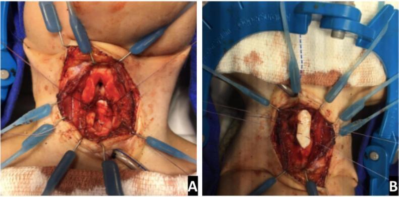
Two clinical photographs (A and B) showing an intraoperative view of a Laryngotracheal Reconstruction (LTR) for subglottic stenosis. The surgical field is exposed via a midline neck incision, with skin and subcutaneous tissues held open by a Lone Star retractor system with multiple blue elastic stays. Image A demonstrates the posterior expansion of the cricoid cartilage; a vertical incision is visible through the posterior cricoid lamina to enlarge the airway diameter. Image B shows the anterior expansion phase, where a white, rectangular costal cartilage graft (rib graft) has been carved and securely placed into the anterior cricoid split to provide structural support and widen the lumen. The surgical site displays anatomical landmarks of the larynx and upper trachea. This visual illustrates the surgical management of complex airway narrowing through both posterior and anterior grafting techniques, a common procedure in pediatric otolaryngology.
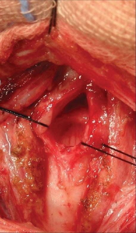
This clinical photograph captures an intraoperative view of an anterior cricoid split (ACS), a reconstructive surgical procedure used to treat subglottic stenosis. The surgical field reveals the laryngeal and upper tracheal airway structures after a vertical midline incision. Centrally, the cricoid and subglottic rings are visibly divided. A white, firm costal cartilage graft is positioned between the split edges to expand the airway lumen. The graft and surrounding cartilaginous tissue are secured with prominent black non-absorbable stay sutures to maintain the expanded position. The surrounding soft tissue is erythematous and vascularized, with evidence of minor cauterization and surgical trauma. This image serves as an educational example of laryngotracheal reconstruction, illustrating the use of structural grafts to increase airway diameter in patients with grade-dependent stenosis. The anatomical focus is the subglottic region, which is the narrowest portion of the airway and a common site for scarring after prolonged endotracheal intubation.
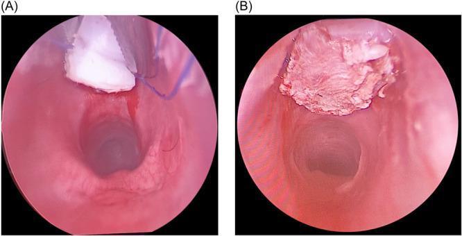
Two endoscopic images showing a laryngoscopic view of an anterior laryngeal cricoid split and graft placement, likely for laryngotracheal reconstruction. Image (A) illustrates the intraoperative phase where a white, carved rib cartilage graft is being positioned into an anterior cricoid and first tracheal ring split. Blue monofilament sutures are visible, used for securing and suspending the graft, alongside surgical instruments. The surrounding laryngeal mucosa appears erythematous and edematous with fresh hemorrhage at the surgical margins. Image (B) shows the final postoperative position of the graft integrated into the anterior wall. The graft surface appears more textured and slightly hyperemic compared to the initial placement, suggesting early mucosal integration or fibrin deposition. The subglottic airway lumen is clearly patent in both views. This sequence demonstrates the surgical management of subglottic stenosis using a costal cartilage graft to expand the airway diameter.
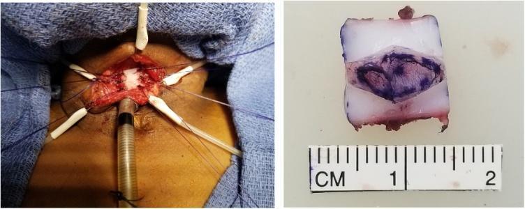
This composite clinical photograph illustrates key stages of a double-stage laryngotracheoplasty for grade III subglottic stenosis. The left image shows the intraoperative surgical field in the anterior neck region. Retractors and stay sutures are used to maintain exposure of a midline sagittal incision through the cricoid cartilage and proximal trachea. An autologous anterior costal cartilage graft, appearing as a white rectangular structure, is seen sutured into the laryngotracheal split to expand the airway lumen. A tracheostomy tube or laryngeal stent is visible inferior to the graft site. The right image is a close-up of the harvested and carved costal cartilage graft on a sterile back table. The graft is approximately 1.5 cm in length and 1 cm in width, as indicated by the adjacent centimeter ruler. It features a diamond-shaped purple surgical ink marking on its semi-translucent, whitish-blue surface to guide orientation and placement. This visual documentation highlights surgical techniques for pediatric airway reconstruction and graft preparation.
Laryngotracheal Reconstruction (LTR)
For MS ENT University Long Question Answer
QUICK OVERVIEW
I. ANATOMY RELEVANT TO LTR
- Cricoid cartilage - the only complete ring in the airway; its anterior arch and posterior lamina are the targets for splitting
- Thyroid cartilage - roof of the subglottis
- First 2-3 tracheal rings - often involved in stenosis
- Recurrent laryngeal nerve - enters larynx posterolaterally; at risk in posterior dissection
- Costal cartilage (6th rib preferred) - primary graft source
II. ETIOLOGY OF LARYNGOTRACHEAL STENOSIS (LTS)
| Cause | Frequency |
|---|---|
| Acquired LTS (post-intubation subglottic + tracheal) | ~50% |
| Bilateral vocal cord mobility impairment | ~17% |
| Wegener's granulomatosis (GPA) | ~10% |
| Idiopathic subglottic stenosis (ISS) | ~8.7% |
| Supraglottic stenosis (sarcoid + other) | ~5.7% |
| Previous papillomatosis treatment | ~2.3% |
| Glottic web | ~2.3% |
| Tracheomalacia | ~1.3% |
III. MYER-COTTON GRADING OF SUBGLOTTIC STENOSIS
| Grade | % Stenosis | Description |
|---|---|---|
| Grade I | 0-50% | Mild; often no intervention needed |
| Grade II | 51-70% | Moderate |
| Grade III | 71-99% | Severe; "pinhole" airway |
| Grade IV | 100% | Complete obstruction; no lumen |
IV. CLINICAL FEATURES
- Biphasic stridor (inspiratory + expiratory) - hallmark
- Exertional dyspnea (progressive; patient compensates by reducing activity)
- Dysphonia, chronic cough
- Dysphagia (if laryngeal function impaired)
- Recurrent "croup" in children with congenital SGS
- Failure to extubate in neonates
V. INVESTIGATIONS
- Rigid laryngoscopy and bronchoscopy (MLB) - gold standard; measures exact location, length, and grade of stenosis
- CT scan of neck and chest (high-resolution/CT virtual bronchoscopy) - assess extent, cartilage involvement; can underestimate if cuts miss apex of stenosis
- Flexible nasopharyngolaryngoscopy - vocal cord mobility, supraglottic anatomy, reflux
- Flow-volume loops (spirometry) - characteristic flat inspiratory-expiratory pattern in fixed obstruction; differentiates from asthma; monitors response to treatment
- Cardiopulmonary exercise testing - severity of functional impairment
- GERD workup - pH study, esophagoscopy (GERD worsens SGS; must be treated before surgery)
- ANCA, ACE levels - rule out GPA, sarcoidosis
- Histopathology - tissue from stenosis at each procedure to rule out neoplasm
VI. PRINCIPLES OF MANAGEMENT
A. Non-surgical / Endoscopic (first line, particularly in adults)
- Scar dilation (balloon dilatation) - balloon inflated to diameter of adjacent normal airway
- CO2 laser radial incisions - 3-4 radial cuts at 8-10W; disrupts circumferential scar
- Intralesional steroid injection - Depo-Medrone (triamcinolone); reduces fibrosis
- Mitomycin-C (MMC) application - antifibrotic; inhibits fibroblast proliferation; applied topically post-laser
- Airway stenting - for patients unfit for open surgery
B. Open Surgery (LTR and related procedures)
- Endoscopic management fails
- Grade III or IV SGS
- Long-segment stenosis
- Cartilaginous framework damage
VII. OPEN SURGICAL PROCEDURES
1. ANTERIOR CRICOID SPLIT (ACS)
- Midline vertical incision through lower thyroid cartilage, entire cricoid ring, and upper 2 tracheal rings
- The ETT acts as a stent for 5-7 days post-op
- No graft required
- Failed extubation x 2 in absence of other causes
- Weight >1.5 kg
- No assisted ventilation / FiO2 <30%
- No congestive heart failure
- No acute respiratory infection
2. LARYNGOTRACHEAL RECONSTRUCTION WITH CARTILAGE GRAFT (LTR)
- Costal cartilage (6th rib) - most common; provides adequate size, easy harvest
- Thyroid ala cartilage
- Auricular cartilage
- Hyoid bone (hyoid-sternohyoid pedicle graft)
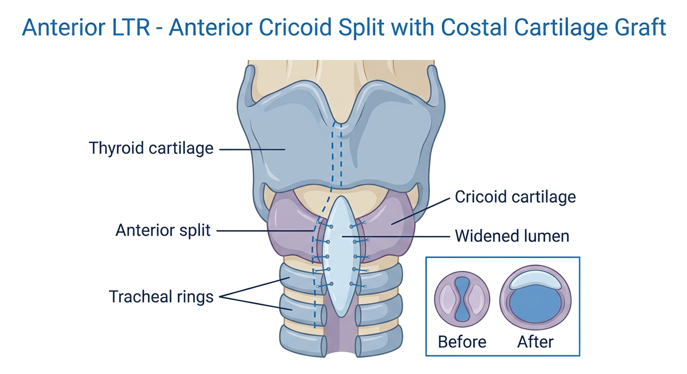
a. Anterior LTR (Anterior Graft)
- Indication: Grade II, mild Grade III SGS
- Technique:
- Horizontal neck incision, subplatysmal flaps
- Strap muscles retracted
- Midline vertical incision through lower thyroid cartilage, cricoid ring, upper tracheal rings
- Scar excised (laryngofissure approach)
- Costal cartilage graft carved as a boat/keel shape, sutured into the anterior split to widen lumen
- Graft held in place with 4/0 PDS or similar suture
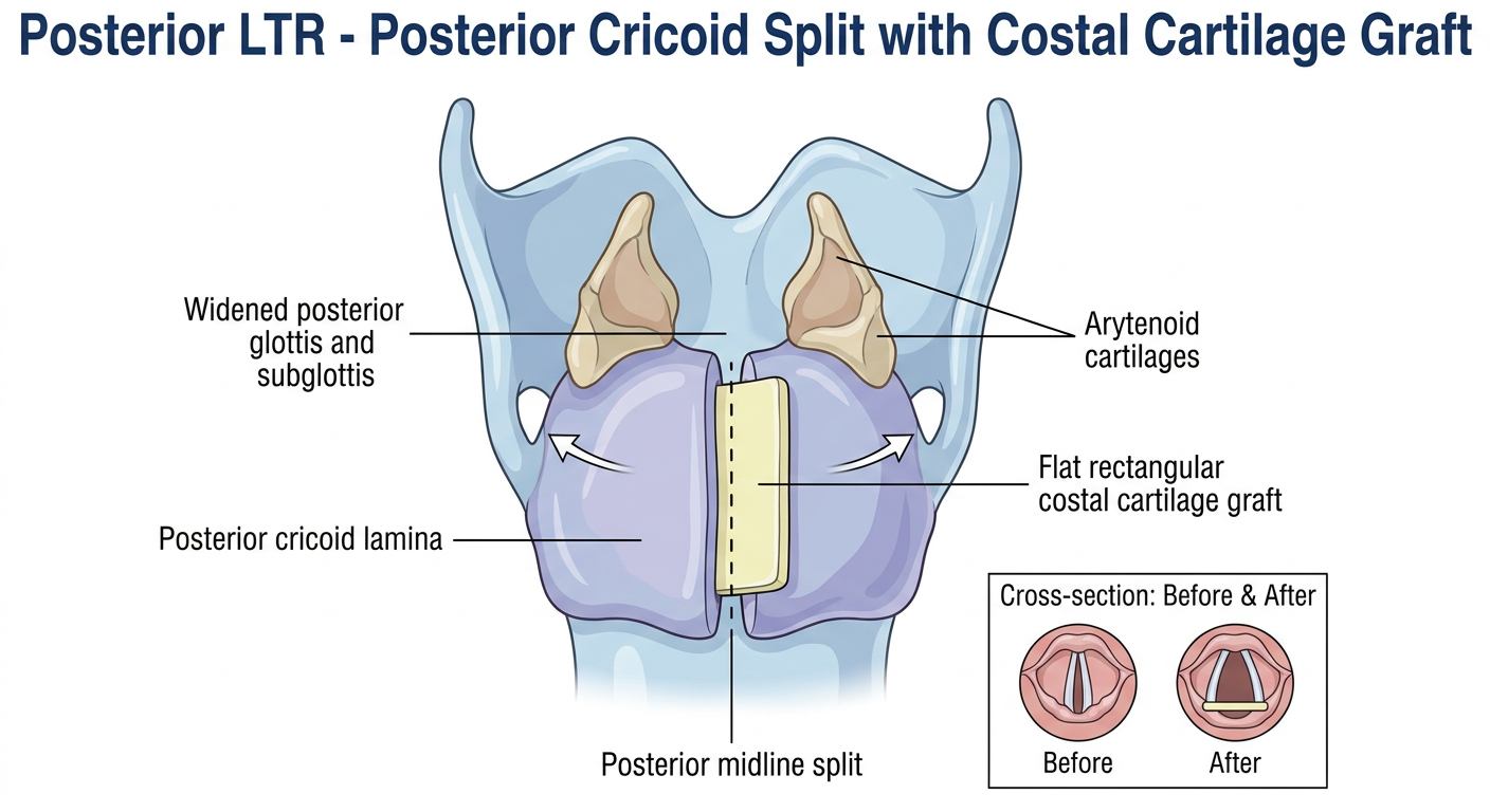
b. Posterior LTR (Posterior Graft)
- Indication: Posterior glottic stenosis, severe SGS (Grade III-IV) where anterior graft alone is insufficient
- Technique:
- Posterior cricoid lamina split in midline
- Costal cartilage graft (flat/rectangular) interposed into posterior split
- The posterior cricoid is the key to interarytenoid distance and posterior glottic width
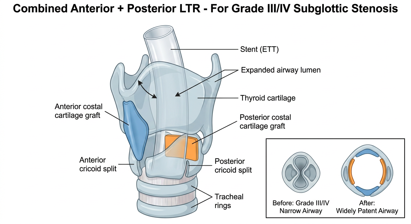
c. Combined Anterior + Posterior LTR
- Indication: Severe Grade III and Grade IV SGS
- Both anterior and posterior grafts placed in same or staged procedures
3. PARTIAL CRICOTRACHEAL RESECTION (PCTR) / CRICOTRACHEAL ANASTOMOSIS
- Introduced by Monnier et al. (early 1990s) into the pediatric age group
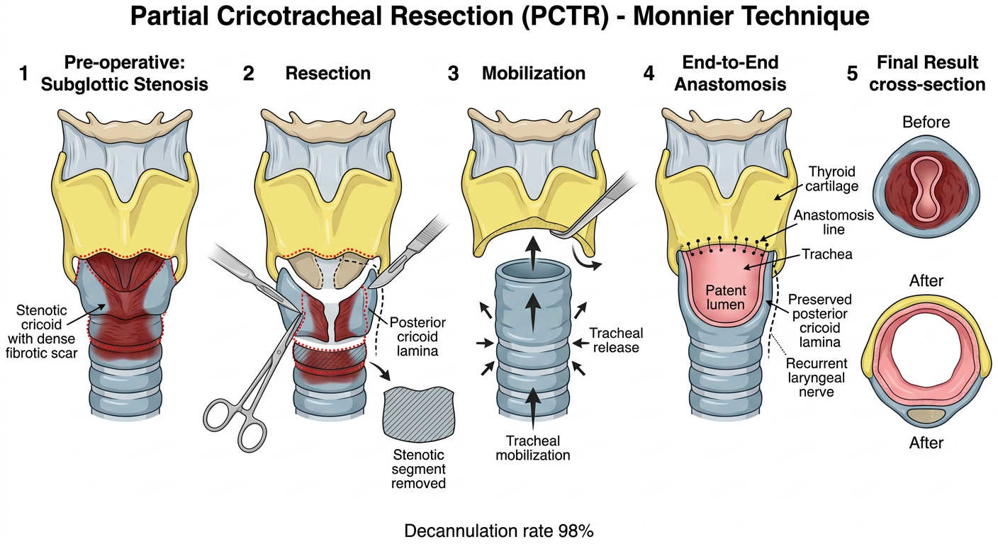
- Principle: Complete resection of the stenotic segment + end-to-end tracheo-thyroid anastomosis
- Advantage over LTR: Removes all diseased tissue; does not depend on grafts
- Indication: Severe Grade III and Grade IV SGS; also adult post-intubation LTS where tracheal resection is feasible
- Maximum resectable length in adults: 4-6 cm (with laryngeal and pulmonary release maneuvers)
- Decannulation rate: 98% (primary PCTR), 94% (salvage PCTR) in leading centers
4. TRACHEAL RESECTION AND ANASTOMOSIS (TRA)
- For isolated tracheal (not subglottic) stenosis
- Mid-tracheal resection with end-to-end anastomosis
- Maximum resectable length: 4-6 cm with release maneuvers (hyoid release, suprahyoid release, pulmonary release)
VIII. STAGING OF LTR: SINGLE vs. DOUBLE STAGE
| Feature | Single-Stage LTR | Double-Stage LTR |
|---|---|---|
| Definition | No tracheotomy at end of procedure; ETT acts as stent | Tracheotomy present; stent placed, decannulated at second operation |
| Advantages | Avoids tracheotomy; shorter hospitalization; better speech/swallow development | Can address multi-level obstruction; safer in difficult-to-intubate, poor lung function, neurologic deficits, prior failed reconstruction |
| Disadvantages | Requires ICU; risk of accidental extubation | Granulation tissue around stent; stent complications; second admission |
| Stent | ETT remains as stent for 5-14 days post-op | T-tube or other airway stent for weeks-months |
| Best for | Grade II, selected Grade III | Grade III-IV, failed previous procedures, comorbidities |
IX. SURGICAL TECHNIQUE OF LTR (Step-by-Step)
- Pre-operative: Treat GERD aggressively (PPI); MRSA swab and eradication; optimize nutrition; flexible nasolaryngoscopy; CT scan; MLB under GA with sizing
- Positioning: Supine, neck extended (shoulder roll)
- Incision: Horizontal skin crease incision at level of cricoid (or use existing tracheostomy incision)
- Exposure: Subplatysmal dissection, vertical split of strap muscles, exposure of thyroid cartilage, cricoid, and proximal trachea
- Airway entry: If tracheostomy present, ventilate through it. If not, suspend laryngoscopy or intubate from above
- Anterior split: Midline vertical incision from lower 1/3 thyroid cartilage through entire cricoid and 2 tracheal rings
- Scar excision: Submucosal resection of fibrotic scar; re-line with mucosa or graft
- Graft harvest: 6th rib, ~2 cm segment; carve to "keel" or "boat" shape for anterior; flat rectangle for posterior
- Graft placement: Suture with 4/0 PDS or Vicryl, interrupted; graft held in place, slightly proud of cartilage surface
- Posterior split (if needed): Patient prone (or through laryngofissure); split posterior cricoid lamina in midline; costal cartilage graft interposed
- Stenting: In single-stage, ETT left as stent for 5-14 days; in double-stage, T-tube or silicone stent placed
- Closure: Strap muscles reapproximated, drain, layered skin closure
- Post-op: ICU monitoring, humidified air, nebulizers, anti-reflux therapy, speech therapy, serial endoscopic surveillance
X. MANAGEMENT BASED ON GRADE
| Grade | Surgery |
|---|---|
| Grade I | Usually no surgery; observation; treat GERD |
| Grade II | LTR with anterior cartilage graft ± posterior cricoid split |
| Grade III (mild) | Anterior graft + posterior cricoid split ± posterior graft |
| Grade III (severe - pinhole) | Both anterior AND posterior grafts; or PCTR |
| Grade IV | Anterior + posterior grafts with prolonged stenting; or PCTR |
XI. OUTCOMES
| Procedure | Decannulation / Success Rate |
|---|---|
| LTR, Grade I-II | >90% |
| LTR, Grade III-IV | 80-90% |
| PCTR (primary) | ~98% |
| PCTR (salvage) | ~94% |
| Endoscopic (post-intubation LTS, adults) | ~66% |
XII. COMPLICATIONS OF LTR
| Complication | Notes |
|---|---|
| Graft failure | Resorption, displacement, infection |
| Re-stenosis | Granulation tissue at graft edges |
| Aspiration / dysphagia | Especially with posterior graft disrupting cricoarytenoid function |
| Dysphonia | Voice change due to laryngofissure or posterior glottic scarring |
| Re-tracheostomy | Required in ~10-20% of severe cases |
| Wound infection | Prophylactic antibiotics required |
| RLN injury | Rare if posterior dissection careful |
| Pneumothorax | Rib graft harvest complication |
| Suprastomal collapse | In double-stage procedures |
XIII. SPECIAL SITUATIONS
Idiopathic Subglottic Stenosis (ISS)
- Predominantly postpubertal Caucasian women; diagnosis of exclusion
- ANCA and ACE must be negative; tissue histology at each procedure
- Most managed with repeated endoscopic surgery (CO2 laser + dilatation + steroid injection) 1-2 times/year
- Cricotracheal resection for severe/refractory cases - Scott-Brown's Ch 76
Wegener's Granulomatosis (GPA) - SGS
- 10% of LTS referrals
- SGS may be the presenting feature
- Medical treatment with cyclophosphamide/methotrexate + steroids
- Endoscopic dilatation + intralesional steroids preferred; surgery only when medical treatment fails
- Risk of anastomotic breakdown if systemic disease active
Post-tracheostomy Stenosis
- "Lambda-shaped" stenotic deformity due to scar contracture at stoma site
- Usually extends 1-2 tracheal rings; tracheal resection preferred
XIV. SIMPLIFIED MEMORY AID (Exam Recall)
- Grade I = Observe
- Grade II = Anterior graft (ACS + graft or LTR)
- Grade III = Anterior + Posterior graft (both)
- Grade IV = Both grafts + prolonged stent OR PCTR
- LTR = augmentation (graft adds to existing structure)
- PCTR = resection (diseased segment removed, anastomosis)
- PCTR better outcomes but needs adequate trachea above and below
- Single = no tracheotomy, ETT stent for 5-14d → ideal
- Double = tracheostomy maintained, stent for weeks → safer in comorbid/failed cases
XV. KEY POINTS FOR EXAM
- LTS most commonly caused by post-intubation injury (~50%); second is bilateral vocal cord mobility impairment
- Myer-Cotton grading - Grade I (<50%) to Grade IV (complete) - guides management
- Cotton's ACS criteria must be met before attempting single-stage in neonates
- Costal cartilage from 6th rib is the preferred graft - adequate bulk, ease of harvest, autologous
- LTR was devised by Robin Cotton; PCTR introduced by Monnier et al. in children
- GERD must be treated aggressively before and after LTR - it is the most common modifiable risk factor for failure
- Congenital cartilaginous SGS - dilatation is absolutely contraindicated
- Decannulation rate is the primary outcome measure for success
- Single-stage LTR avoids tracheotomy and is preferred in Grade II, selected Grade III
- In adults, the maximum tracheal segment resectable is 4-6 cm (with release maneuvers)
Generate image for anterior LTR and posterior LTR and Combined separately
Medical surgical diagram of Anterior Laryngotracheal Reconstruction (Anterior LTR). Clean anatomical illustration showing: 1) A frontal cross-sectional view of the larynx and upper trachea with the cricoid cartilage and thyroid cartilage labeled. 2) A midline vertical incision (anterior cricoid split) through the lower thyroid cartilage, entire cricoid ring, and upper 2 tracheal rings shown with dotted cut lines. 3) The split cricoid held open with a boat-shaped or keel-shaped costal cartilage graft sutured into the anterior gap, widening the airway lumen anteriorly. 4) Labels: Thyroid cartilage, Cricoid cartilage, Anterior split, Costal cartilage graft, Tracheal rings, Widened lumen. 5) A small inset showing the airway cross-section before (narrow) and after (wide) graft placement. Title at top: "Anterior LTR - Anterior Cricoid Split with Costal Cartilage Graft". White background, professional medical illustration style with blue and anatomical color tones.
Medical surgical diagram of Posterior Laryngotracheal Reconstruction (Posterior LTR). Clean anatomical illustration showing: 1) A posterior view of the cricoid cartilage with the posterior cricoid lamina prominently displayed. 2) A midline vertical incision through the posterior cricoid lamina (posterior cricoid split) shown with cut lines. 3) A flat rectangular costal cartilage graft interposed into the posterior split, pushing the posterior lamina outward and widening the interarytenoid distance and posterior glottic width. 4) The arytenoid cartilages shown on either side, now separated wider after graft. 5) Labels: Posterior cricoid lamina, Posterior midline split, Flat rectangular costal cartilage graft, Arytenoid cartilages, Widened posterior glottis and subglottis. 6) Small cross-sectional inset before and after showing posterior expansion. Title at top: "Posterior LTR - Posterior Cricoid Split with Costal Cartilage Graft". White background, professional medical textbook illustration style, anatomical color scheme.
Medical surgical diagram of Combined Anterior and Posterior Laryngotracheal Reconstruction (Combined LTR). Clean anatomical illustration showing both grafts together: 1) A lateral or combined view of the larynx showing BOTH anterior and posterior splits of the cricoid cartilage simultaneously. 2) Anteriorly: a keel-shaped costal cartilage graft in the anterior midline split of the cricoid and lower thyroid cartilage. 3) Posteriorly: a flat rectangular costal cartilage graft in the posterior cricoid lamina split. 4) The overall airway lumen is shown significantly expanded in all directions (360 degrees). 5) An endotracheal tube or T-tube stent shown inside the lumen acting as a stent. 6) Labels: Anterior costal cartilage graft, Posterior costal cartilage graft, Anterior cricoid split, Posterior cricoid split, Stent (ETT), Expanded airway lumen, Thyroid cartilage, Tracheal rings. 7) Cross-sectional view inset showing Grade III/IV narrow airway before vs. widely patent airway after combined grafting. Title at top: "Combined Anterior + Posterior LTR - For Grade III/IV Subglottic Stenosis". White background, professional medical illustration style, color-coded grafts (anterior in blue, posterior in orange).
1. Anterior LTR - Anterior Cricoid Split with Costal Cartilage Graft
2. Posterior LTR - Posterior Cricoid Split with Costal Cartilage Graft
3. Combined Anterior + Posterior LTR
Quick Comparison Table
| Feature | Anterior LTR | Posterior LTR | Combined LTR |
|---|---|---|---|
| Site of split | Anterior cricoid | Posterior cricoid lamina | Both |
| Graft shape | Keel/boat-shaped | Flat rectangle | Both shapes |
| Grade | II, mild III | Posterior glottic stenosis | Severe III, IV |
| Primary effect | Anterior expansion | Posterior + interarytenoid widening | 360° expansion |
| Stent required | Optional | Usually yes | Always yes |
Generate image for partial cricotracheal resection
Detailed medical surgical diagram of Partial Cricotracheal Resection (PCTR) shown as a step-by-step sequence. Professional anatomical illustration on white background with clear labels. Panel 1 - "Pre-operative: Subglottic Stenosis": Frontal view of larynx showing thyroid cartilage, stenotic cricoid with dense fibrotic scar narrowing the subglottis (shaded dark), first and second tracheal rings below. Red dotted lines marking the planned resection zone - from the lower margin of the thyroid cartilage through the entire cricoid and 1-2 tracheal rings below. Panel 2 - "Resection": The stenotic segment (cricoid arch anteriorly + scarred tracheal rings) is being removed. The posterior cricoid lamina is partially preserved to protect the recurrent laryngeal nerve. Scissors or scalpel shown cutting. The resected specimen shown separately as a grey hatched block labeled "Stenotic segment removed". Panel 3 - "Mobilization": The trachea is mobilized upward (arrows showing upward pull of trachea) to reduce tension on anastomosis. The inferior thyroid cartilage margin is trimmed and shaped. Small arrows indicating tracheal release. Panel 4 - "End-to-End Anastomosis": The tracheal stump is sutured directly to the lower margin of the thyroid cartilage (tracheo-thyroid anastomosis) with interrupted absorbable sutures shown as small black dots. The airway lumen is now wide and patent. Labels: Thyroid cartilage, Anastomosis line, Trachea, Patent lumen, Preserved posterior cricoid lamina, Recurrent laryngeal nerve (dashed line). Panel 5 - "Final Result cross-section": Before vs After cross sections showing narrow stenotic lumen vs wide patent lumen. Title at top: "Partial Cricotracheal Resection (PCTR) - Monnier Technique". Color scheme: thyroid cartilage in yellow, trachea in blue-grey, scar in dark red, sutures in black, normal mucosa in pink. Decannulation rate 98% noted at bottom.
Partial Cricotracheal Resection (PCTR)
Step-by-Step Explanation
- The anterior cricoid arch and stenotic tracheal rings are completely excised
- The posterior cricoid lamina is partially preserved - this is critical to protect the recurrent laryngeal nerves (RLN) which run postero-laterally
- The entire diseased segment is removed (unlike LTR which augments around it)
- The trachea is mobilized superiorly by releasing pretracheal fascia, and sometimes with hyoid or pulmonary release maneuvers
- This reduces tension on the anastomosis - tension is the #1 cause of anastomotic dehiscence
- The tracheal stump is sutured end-to-end directly to the inferior margin of the thyroid cartilage
- Interrupted absorbable sutures (e.g., PDS 3/0)
- The neck is kept slightly flexed post-operatively for 1-2 weeks to reduce anastomotic tension
PCTR vs LTR - Key Distinction for Exams
| Feature | LTR | PCTR |
|---|---|---|
| Principle | Augmentation with graft | Complete resection + anastomosis |
| Devised by | Robin Cotton | Monnier et al. |
| Graft needed | Yes (costal cartilage) | No |
| Recurrence risk | Higher (graft resorption) | Lower (all disease removed) |
| Decannulation rate | 80-90% (Gr III/IV) | 98% primary, 94% salvage |
| Grade suitability | II, III | Severe III, IV |
| Max resection | - | 4-6 cm (adults) |
| Post-op position | Normal | Neck flexion for 1-2 weeks |
PCTR is the procedure of choice for severe Grade III and Grade IV SGS, and LTR is the last resort after failed PCTR if further resection is not possible.