
Please give the details in systemic wise neet Pg
I need a bit more clarity before diving in. "Systemic wise NEET PG" could mean several things:
Asking for Preferences
Which subject or topic do you want system-wise details for? · What kind of details are you looking for?
Searching the Web
NEET PG 2025 2026 full syllabus system wise all subjects NBE official
Reading Web Page
https://www.prepladder.com/neet-pg-study-material/syllabus/n…
Here is the complete NEET PG 2026 - Full System-wise / Subject-wise Syllabus Breakdown:
NEET PG 2026 - Complete System-wise Syllabus
Exam Overview
| Detail | Info |
|---|---|
| Conducted by | NBEMS (National Board of Examinations in Medical Sciences) |
| Syllabus Authority | National Medical Commission (NMC) |
| Total Questions | 200 (180 scorable) |
| Total Marks | 800 |
| Duration | 3 hours 30 minutes |
| Mode | Online (CBT) |
| Question Type | MCQs |
| Official Website | natboard.edu.in |
PHASE I - PRE-CLINICAL SUBJECTS
1. ANATOMY
| System | Topics |
|---|---|
| General Embryology | Gametogenesis, Developmental Timeline (Week 1-4), Germ layer derivatives (Ectoderm, Mesoderm, Endoderm), Neural crest cells, Placenta formation |
| Histology | Epithelial tissue, Glands, Connective tissue, Cartilage, Lymphoid tissue, Muscular tissue, Integumentary system, Cell junctions |
| Histology - Organ Systems | Respiratory system, Digestive system, Urinary system, Genital system |
| Osteology & Arthrology | Bones, Joints classification and types |
| Upper Limb | Axilla, Brachial plexus, Arm, Elbow, Forearm, Wrist, Hand, Dermatomes |
| Lower Limb | Femoral triangle, Hip joint, Knee joint, Leg, Foot, Lumbosacral plexus |
| Thorax | Heart, Lungs, Mediastinum, Great vessels, Intercostal spaces |
| Abdomen | Peritoneum, Stomach, Intestines, Liver, Pancreas, Spleen, Kidneys, Retroperitoneum |
| Pelvis & Perineum | Pelvic floor, Bladder, Rectum, Reproductive organs, Perineal spaces |
| Head & Neck | Skull, Cranial nerves (I-XII), Scalp, Face, Parotid, TMJ, Orbit, Nasal cavity, Pharynx, Larynx |
| Neuroanatomy | Brain stem, Cerebellum, Cerebrum, Basal ganglia, Limbic system, Thalamus, Spinal cord tracts, Meninges, Ventricles & CSF |
| Surface & Radiological Anatomy | Clinical surface landmarks, X-ray/CT anatomy |
2. PHYSIOLOGY
| System | Topics |
|---|---|
| General Physiology | Cell membrane, Transport mechanisms, Resting membrane potential, Action potential |
| Blood | Composition, RBC, WBC, Platelets, Hemoglobin, Erythropoiesis, Blood groups, Coagulation cascade, Bleeding/Clotting tests |
| Cardiovascular System | Cardiac cycle, Heart sounds, ECG, Cardiac output, Blood pressure regulation, Coronary circulation, Microcirculation |
| Respiratory System | Lung volumes & capacities, Mechanics of breathing, Gas exchange (V/Q ratio), Oxygen & CO2 transport, Regulation of respiration, Hypoxia |
| Gastrointestinal System | Salivation, Deglutition, Gastric secretion, Pancreatic & Bile secretion, Absorption, GI motility, GI hormones |
| Excretory (Renal) System | Glomerular filtration (GFR), Tubular reabsorption/secretion, Concentration & dilution of urine, Acid-base balance, Micturition |
| Endocrine System | Pituitary-hypothalamic axis, Thyroid, Parathyroid, Adrenal cortex & medulla, Pancreatic hormones (Insulin/Glucagon), Sex hormones |
| Reproductive System | Male reproductive physiology, Female reproductive cycle, Menstrual cycle, Pregnancy physiology, Lactation |
| Nervous System | Sensory system (touch, pain, proprioception), Motor system, Reflexes, Cerebellum, Basal ganglia, Higher functions, Autonomic nervous system |
| Special Senses | Vision (eye physiology), Audition (ear physiology), Taste, Smell |
| Muscle Physiology | Skeletal, Cardiac, and Smooth muscle - contraction mechanisms |
| Exercise Physiology | VO2 max, adaptations |
3. BIOCHEMISTRY
| System | Topics |
|---|---|
| Carbohydrate Metabolism | Glycolysis, TCA cycle, Pentose phosphate pathway, Glycogenesis/Glycogenolysis, Gluconeogenesis, HMP shunt |
| Lipid Metabolism | Beta-oxidation, Ketone bodies, Cholesterol synthesis, Lipoproteins (LDL/HDL/VLDL), Fatty acid synthesis |
| Protein & Amino Acid Metabolism | Transamination, Urea cycle, Amino acid disorders (PKU, Alkaptonuria, Homocystinuria) |
| Enzymes | Classification, Kinetics (Km, Vmax, Michaelis-Menten), Inhibitors |
| Vitamins | Fat-soluble (A, D, E, K) and Water-soluble (B-complex, C) - deficiencies and excess |
| Minerals & Trace Elements | Iron, Calcium, Zinc, Copper, Iodine metabolism |
| Nucleotide Metabolism | Purine/Pyrimidine synthesis and degradation, Gout |
| Molecular Biology | DNA replication, Transcription, Translation, PCR, Recombinant DNA technology, Gene mutations |
| Hemoglobin & Porphyrin | Hb structure, variants (HbS, HbA2), Heme synthesis, Porphyrias, Jaundice types |
| Nutrition | Proteins, Carbohydrates, Fats - daily requirements, Caloric values, PEM |
| Plasma Proteins | Albumin, Globulins, Acute phase reactants |
PHASE II - PARA-CLINICAL SUBJECTS
4. PATHOLOGY
| System | Topics |
|---|---|
| General Pathology | Cell injury & necrosis, Apoptosis, Amyloidosis, Inflammation (acute & chronic), Granulomas, Healing & repair, Hemodynamic disorders (edema, thrombosis, embolism, infarction, shock) |
| Neoplasia | Benign vs malignant, Carcinogenesis, Tumor markers, Oncogenes & tumor suppressor genes, TNM staging, Paraneoplastic syndromes |
| Immunopathology | Hypersensitivity (Type I-IV), Autoimmune diseases, Immunodeficiency disorders, Amyloid |
| Hematopathology | Anemias (iron deficiency, megaloblastic, hemolytic, aplastic), Leukemias (ALL, AML, CLL, CML), Lymphomas (Hodgkin/Non-Hodgkin), Multiple myeloma, Bleeding disorders |
| Cardiovascular Pathology | Atherosclerosis, IHD, MI, Cardiomyopathies, Rheumatic heart disease, Endocarditis |
| Respiratory Pathology | Pneumonia, TB, COPD, Lung cancer, Mesothelioma, Pulmonary fibrosis |
| GIT Pathology | Esophageal disorders, Peptic ulcer, Gastric cancer, IBD (Crohn's, UC), Colon polyps, Colorectal cancer, Appendicitis |
| Hepatobiliary Pathology | Hepatitis (viral types), Cirrhosis, Hepatocellular carcinoma, Cholestasis, Gallstones |
| Renal Pathology | Glomerulonephritis (all types), Nephrotic/Nephritic syndromes, Pyelonephritis, Renal cell carcinoma, Wilms tumor |
| Reproductive Pathology | Breast (fibrocystic disease, carcinoma), Cervix (dysplasia, carcinoma), Ovary, Endometrium, Prostate, Testicular tumors |
| CNS Pathology | Brain tumors (Gliomas, Meningioma, Medulloblastoma), Demyelinating diseases, Neurodegenerative diseases |
| Endocrine Pathology | Thyroid tumors (Papillary, Follicular, Medullary, Anaplastic), Pheochromocytoma, Adrenal tumors, MEN syndromes |
| Bone & Soft Tissue Pathology | Osteosarcoma, Ewing's sarcoma, Osteomyelitis, Paget's disease |
| Skin Pathology | Melanoma, BCC, SCC, Psoriasis, Bullous diseases |
| Genetic Diseases | Chromosomal disorders (Down, Turner, Klinefelter), Mendelian disorders, Mitochondrial diseases |
| Cytology | FNAC basics, Pap smear interpretation |
5. PHARMACOLOGY
| System | Topics |
|---|---|
| General Pharmacology | Pharmacokinetics (ADME), Pharmacodynamics, Dose-response, Drug interactions, Adverse drug reactions, Drug monitoring |
| Autonomic Nervous System | Cholinergics/Anticholinergics, Adrenergics/Antiadrenergics, Ganglion blockers, Neuromuscular blockers |
| Cardiovascular System | Anti-hypertensives, Anti-anginals, Anti-arrhythmics (Vaughan-Williams classification), Diuretics, Cardiac glycosides, Anti-heart failure drugs |
| Respiratory System | Bronchodilators, Anti-asthma drugs, Cough suppressants, Decongestants |
| GIT | Antiemetics, Prokinetics, Antacids, PPIs, H2 blockers, Laxatives, Anti-diarrheal, Drugs for IBD |
| CNS | Sedative-hypnotics (BZDs, barbiturates), Antiepileptics, Anti-Parkinson drugs, Antipsychotics, Antidepressants (SSRIs, TCAs, MAOIs), Anxiolytics, Opioid analgesics, NSAIDs, Drugs of abuse |
| Endocrine System | Insulin types & analogs, Oral hypoglycemics, Thyroid drugs, Corticosteroids, Oral contraceptives, Sex hormones, Osteoporosis drugs |
| Hematology | Anticoagulants (Heparin, Warfarin, NOACs), Thrombolytics, Antiplatelet drugs, Hematinics |
| Antimicrobials | Beta-lactams (Penicillins, Cephalosporins), Carbapenems, Macrolides, Aminoglycosides, Fluoroquinolones, Tetracyclines, Glycopeptides (Vancomycin), Chloramphenicol |
| Anti-TB & Anti-leprosy | RHEZ regimens, Drug resistance mechanisms, MDR/XDR TB |
| Antiviral | HIV drugs (NRTIs, NNRTIs, PIs, Integrase inhibitors), Antiherpes, Antihepatitis, Influenza drugs |
| Antifungal | Azoles, Amphotericin B, Echinocandins |
| Anti-parasitic | Antimalarials, Antihelmintics, Antiprotozoals |
| Chemotherapy (Anticancer) | Alkylating agents, Antimetabolites, Vinca alkaloids, Taxanes, Monoclonal antibodies, Targeted therapy |
| Renal & Immunosuppressant | Diuretics (detailed), Immunosuppressants (Cyclosporine, Tacrolimus, Azathioprine) |
| Clinical Pharmacology | Prescription writing, Drug safety in pregnancy (FDA categories), Drugs in lactation, Pediatric doses |
6. MICROBIOLOGY
| System | Topics |
|---|---|
| General Microbiology | Bacterial structure, Growth, Sterilization & disinfection, Staining techniques, Bacterial genetics (plasmids, conjugation, transformation) |
| Immunology | Innate vs Adaptive immunity, T cells/B cells, Complement system, Immunoglobulins, MHC (HLA), Hypersensitivity, Vaccines, Immunodeficiency |
| CVS & Blood Infections | Endocarditis organisms, Bacteremia/Septicemia, Blood-borne infections |
| Respiratory Infections | URTI organisms, Pneumonia (typical/atypical), TB (Mycobacterium tuberculosis - in detail), Legionella, Influenza, COVID-19 |
| GIT & Liver Infections | E. coli types, Salmonella, Shigella, Cholera, H. pylori, Hepatitis viruses (A, B, C, D, E) |
| CNS Infections | Bacterial meningitis (N. meningitidis, H. influenzae, S. pneumoniae), Viral encephalitis, Fungal meningitis (Cryptococcus), Neurocysticercosis, Rabies |
| Urinary Tract Infections | E. coli, Klebsiella, Proteus - mechanisms and resistance |
| Pelvic & STIs | Neisseria gonorrhoeae, Chlamydia, Syphilis (T. pallidum), HSV, HIV, HPV, Trichomonas |
| Skin & Soft Tissue Infections | S. aureus (MRSA), Group A Strep, Clostridium (gas gangrene, tetanus, botulism) |
| Zoonoses | Brucella, Leptospira, Anthrax, Plague, Rabies, Rickettsial infections |
| Fungi | Candida, Aspergillus, Cryptococcus, Histoplasma, Dermatophytes, Mucor |
| Parasitology | Malaria (Plasmodium species), Filaria, Kala-azar (Leishmania), Amoebiasis, Giardia, Cryptosporidiosis, Toxoplasma, Tapeworms, Roundworms |
| Virology | DNA viruses (Herpes group, Adenovirus, Poxvirus, HPV), RNA viruses (Influenza, Measles, Mumps, Polio, Dengue, HIV), Hepatitis viruses |
| Hospital Infections | Nosocomial infections, Hand hygiene, Biomedical waste management, Biosafety levels |
7. FORENSIC MEDICINE & TOXICOLOGY
| System | Topics |
|---|---|
| Medical Jurisprudence | MCI/NMC Act, IPC sections relevant to doctors, POCSO, Mental Health Act, Consent, Dying declaration |
| Thanatology | Signs of death, Postmortem changes, Time since death estimation |
| Wounds | Types (incised, lacerated, contused, gunshot), Wound age estimation |
| Asphyxia | Hanging, Strangulation, Drowning, Suffocation - differences |
| Sexual Offences | Rape examination, Virginity, Potency, Pregnancy |
| Identification | Age estimation (teeth, bone), Identification methods |
| Toxicology | Corrosives (acid/alkali), Metallic poisons (Arsenic, Mercury, Lead), Organic poisons (Organophosphates, Cyanide, Snake venom), CNS poisons (Alcohol, Opioids, Barbiturates, Cannabis), Food poisoning |
8. PREVENTIVE & SOCIAL MEDICINE (PSM / Community Medicine)
| System | Topics |
|---|---|
| Epidemiology | Study designs (cohort, case-control, cross-sectional, RCT), Measures of disease frequency (incidence, prevalence), Bias and confounding, Screening tests (sensitivity, specificity, PPV, NPV) |
| Biostatistics | Measures of central tendency, Normal distribution, Statistical tests (t-test, chi-square, ANOVA), p-value, Confidence interval, Sample size |
| National Health Programs | RNTCP/NTEP (TB), NVBDCP (Malaria, Dengue, Filaria), Universal Immunization Programme, ICDS, NHM, Janani Suraksha Yojana, PMSMA |
| Immunization | Cold chain, Vaccine schedules (UIP), Types of vaccines, Contraindications |
| Nutrition & PEM | Protein-Energy Malnutrition, Nutritional surveys, Mid-day meal, PEM programs |
| Communicable Diseases | Eradicated diseases (Smallpox), Elimination targets (Polio, Kala-azar, Leprosy, Filaria), Disease surveillance |
| Environment Health | Air/Water/Soil pollution, Water purification methods, Sanitation |
| Occupational Health | Pneumoconioses, Noise-induced hearing loss, Occupational exposures |
| Demography | Census, Vital statistics, Fertility/Mortality rates, Demographic transition |
| Family Planning | Contraceptive methods, MTP Act, Family planning programs |
| Health Administration | Health planning (Five Year Plans), PHC, CHC, District Hospital levels, AYUSHMAN Bharat |
| Mental Health | Mental Health Act 2017, Community psychiatry |
| COVID-19 | Pandemic management, PPE donning/doffing, Vaccines, Biomedical waste |
PHASE III - CLINICAL SUBJECTS
9. GENERAL MEDICINE
| System | Topics |
|---|---|
| Infectious Diseases | Typhoid, Malaria, Dengue, Leptospirosis, Meningitis, Encephalitis, HIV/AIDS, Tuberculosis, Infective Endocarditis, Brucellosis |
| Cardiovascular System | Hypertension, Coronary artery disease, Heart failure, Arrhythmias, Valvular heart diseases, Pericardial diseases, Cardiomyopathies |
| Respiratory System | Pneumonia, COPD, Asthma, Pleural effusion, Pneumothorax, ILD, Lung cancer, Sleep apnea, Pulmonary hypertension |
| Gastroenterology | GERD, Peptic ulcer disease, IBD (Crohn's, UC), Liver cirrhosis, Portal hypertension, Acute/Chronic hepatitis, Pancreatitis, Malabsorption syndromes, GI bleeding |
| Nephrology | Acute kidney injury, CKD, Glomerulonephritis, Nephrotic syndrome, Nephrolithiasis, UTI, Electrolyte disorders (Na, K, Ca, Mg, Phosphate) |
| Neurology | Stroke (ischemic/hemorrhagic), Epilepsy, Meningitis, Peripheral neuropathy, Myasthenia gravis, Guillain-Barre syndrome, Multiple sclerosis, Parkinson's disease, Dementia, Headache syndromes |
| Endocrinology | Diabetes mellitus (Type 1 & 2), Diabetic complications, Thyroid disorders (hypo/hyper), Adrenal disorders (Addison, Cushing), Pituitary disorders, Hyper/Hypocalcemia |
| Hematology | Anemias, Leukemias, Lymphomas, Multiple myeloma, Coagulation disorders (Hemophilia, ITP, DIC), Polycythemia vera, Sickle cell disease, Thalassemia |
| Rheumatology | Rheumatoid arthritis, SLE, Ankylosing spondylitis, Gout, Pseudogout, Vasculitis, Scleroderma, Sjogren's syndrome |
| Dermatology | Psoriasis, Eczema/Dermatitis, Acne, Urticaria, SJS/TEN, Pemphigus, Bullous pemphigoid, Skin infections (bacterial, fungal, viral), Leprosy, Scabies, Pigmentary disorders, Skin cancers |
| Venereology / STDs | Syphilis, Gonorrhea, Genital herpes, Chancroid, LGV, Condyloma acuminata, HIV |
| Psychiatry | Schizophrenia, Mood disorders (Bipolar, Depression), Anxiety disorders (GAD, OCD, Panic, PTSD), Somatoform disorders, Substance use disorders, Sleep disorders, Child psychiatry (ADHD, Autism) |
| Poisoning & Emergencies | Organophosphate, Paracetamol, Salicylate, Iron poisoning, Snake bite management, ICU basics, Acid-base disorders |
10. GENERAL SURGERY
| System | Topics |
|---|---|
| Surgical Basics | Wound healing, Fluid and electrolytes, Blood transfusion, Surgical infections, Pre/Post-operative care, Nutritional support |
| Trauma | ATLS protocol, Burns (degrees, fluid resuscitation - Parkland formula), Head injury, Abdominal trauma, Chest trauma |
| Head & Neck | Thyroid (goiter, carcinoma, thyroiditis), Parathyroid, Salivary gland tumors, Neck swellings, Oral cavity cancer |
| Breast | Breast carcinoma (TNM, types, management), FNAC, Fibroadenoma, Fibrocystic disease |
| Abdomen | Hernia (inguinal, femoral, umbilical, incisional), Appendicitis, Intestinal obstruction, Peritonitis, Peptic ulcer complications (perforation, bleeding), GI cancers (gastric, colon), Fistulas |
| Hepatobiliary & Pancreas | Gallstones (cholelithiasis, cholecystitis, choledocholithiasis, ERCP), Pancreatitis (acute/chronic), Hepatocellular carcinoma, Pancreatic cancer |
| Vascular Surgery | Peripheral vascular disease, Varicose veins, DVT, Aortic aneurysm, Arterial occlusion |
| Urology | BPH, Prostate cancer, Renal cell carcinoma, Urothelial carcinoma, Urolithiasis, Hydronephrosis, Testicular tumors (NSGCT, Seminoma), Phimosis, Hypospadias |
| Paediatric Surgery | Intussusception, Hirschsprung's disease, Pyloric stenosis, TEF/Esophageal atresia, CDH, Imperforate anus |
| Orthopedics | Fractures (types, healing, complications), Dislocations (hip, shoulder), Spine disorders (IVDP, spondylosis), Bone tumors, Osteomyelitis, Joint replacement, Congenital deformities (CTEV, DDH, torticollis) |
| Plastic Surgery | Skin grafts (split/full thickness), Flaps, Keloids, Burns management |
| Surgical Oncology | Principles, Staging, Margins, Common GI/Hepatobiliary/Breast oncology |
| Anaesthesia | Preoperative assessment, GA vs regional (spinal, epidural), Muscle relaxants, Inhalational & IV agents, Complications, Monitoring |
11. OBSTETRICS & GYNECOLOGY
| System | Topics |
|---|---|
| Obstetrics | Normal pregnancy (ANC, fetal development), Diagnosis of pregnancy, Abnormal lie/presentation, APH (Placenta previa, Abruption), Hypertensive disorders (PIH, Preeclampsia, Eclampsia), Gestational diabetes, Anemia in pregnancy, PROM, Preterm labor |
| Labor & Delivery | Normal labor (stages), Induction, Augmentation, Instrumental delivery (forceps, vacuum), C-section indications, Third stage management, PPH management |
| Puerperium | Normal & abnormal puerperium, Lactation, Puerperal sepsis |
| Gynecology | Menstrual cycle disorders (Amenorrhea, DUB/AUB, Dysmenorrhea), Menopause, Endometriosis, PCOS, Fibroids (leiomyoma), Ovarian cysts/tumors |
| Infertility | Male & female factors, Investigation, ART (IVF, IUI, ICSI) |
| Cervical & Uterine Cancers | Cervical carcinoma (HPV, Pap smear, LEEP, Colposcopy, Stages), Endometrial carcinoma |
| Ovarian Tumors | Tumor markers (CA-125, AFP, hCG), Teratoma, Serous/Mucinous cystadenocarcinoma |
| Vulva & Vagina | Vulvar carcinoma, Vaginitis (BV, Trichomonas, Candida) |
| Contraception | IUCD, Oral contraceptive pills, Emergency contraception, Permanent sterilization |
| MTP & Legal | MTP Act 2021, POCSO, Pre-conception and prenatal diagnostic techniques (PCPNDT) Act |
12. PAEDIATRICS
| System | Topics |
|---|---|
| Growth & Development | Milestones (gross motor, fine motor, language, social), Anthropometry, Nutrition |
| Neonatology | Normal newborn, Neonatal jaundice, Birth asphyxia (APGAR), Respiratory distress syndrome, NEC, Sepsis neonatorum, IUGR, Prematurity complications |
| Immunization | UIP schedule, New vaccines (Rota, PCV, IPV, MR), Cold chain |
| Nutrition | Breast feeding, Weaning, PEM (Kwashiorkor, Marasmus), Vitamin deficiencies (Rickets, Scurvy, Night blindness) |
| Respiratory | Pneumonia, Croup, Bronchiolitis, Asthma in children, Foreign body aspiration, Cystic fibrosis |
| Cardiovascular | Congenital heart diseases - Acyanotic (ASD, VSD, PDA, Coarctation) and Cyanotic (TOF, TGA, Truncus, TAPVC), Rheumatic fever |
| Gastroenterology | Diarrheal diseases & ORS, Typhoid in children, Intussusception, Pyloric stenosis, Celiac disease |
| Hematology | Iron deficiency anemia, Hemolytic anemias (G6PD, Sickle cell, Thalassemia), Hemophilia, ITP, ALL (most common childhood leukemia) |
| Neurology | Febrile seizures, Epilepsy in children, Meningitis, Cerebral palsy, NMDs (Duchenne, SMA) |
| Genetics & Metabolic | Down syndrome, Turner, Klinefelter, PKU, Galactosemia, Mucopolysaccharidoses, Wilson disease |
| Infectious Diseases | Measles, Chickenpox, Mumps, Pertussis, Diphtheria, Polio, Meningococcemia, Kawasaki disease |
| Oncology | ALL, Wilms tumor (Nephroblastoma), Neuroblastoma, Medulloblastoma, Retinoblastoma |
| Emergencies | Fluid resuscitation, Shock, Sepsis management, PALS basics |
13. OPHTHALMOLOGY
| System | Topics |
|---|---|
| Anatomy of Eye | Coats of eyeball, Chambers, Lens, Retina layers, Visual pathway |
| Eyelid & Lacrimal | Chalazion, Hordeolum, Ectropion, Entropion, Ptosis, Blepharitis, Dacryocystitis |
| Conjunctiva | Conjunctivitis (bacterial, viral, allergic, trachoma), Subconjunctival hemorrhage, Pterygium |
| Cornea | Keratitis, Corneal ulcer (bacterial, viral - herpetic, fungal, Acanthamoeba), Corneal transplant (PKP) |
| Uvea | Uveitis (anterior, posterior, pan-uveitis), Hyphema |
| Glaucoma | Types (POAG, PACG, secondary), Tonometry, Visual field changes, Medical & surgical management |
| Lens | Cataract (types, Morgagnian, Brunescent), Surgical techniques (ECCE, phacoemulsification), IOL, Ectopia lentis |
| Retina | Diabetic retinopathy (NPDR/PDR), Hypertensive retinopathy (Keith-Wagener classification), CRVO, CRAO, RD (rhegmatogenous/tractional/exudative), Macular degeneration, Retinoblastoma, Retinitis pigmentosa |
| Strabismus | Types, Amblyopia, Management |
| Refractive Errors | Myopia, Hypermetropia, Astigmatism, Presbyopia, LASIK |
| Optic Nerve | Papilledema vs optic neuritis, AION, Optic atrophy |
| Trauma | Chemical burns (acid/alkali first aid), Blunt/penetrating trauma, Hyphema |
| Drugs in Ophthalmology | Miotics, Mydriatics, Anti-glaucoma drugs, Steroid eye drops |
14. ENT (Ear, Nose & Throat)
| System | Topics |
|---|---|
| Ear | Anatomy (outer, middle, inner ear), Otitis media (acute, chronic - CSOM types: tubotympanic vs atticoantral), Cholesteatoma, Otosclerosis, SNHL vs Conductive HL, Audiometry (PTA, impedance), Meniere's disease, BPPV, Acoustic neuroma, Otoscopic findings |
| Nose & PNS | Rhinitis (allergic, vasomotor), DNS (deviated nasal septum), Nasal polyps, Sinusitis (acute/chronic, complications), Epistaxis (Kiesselbach's area, management), Juvenile nasopharyngeal angiofibroma, Nasopharyngeal carcinoma |
| Throat & Larynx | Tonsillitis, Peritonsillar abscess (Quinsy), Retropharyngeal abscess, Parapharyngeal abscess, Laryngitis, Epiglottitis, Hoarseness (causes), Vocal cord palsy, Laryngeal carcinoma |
| Salivary Glands | Parotid tumors (Pleomorphic adenoma, Warthin, Mucoepidermoid), Parotitis |
| Neck Swellings | Branchial cyst, Thyroglossal cyst, Cystic hygroma |
| Tracheostomy | Indications, Complications |
| Hearing Loss in Children | Congenital deafness, Cochlear implant |
| Audiological Tests | Rinne test, Weber test, Schwabach, Pure tone audiometry, Tympanometry |
15. RADIODIAGNOSIS
| System | Topics |
|---|---|
| Physics | X-ray production, CT principles, MRI physics (T1/T2 weighting), Ultrasound (Doppler), Nuclear medicine (PET/SPECT), Radiation protection |
| Chest Radiology | Normal CXR, Consolidation, Pleural effusion signs, Pneumothorax, Cardiomegaly, Mediastinal masses |
| Abdomen | Plain X-ray abdomen (air under diaphragm, bowel obstruction patterns), USG abdomen, CT abdomen findings |
| CNS | CT head (acute hemorrhage - hyperdense, old - hypodense), MRI brain (stroke, tumors, demyelination), Spinal imaging |
| MSK Radiology | Fracture patterns, Bone tumors on X-ray, Arthritis changes |
| USG | FAST exam (trauma), Obstetric USG, Thyroid USG, Color Doppler |
| Interventional Radiology | ERCP, Angiography, CT-guided biopsy |
| Radiation Hazards & Protection | Units (Gy, Sv), ALARA principle, Radiation-induced malignancies |
SYSTEM-WISE QUICK REFERENCE MATRIX
| Body System | Anatomy | Physiology | Pathology | Medicine | Surgery |
|---|---|---|---|---|---|
| CVS | Heart anatomy, Coronaries | Cardiac cycle, ECG, CO | Atherosclerosis, MI, RHD | HTN, IHD, Heart failure, Arrhythmias | Cardiac surgery basics |
| Respiratory | Lungs, Bronchi, Pleura | V/Q ratio, Gas transport | Pneumonia, TB, Lung CA | COPD, Asthma, ILD, PE | Chest trauma, Thoracotomy |
| GIT | Peritoneum, Bowel anatomy | GI secretions, Motility | Peptic ulcer, IBD, CRC | Hepatitis, Cirrhosis, IBD | Appendicitis, Hernia, Bowel CA |
| Renal | Kidney anatomy, Nephron | GFR, Tubular function | GN types, Nephrotic | AKI, CKD, Electrolytes | Renal CA, BPH, Urolithiasis |
| CNS | Brain, Spinal cord tracts | Reflexes, Motor/Sensory | Brain tumors, Demyelination | Stroke, Epilepsy, GBS | Head injury, Spine surgery |
| Endocrine | Gland anatomy | Hormones & feedback | Thyroid CA, Adrenal | DM, Thyroid disorders | Thyroidectomy, Adrenalectomy |
| Hematology | Bone marrow, Spleen | Erythropoiesis, Coag | Leukemias, Lymphomas | Anemias, Coag disorders | Splenectomy |
| Reproductive | Pelvic anatomy | Menstrual cycle | Cervical CA, Ovarian tumors | - | O&G full section |
| MSK | Bones, Joints, Muscles | Muscle contraction | Osteosarcoma, Osteomyelitis | Rheumatoid, Gout, SLE | Fractures, Orthopedics |
EXAM PATTERN & SUBJECT WEIGHTAGE
| Category | Subjects | Approx. % Weightage |
|---|---|---|
| Pre-Clinical | Anatomy, Physiology, Biochemistry | ~20-25% |
| Para-Clinical | Pathology, Pharmacology, Microbiology, FMT, PSM | ~30-35% |
| Clinical | Medicine, Surgery, OBG, Peds, Ophtha, ENT, Radiology, Psychiatry, Dermatology | ~40-45% |
High-Yield Clinical Subjects (most questions):
- General Medicine (including Dermatology & Psychiatry)
- General Surgery (including Orthopedics & Anaesthesia)
- Obstetrics & Gynecology
- Pathology
- Pharmacology
PREPARATION TIPS (System-wise approach)
- Cardiovascular system: Study together across Anatomy + Physiology + Pathology + Medicine + Surgery for integrated understanding
- Respiratory system: Link Physiology (V/Q) + Pathology (TB, lung CA) + Medicine (COPD, Asthma)
- GIT: Pathology (IBD, CA) + Medicine (hepatitis, cirrhosis) + Surgery (appendicitis, hernia) go hand-in-hand
- Neurology: Neuroanatomy + Neurophysiology + Neuropathology + Medicine neurology are heavily integrated in NEET PG
- Endocrinology: Biochemistry + Physiology + Pathology + Medicine all tested together
- PSM/Biostatistics: Non-negotiable - always 15-20 questions on biostatistics and national programs
This covers the complete NEET PG 2026 syllabus broken down system-wise and subject-wise. For official confirmation, always check the information brochure at natboard.edu.in. Good luck with your preparation!
How to study system wise in neet pg please give details
Searching the Web
how to study system wise NEET PG 2026 strategy integrated approach topper tips
Here is a complete, practical guide on how to study system-wise for NEET PG - the most effective approach used by toppers.
How to Study System-Wise for NEET PG - Complete Guide
What is "System-wise Study" and Why it Works
In NEET PG, questions are integrated - a single MCQ on "pulmonary embolism" may test Anatomy (lung vasculature), Physiology (V/Q mismatch), Pathology (mechanism), Medicine (diagnosis), and Radiology (CTPA) - all at once.
Studying system-wise means you study one body system across ALL subjects simultaneously, instead of finishing one full subject before moving to the next. This builds deep, interconnected understanding that is exactly how NEET PG tests you.
The Core Principle: Horizontal Integration
Instead of:
Anatomy (3 months) → Physiology (2 months) → Pathology (2 months) → Medicine (3 months)...
Do this:
Cardiovascular System - Anatomy + Physiology + Pathology + Pharmacology + Medicine + Surgery (2-3 weeks) → Respiratory System (2-3 weeks) → and so on...
Step-by-Step System-wise Study Method
STEP 1: Divide the Body into Systems (Study Units)
Study these 12 systems one by one:
| # | System | Duration (approx.) |
|---|---|---|
| 1 | Cardiovascular System (CVS) | 3 weeks |
| 2 | Respiratory System | 2.5 weeks |
| 3 | Gastrointestinal System (GIT) | 3 weeks |
| 4 | Renal / Nephrology | 2 weeks |
| 5 | Central Nervous System (CNS) | 3 weeks |
| 6 | Endocrine System | 2.5 weeks |
| 7 | Hematology | 2 weeks |
| 8 | Reproductive System (OBG) | 3 weeks |
| 9 | Musculoskeletal / Orthopedics | 2 weeks |
| 10 | Skin / Dermatology | 1.5 weeks |
| 11 | Head & Neck (Eye, ENT, Throat) | 2.5 weeks |
| 12 | General / Cross-cutting (FMT, PSM, Biostatistics) | 2.5 weeks |
Total first pass: ~30 weeks (7 months)
STEP 2: For Each System, Study These Layers in Order
For every system, follow this 5-layer approach:
Layer 1 - ANATOMY (Day 1-2 of each system)
- Start with the structural foundation
- Focus on: key relations, blood supply, nerve supply, lymphatics, applied anatomy
- What to do:
- Read relevant anatomy chapter (BD Chaurasia / Snell)
- Mark clinically important points (things surgeons and clinicians use)
- Draw 1 diagram/flowchart of the region
- Practice 10-15 anatomy MCQs from that system
Example for CVS: Chambers of heart, coronary arteries (LAD, RCA, LCX), conducting system (SA node, AV node, Bundle of His), pericardium layers, great vessels
Layer 2 - PHYSIOLOGY (Day 2-3)
- Understand how the system works normally
- Focus on: mechanisms, feedback loops, clinical correlations
- What to do:
- Read Ganong or AK Jain / notes
- Make a 1-page flowchart of the key mechanism
- Link physiology directly to the pathology you'll study next
Example for CVS: Cardiac cycle, heart sounds (S1-S4), ECG waves and intervals, cardiac output (Starling's law), blood pressure regulation (baroreceptors, RAAS)
Layer 3 - PATHOLOGY (Day 3-5)
- Understand what goes wrong
- Focus on: mechanisms of disease, gross/histological findings, important investigations
- What to do:
- Read Harsh Mohan / Robbins (relevant chapters)
- Link every disease to the physiology you just studied
- Make a comparison table (e.g., different types of MI, or types of cardiomyopathy)
Example for CVS: Atherosclerosis (foam cells, fatty streaks), MI (zones of necrosis - coagulative, troponin rise timeline), Rheumatic heart disease (Aschoff bodies), Endocarditis (vegetations - types), Cardiomyopathies (dilated vs hypertrophic vs restrictive)
Layer 4 - PHARMACOLOGY (Day 5-6)
- Learn the drugs used to treat diseases of that system
- Focus on: mechanism of action, side effects, contraindications, drug of choice (DOC)
- What to do:
- Study only drugs relevant to the current system
- Make a drug comparison table
- Mark unique/high-yield side effects
Example for CVS: Beta-blockers (mechanisms, contraindications), ACE inhibitors vs ARBs, Diuretics (loops, thiazides, K-sparing), Digoxin (toxicity signs), Antiarrhythmics (Vaughan-Williams classes), Antiplatelets (Aspirin, Clopidogrel), Anticoagulants
Layer 5 - CLINICAL (Day 6-10)
- Apply everything to clinical diagnosis and management
- Focus on: presenting symptoms, key investigations, treatment protocols, complications
- This is where Medicine, Surgery, OBG, Pediatrics come in
- What to do:
- Read the relevant clinical chapter
- Solve 50-100 system-wise MCQs
- Attempt previous year NEET PG questions for that system
Example for CVS: Hypertension management (JNC/ACC/AHA guidelines), Acute MI management (STEMI vs NSTEMI), Heart failure (HFrEF vs HFpEF, NYHA classification), Infective endocarditis (Duke's criteria), Arrhythmias (AF management, AVRT vs AVNRT)
STEP 3: After Each System - Consolidation Day
After completing all 5 layers of a system:
- Solve 100 mixed MCQs from that system (across all subjects)
- Review your notes and highlight the points you got wrong
- Make a 1-page summary sheet (also called a "rapid revision sheet") for that system
- Attempt a system-wise test if your coaching/app offers one
STEP 4: Parallel Subjects (Never Stop These)
While studying system-wise, keep these going in parallel every day:
| Subject | Daily Time | Why |
|---|---|---|
| PSM/Biostatistics | 30 min | 15-20 questions every exam, standalone subject |
| Forensic Medicine | 20 min | IPC sections, toxicology - not system-integrated |
| Microbiology (General) | 20 min | Immunology basics, staining methods |
| MCQ Practice | 1-2 hours | Non-negotiable daily habit |
System-wise Study Schedule (12-Month Plan)
Phase 1: Foundation (Months 1-7) - First Pass
Month 1-2: CVS + Respiratory
- Week 1-3: CVS - Anatomy → Physiology → Pathology → Pharma → Medicine/Surgery
- Week 4-7: Respiratory - same 5-layer approach
Month 3-4: GIT + Renal
- GIT: Esophagus to Rectum, Liver, Pancreas, Gallbladder
- Renal: Glomerular diseases, tubular disorders, AKI vs CKD
Month 5: CNS
- Longest and most complex system
- Neuroanatomy (tracts!) → Neurophysiology → Neuropathology → Neuro-pharma → Neurology + Neurosurgery
Month 6: Endocrine + Hematology
- Endocrine: Hypothalamus-pituitary axis down to adrenals
- Hematology: Anemias, leukemias, coagulation cascade - very high yield
Month 7: OBG + MSK + Dermatology
- OBG needs dedicated time - treat it as a system on its own
- Ortho + Derma can be done simultaneously
Phase 2: Revision + Integration (Months 8-10)
Month 8-9: Second Pass (Rapid Revision)
- Go through your 1-page summary sheets for each system
- Solve system-wise PYQs (NEET PG previous 5 years)
- Cover Head & Neck systems: Eye, ENT, Throat
Month 10: High-yield focus
- Identify weak systems from your MCQ performance
- Re-study those systems only - targeted revision
- Focus on "repeated topics" across exam years
Phase 3: Mock Tests + Final Revision (Months 11-12)
Month 11:
- Minimum 2 full-length mock tests per week (200 questions, timed)
- Analyze every wrong answer - link it back to the system
- Do rapid revision of all 12 summary sheets
Month 12 (Final):
- One mock test every 2-3 days
- Only revise from notes - NO new topics
- Focus on PSM, Biostatistics, FMT (easy marks often lost)
- Rest well in the last 2 days before exam
System-wise Study for Each Subject - Key Focus Areas
CARDIOVASCULAR SYSTEM - What to Master
| Subject | Must-Know Topics |
|---|---|
| Anatomy | Coronary artery territories (LAD = anterior wall), Conducting system, Fossa ovalis, Coronary sinus |
| Physiology | Cardiac cycle, Frank-Starling law, ECG intervals, JVP waveforms, Cardiac output regulation |
| Pathology | MI zones & timeline (0-6h, 6-24h, 1-3d, 3-7d, >3wk), Atherosclerosis stages, RHD (Aschoff bodies, Macallum's patch) |
| Pharmacology | Anti-anginals (nitrates, beta-blockers, CCBs), ACE inhibitors, Digoxin toxicity, Antiarrhythmic classes |
| Medicine | STEMI vs NSTEMI management, JNC-8 HTN guidelines, Heart failure (BNP, NYHA staging), Duke's criteria for IE |
RESPIRATORY SYSTEM - What to Master
| Subject | Must-Know Topics |
|---|---|
| Anatomy | Lung segments (bronchopulmonary), Hilum relations, Pleura layers, Lymphatic drainage |
| Physiology | Lung volumes (TLC, RV, FRC, etc.), FEV1/FVC ratio (obstructive vs restrictive), V/Q ratio, Hypoxic pulmonary vasoconstriction, O2-Hb dissociation curve |
| Pathology | Lobar vs Bronchopneumonia, TB (Ghon focus, Ranke complex, Caseous necrosis), Lung carcinoma types (squamous, adeno, small cell - location + features), Mesothelioma (asbestosis) |
| Pharmacology | Bronchodilators (SABA, LABA, SAMA, LAMA), ICS (inhaled corticosteroids), Anti-TB (RHEZ - mechanisms + side effects) |
| Medicine | COPD GOLD staging, Asthma stepwise management, Pleural effusion (exudate vs transudate - Light's criteria), Pulmonary embolism (Well's score, D-dimer, CTPA) |
GIT SYSTEM - What to Master
| Subject | Must-Know Topics |
|---|---|
| Anatomy | Duodenal relations, Portal circulation & portosystemic anastomoses, Peritoneal folds and fossae, Meckel's diverticulum |
| Physiology | Gastric acid secretion (parietal cells, chief cells), GI hormones (Gastrin, Secretin, CCK, GIP), Bile composition & circulation |
| Pathology | Peptic ulcer (H. pylori association, complications), IBD comparison table (Crohn's vs UC), Colorectal cancer (FAP, Lynch syndrome), Cirrhosis (causes, complications) |
| Pharmacology | PPIs vs H2 blockers, Antiemetics (mechanism - ondansetron, metoclopramide, domperidone), Lactulose in hepatic encephalopathy |
| Medicine + Surgery | Child-Pugh score, MELD score, Barrett's esophagus, Management of UGI bleed (Rockall score), Appendicitis (Alvarado score), Hernia types |
CNS SYSTEM - What to Master
| Subject | Must-Know Topics |
|---|---|
| Anatomy | Spinal cord tracts (dorsal column, spinothalamic, corticospinal - decussation levels), Cranial nerve foramina, Circle of Willis, Cerebral lobes - functions, Internal capsule |
| Physiology | Motor pathways (UMN vs LMN), Reflex arcs, Cerebellar functions (PAST - dysdiadochokinesia, ataxia), EEG basics, Sleep stages |
| Pathology | Cerebral infarction vs hemorrhage on CT, Herniation syndromes, Brain tumors (Glioblastoma - butterfly pattern, Meningioma, Medulloblastoma, Acoustic neuroma) |
| Pharmacology | Antiepileptics (drug of choice for each seizure type), Antipsychotics (EPS, tardive dyskinesia), Antidepressants (SSRIs, TCA - side effects), Anti-Parkinson drugs |
| Medicine | Stroke (NIHSS, tPA criteria, time window), Meningitis CSF analysis table, GBS (ascending paralysis, albuminocytologic dissociation), MS (demyelination, McDonald criteria) |
Resources for System-wise Study
Books (System-wise approach)
| System/Subject | Recommended Book |
|---|---|
| Anatomy | BD Chaurasia (clinically relevant chapters), Snell's |
| Physiology | AK Jain / Ganong |
| Pathology | Harsh Mohan / Robbins (for clarity) |
| Pharmacology | KD Tripathi / Sparsh Gupta |
| Medicine | Harrison's (selective) / Davidson's / CMDT |
| Surgery | Bailey & Love (selective) / SRB |
| PSM | Park's Textbook of PSM |
Apps & Online Resources
| Resource | Best For |
|---|---|
| PrepLadder | Video lectures + MCQs, system-wise tests |
| DAMS | Test series, grand tests |
| Marrow | Notes + MCQ bank with system-wise filter |
| NEETPG.in | Previous year question bank |
| Anki | Spaced repetition flashcards for facts |
Daily Routine for System-wise Preparation
| Time | Activity |
|---|---|
| 6:00 AM | Wake up, light exercise (30 min) |
| 7:00 - 9:00 AM | Anatomy / Physiology of current system |
| 9:00 - 11:00 AM | Pathology of current system |
| 11:00 AM - 12:00 PM | MCQ solving (50 questions from yesterday's topic) |
| 12:00 - 1:00 PM | Lunch + break |
| 1:00 - 3:00 PM | Pharmacology / Microbiology (system-relevant) |
| 3:00 - 5:00 PM | Clinical subject (Medicine or Surgery of current system) |
| 5:00 - 6:00 PM | Walk / relaxation |
| 6:00 - 8:00 PM | PSM / FMT (parallel subjects) |
| 8:00 - 9:00 PM | Revision of the day + make notes |
| 9:00 - 10:00 PM | Dinner + light reading / flashcards |
| 10:30 PM | Sleep |
Daily minimum: 8-10 hours study + 100-200 MCQs
Common Mistakes to Avoid
| Mistake | What to Do Instead |
|---|---|
| Finishing one full subject before moving to next | Switch to system-wise integrated study |
| Reading without solving MCQs | Solve MCQs after every topic (same day) |
| Ignoring PSM and Biostatistics | Study 20-30 min every single day |
| Over-reading theory books | Use notes + MCQs - don't read Harrison's cover to cover |
| No revision plan | Make 1-page system summaries and revise weekly |
| Skipping mock tests | Minimum 2 full mocks per month from Month 6 onwards |
| Studying in isolation (only one subject) | Always link Pathology to Pharmacology to Medicine |
| Ignoring PYQs (previous year questions) | Last 5 years PYQs are gold - many questions repeat |
High-Yield Systems (Maximum ROI)
Ranked by marks potential per hour of study:
- Hematology - Very formulaic, high repetition, easy marks
- Endocrinology - Physiological base helps, MCQ patterns predictable
- CVS - High weightage across all subjects
- Renal/Nephrology - Glomerulonephritis table is repeatedly tested
- PSM/Biostatistics - 15-20 guaranteed questions, very scorable
- Pharmacology - Learn DOC, side effects, mechanisms = instant marks
Summary: The System-wise Formula
For each system:
Anatomy → Physiology → Pathology → Pharmacology → Clinical Application
             +
Daily MCQ practice (100-200/day)
             +
Weekly revision of completed systems
             +
Monthly full mock tests
             +
Daily 30 min PSM + FMT (parallel tracks)
This approach ensures that when a NEET PG question asks about "a 55-year-old with crushing chest pain, ST elevation in leads II, III, aVF" - you already know the anatomy (RCA territory), physiology (wall motion abnormality), pathology (coagulative necrosis, troponin timeline), pharmacology (thrombolytics vs PCI, antiplatelet loading) and clinical management (STEMI protocol) - all as one integrated memory block.
That is the real advantage of system-wise study for NEET PG.
Generate sample study notes for Cardiovascular system (integrated approach)
Reading File
Now let me search the medical library across all relevant CVS topics simultaneously:
Finding Sources
Finding Sources
Finding Sources
Finding Sources
Good - I found key textbooks. Now let me pull content from the most relevant sources across all CVS layers:
Finding Sources
Finding Sources
Finding Sources
Finding Sources
Now let me read the key sections from the textbooks:
Reading File
Reading File
Reading File
Excellent - rich content from Guyton, Ganong, and Robbins. Now let me fetch the key cardiac cycle diagram and pull MI/pharmacology data:
Loading Image
Finding Sources
Finding Sources
I have rich textbook content from Guyton, Ganong, Robbins, Goldman-Cecil, Goodman & Gilman, and Braunwald's. Let me now compile the full integrated notes.Here are your complete Integrated NEET PG Study Notes - Cardiovascular System, compiled from Guyton & Hall, Ganong's Physiology, Robbins Pathology, Goodman & Gilman's Pharmacology, Goldman-Cecil Medicine, and Braunwald's Heart Disease.
CARDIOVASCULAR SYSTEM - Integrated NEET PG Notes
How to Read These Notes
Every section follows the Anatomy → Physiology → Pathology → Pharmacology → Clinical chain. MCQ-high-yield points are marked 🔑. Mnemonics are marked 💡.
MODULE 1: ANATOMY OF THE HEART
Coronary Arteries - The Most Tested Anatomy
| Vessel | Territory Supplied | Clinical Relevance |
|---|---|---|
| Left Anterior Descending (LAD) | Anterior LV wall, Anterior 2/3 of IVS, Apex | Occlusion = Anterior STEMI (leads V1-V4) |
| Left Circumflex (LCX) | Lateral & posterior LV wall, LA | Occlusion = Lateral STEMI (leads I, aVL, V5-V6) |
| Right Coronary Artery (RCA) | Right ventricle, Posterior LV (80-90% people), SA node, AV node | Occlusion = Inferior STEMI (leads II, III, aVF) |
🔑 LAD is the "widow maker" - supplies largest territory; its occlusion is the most lethal MI.
🔑 SA node blood supply: From RCA in 60%, LCX in 40%.
🔑 AV node blood supply: From RCA in 80-90%, LCX in 10-20%.
Guyton & Hall Textbook of Medical Physiology - Coronary blood flow in resting adults averages 70 mL/min/100g heart weight (~225 mL/min = 4-5% of total cardiac output).
Key coronary anatomy facts:
- Coronary blood flow occurs mainly in diastole (systole compresses intramyocardial vessels - especially left ventricle)
- Coronary venous return: 75% via coronary sinus into RA; right ventricular blood via anterior cardiac veins directly into RA
- Thebesian veins drain small amount directly into all cardiac chambers
💡 Mnemonic - Coronary dominance: "Right is Right (dominant in 80-90%) - RCA gives posterior descending artery (PDA)"
Conducting System
SA Node (Pacemaker, 60-100/min)
    ↓ (internodal tracts)
AV Node (delay 0.1 sec - allows ventricular filling)
    ↓
Bundle of His
    ↓
Right Bundle Branch + Left Bundle Branch (anterior + posterior fascicles)
    ↓
Purkinje fibers → Ventricular myocardium
🔑 Intrinsic rates:
- SA node: 60-100/min
- AV node: 40-60/min
- Ventricular (Purkinje): 20-40/min
🔑 Blood supply of conduction system:
- SA node: RCA (60%), LCX (40%)
- AV node: RCA (80-90%), LAD first septal perforator (10-20%)
- Bundle of His: dual supply (RCA + LAD)
Heart Chambers - Key Relations
| Feature | Right Heart | Left Heart |
|---|---|---|
| Systolic pressure | 25 mmHg | 120 mmHg |
| Diastolic pressure | 0-8 mmHg | 8-12 mmHg |
| Wall thickness | Thinner (3-5 mm) | Thicker (8-12 mm) |
| Failure signs | JVD, Edema, Hepatomegaly | Pulmonary edema, Orthopnea |
MODULE 2: PHYSIOLOGY - CARDIAC CYCLE
The Complete Cardiac Cycle (at HR 75/min = 0.8 sec)
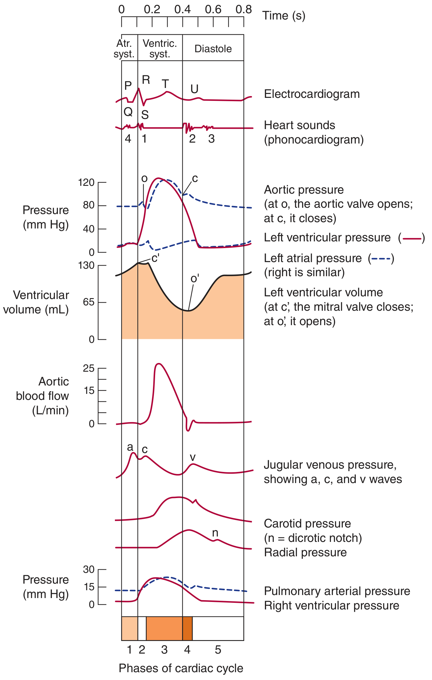
| Phase | Event | Valves | Key Feature |
|---|---|---|---|
| 1 - Atrial Systole | P wave → atrial contraction | MV & TV open | Last 20-30% of ventricular filling (atrial "kick") |
| 2 - Isovolumetric Contraction | QRS → ventricles contract, no flow yet | All 4 valves CLOSED | Highest O2 consumption; pressure ↑ rapidly |
| 3 - Ventricular Ejection | LV pressure > Aortic pressure | Aortic & Pulmonic valves OPEN | Stroke volume ejected; EF = SV/EDV |
| 4 - Isovolumetric Relaxation | T wave → ventricles relax | All 4 valves CLOSED | Pressure falls rapidly |
| 5 - Ventricular Filling (Diastole) | LV pressure < LA pressure | Mitral valve OPENS | Rapid filling (70%), then slow filling, then atrial kick |
🔑 Duration at HR 75: Systole = 0.27 sec | Diastole = 0.53 sec
🔑 At high heart rates, diastole shortens more than systole - compromises coronary filling!
Heart Sounds
| Sound | Cause | Heard Best | Significance |
|---|---|---|---|
| S1 | Closure of MV + TV (beginning of systole) | Apex (mitral area) | Loud in MS; Soft in MR |
| S2 | Closure of AV + PV (end of systole) | Base | Split on inspiration (physiological); Fixed split = ASD |
| S3 | Rapid ventricular filling (early diastole) | Apex, LLD | Pathological in adults > 40 yrs = LV failure; Normal in children |
| S4 | Atrial contraction against stiff ventricle (late diastole) | Apex | Hypertension, AS, LVH - never normal |
🔑 S3 gallop = Heart failure (ventricular overload)
🔑 S4 gallop = Stiff ventricle (diastolic dysfunction, HCM, severe HTN)
🔑 "Tennessee" (S3) vs "Kentucky" (S4) pattern sounds
💡 Mnemonic - Heart sounds: "Many Thanks (S1 = Mitral + Tricuspid closing); Aortic Pulmonic (S2 = Aortic + Pulmonic closing)"
JVP Waveforms
| Wave | Cause | Loss/Exaggeration |
|---|---|---|
| a wave | Atrial contraction | Lost in AF; Giant a = TS, Complete heart block |
| c wave | Tricuspid valve closure (bulging into RA) | Often not visible |
| x descent | Atrial relaxation | Prominent in cardiac tamponade |
| v wave | Venous filling against closed TV | Giant v wave = Tricuspid regurgitation |
| y descent | Tricuspid valve opens, blood rushes into RV | Absent y = Cardiac tamponade; Sharp y = Constrictive pericarditis |
🔑 Kussmaul's sign (JVP rises on inspiration) = Constrictive pericarditis, Cardiac tamponade, RV infarct
Cardiac Output & Frank-Starling Law
Cardiac Output (CO) = Heart Rate × Stroke Volume
- Normal CO = 5 L/min
- Cardiac Index = CO/BSA = 2.2-4.2 L/min/m²
Frank-Starling Law: "The heart pumps out what comes in"
- ↑ Preload (venous return) → ↑ EDV → ↑ fiber stretch → ↑ force of contraction → ↑ SV
- This is the basis of why:
- IV fluids improve CO in hypovolemia
- Failing hearts cannot compensate (flat Starling curve)
Determinants of Stroke Volume:
- Preload (EDV - filling pressure)
- Afterload (aortic pressure - resistance to ejection)
- Contractility (inotropy - intrinsic force of contraction)
💡 Mnemonic - Factors ↑ Contractility: "CAT - Catecholamines, Digitalis (↑Ca²⁺), Thyroid hormones"
Blood Pressure Regulation
↓ BP detected by baroreceptors (carotid sinus, aortic arch)
        ↓
Sympathetic activation
        ↓
↑ HR + ↑ Contractility + Vasoconstriction
        ↓
Long-term: RAAS activation (Renin → Angiotensin II → Aldosterone)
        ↓
Na+ & water retention → ↑ blood volume → ↑ BP
🔑 Renin is secreted by juxtaglomerular cells of kidney in response to: ↓ BP, ↓ Na delivery, sympathetic stimulation
🔑 ACE converts Ang I → Ang II (lungs are main site)
🔑 Aldosterone (adrenal cortex) → ↑ Na reabsorption in collecting duct → ↑ BP
MODULE 3: PATHOLOGY
Atherosclerosis - Mechanism
Endothelial injury (HTN, smoking, diabetes, dyslipidemia)
        ↓
LDL enters intima → oxidized LDL
        ↓
Macrophages engulf oxidized LDL → FOAM CELLS
        ↓
Fatty streak (earliest lesion - reversible)
        ↓
Fibroblast + SMC migration → Fibrous plaque (atheroma)
        ↓
Complicated lesion: Calcification, Ulceration, Thrombosis
🔑 Foam cells = Lipid-laden macrophages = Hallmark of early atherosclerosis
🔑 Most common sites: Coronaries, Aorta (especially infrarenal), Carotid bifurcation, Circle of Willis, Popliteal artery
🔑 "Response to injury" hypothesis - Ross (1976/1999)
Myocardial Infarction - The Most Important CVS Topic
Coronary Territory → MI Type
| Artery Occluded | Territory | ECG Leads | MI Type |
|---|---|---|---|
| LAD | Anterior LV, Apex | V1-V4 | Anterior MI |
| LCX | Lateral LV | I, aVL, V5-V6 | Lateral MI |
| RCA | Inferior LV, RV | II, III, aVF | Inferior MI |
| Posterior (PDA from RCA) | Posterior wall | V7-V9 (reciprocal in V1-V2) | Posterior MI |
Timeline of MI Changes (NEET PG favorite!)
| Time | Gross | Microscopy | Cardiac Enzymes |
|---|---|---|---|
| 0-1 hr | None visible | None | Myoglobin ↑ (1st to rise) |
| 1-4 hr | Pallor beginning | Wavy fibers, Early coagulative necrosis | Troponin I & T start rising |
| 4-12 hr | Pallor | Coagulative necrosis begins | Troponin peaks |
| 12-24 hr | Pale, yellow | PMN infiltration begins | CK-MB peaks at 24hr |
| 1-3 days | Yellow-white | Maximum PMN infiltration | CK-MB normalizes by 48-72hr |
| 3-7 days | Yellow-tan (soft) | Macrophage infiltration, Phagocytosis starts | Troponin I normalizes day 7-10 |
| 1-3 weeks | Hyperemic border | Granulation tissue, Neovascularization | Troponin T normalizes week 2 |
| > 6 weeks | White scar | Dense fibrosis, Scar complete | All normal |
🔑 First enzyme to rise: Myoglobin (1-3 hrs)
🔑 Most sensitive & specific: Troponin I & T (rise by 3-6 hrs, peak at 24 hrs, persist 7-10 days Troponin I; 14 days Troponin T)
🔑 CK-MB - Best for re-infarction (normalizes quickly; second rise indicates new MI)
🔑 LDH - Used historically when patient presents late; LDH1 > LDH2 = "flipped LDH" (cardiac)
Zones of MI (Pathology)
Central DEAD zone → Zone of Injury → Zone of Ischemia
(Q wave)           (ST elevation)   (T wave inversion)
Complications of MI (by time)
| Timing | Complication |
|---|---|
| 0-24 hrs | Fatal arrhythmias (VF - #1 cause of death in acute MI), Sudden death |
| 1-3 days | Cardiogenic shock (massive MI >40% LV), Pericarditis (early) |
| 3-7 days | Free wall rupture (soft necrotic muscle) → Hemopericardium → Tamponade |
| 5-7 days | Papillary muscle rupture → Acute severe MR → Flash pulmonary edema |
| 7-14 days | IVS (septal) rupture → VSD |
| Weeks-months | LV aneurysm, Dressler's syndrome (autoimmune pericarditis - 2-10 wks) |
🔑 "Dead men tell no tales" at Day 3-5 - highest risk of rupture (maximum softening)
🔑 Dressler's syndrome = fever + pericarditis + elevated ESR 2-10 weeks post-MI
Infective Endocarditis
Duke's Criteria (2 major OR 1 major + 3 minor OR 5 minor = Definite IE)
Major Criteria:
- Blood cultures: typical organism ×2 (S. aureus, Viridans streptococci, HACEK group)
- Echo evidence: vegetation, abscess, new valvular regurgitation
Minor Criteria:
- Predisposing heart condition or IVDU
- Fever >38°C
- Vascular phenomena (emboli, Janeway lesions, splinter hemorrhages)
- Immunological phenomena (Osler nodes, Roth spots, glomerulonephritis, ↑ RF)
- Blood culture positive (not meeting major criterion)
💡 Mnemonic for peripheral signs of IE:
- Janeway lesions = painless, palms/soles (Janeway = J for "Just painless")
- Osler nodes = painful, finger/toe pads (Osler = "Ouch!")
- Roth spots = retinal hemorrhages with pale center
- Splinter hemorrhages = under nails
Organisms in IE by Setting
| Setting | Most Likely Organism |
|---|---|
| Native valve (normal) | Viridans streptococci (S. viridans) - after dental procedure |
| Native valve (abnormal/RHD) | Viridans streptococci |
| IV drug use | S. aureus (right-sided, tricuspid valve) |
| Prosthetic valve < 2 months | S. epidermidis (CoNS) |
| Prosthetic valve > 2 months | Viridans streptococci |
| Colon cancer | Streptococcus bovis (S. gallolyticus) |
| Nosocomial | S. aureus, CoNS |
Rheumatic Heart Disease
Pathogenesis:
- Group A beta-hemolytic streptococcal pharyngitis (Strep pyogenes)
- Molecular mimicry: Anti-streptococcal M protein antibodies cross-react with cardiac proteins (myosin, laminin)
- Immune injury via antibodies + T cells
- 1-3% of streptococcal pharyngitis cases develop rheumatic fever
Robbins & Kumar Basic Pathology - Aschoff bodies are pathognomonic for rheumatic fever; contain Anitschkow cells (caterpillar cells) - activated macrophages with chromatin condensed into a slender wavy ribbon.
🔑 Aschoff body = Pathognomonic of rheumatic fever = Granuloma-like collection of lymphocytes, plasma cells, Anitschkow cells around fibrinoid necrosis
🔑 Anitschkow cells = "Caterpillar cells" (wavy chromatin)
🔑 VALVES AFFECTED (order of frequency): Mitral > Aortic > Tricuspid > Pulmonic
Jones Criteria for Rheumatic Fever
Major (💡 JONES): Joints (migratory polyarthritis), cOrea (Sydenham's), Nodules (subcutaneous), Erythema marginatum, carditis (pancarditis)
Minor: Fever, ↑ ESR/CRP, Prolonged PR interval, Previous rheumatic fever
Requirement: 2 Major OR 1 Major + 2 Minor PLUS evidence of recent strep infection (↑ASO titer, positive culture/rapid antigen test)
Cardiomyopathies
| Feature | Dilated (DCM) | Hypertrophic (HCM) | Restrictive (RCM) |
|---|---|---|---|
| Gross | Large, dilated, flabby | Heavy, hypertrophied (asymmetric septal) | Normal or slightly enlarged |
| Main defect | Systolic dysfunction ↓EF | Diastolic dysfunction (LVOTO) | Diastolic dysfunction (stiff) |
| Causes | Alcohol, Viral (Coxsackie B), Idiopathic, Peripartum, Doxorubicin | AD mutation (Myosin heavy chain β-MYH7, MYBPC3) | Amyloid, Sarcoid, Hemochromatosis, Löffler's |
| ECG | BBB, ST changes | LVH, Septal Q waves | Low voltage (amyloid) |
| Echo finding | ↓EF, dilated chambers | Asymmetric septal hypertrophy (ASH), SAM of mitral valve, LVOTO gradient | Biatrial enlargement, normal/small ventricles |
| Treatment | ACE-I, BB, MRA, SGLT2i, ICD | Avoid vasodilators/diuretics; Beta-blockers, Verapamil; Septal myectomy/TAVI | Treat underlying cause |
🔑 HCM: Most common cause of sudden cardiac death in young athletes
🔑 SAM = Systolic Anterior Motion of mitral valve → causes dynamic LVOTO in HCM
🔑 HCM murmur increases with Valsalva and standing (↓ preload → ↑ obstruction)
🔑 HCM murmur decreases with squatting and passive leg raise (↑ preload → ↓ obstruction)
MODULE 4: PHARMACOLOGY
Antihypertensive Drugs - Integrated Mechanism Map
RAAS System                    Sympathetic System         Direct Vasodilation
     ↓                               ↓                          ↓
ACE Inhibitors                 Beta-blockers              Calcium Channel Blockers
ARBs                           Alpha-1 blockers           Hydralazine (arterioles)
Aliskiren (renin inhibitor)    Central: Clonidine         Nitrates (veins >> arteries)
Aldosterone antagonists                ↓
(Spironolactone, Eplerenone)    ↓ HR, ↓ contractility
                                ↓ Renin release
     ↓
Diuretics (Thiazide, Loop, K-sparing)
↓ Volume → ↓ preload → ↓ BP
Drug Classes - Key Facts
| Class | Example | Mechanism | Key Side Effect | Indication / CI |
|---|---|---|---|---|
| ACE Inhibitors | Ramipril, Enalapril | Block ACE → ↓ Ang II → ↓ Aldosterone | Dry cough (↑ bradykinin), Angioedema, Hyperkalemia | 1st line HF, Post-MI, CKD with DM; CI: Pregnancy, Bilateral RAS |
| ARBs | Losartan, Valsartan | Block AT1 receptor | Hyperkalemia (no cough) | Same as ACEi; alternative if ACEi cough |
| Beta-blockers | Metoprolol, Carvedilol, Bisoprolol | Block β1 (heart) → ↓ HR, ↓ CO, ↓ Renin | Bradycardia, Bronchospasm (non-selective), Mask hypoglycemia | IHD, HF (with ACEi), Post-MI; CI: Asthma, AV block |
| Calcium Channel Blockers | Amlodipine (DHP), Verapamil (non-DHP) | Block L-type Ca²⁺ channels → Vasodilation / ↓HR | Peripheral edema (DHP), Constipation (Verapamil), Bradycardia (non-DHP) | HTN, Angina; Verapamil + BB = dangerous AV block |
| Thiazide diuretics | Hydrochlorothiazide, Chlorthalidone | Block NCC in DCT → ↓ Na reabsorption | Hypokalemia, Hyponatremia, Hyperuricemia, Hyperglycemia, Hypercalcemia | 1st line HTN (esp. African Americans with CCB) |
| Loop Diuretics | Furosemide, Torsemide | Block NKCC2 in loop of Henle | Hypokalemia, Hyponatremia, Ototoxicity (Furosemide), Hypocalcemia | Acute heart failure, Pulmonary edema, Hypertensive urgency |
| Aldosterone antagonists | Spironolactone, Eplerenone | Block aldosterone receptor → ↑ K, ↑ Mg | Hyperkalemia; Spironolactone: Gynecomastia, Menstrual irregularities | HF (HFrEF), Secondary HTN from hyperaldosteronism; CI: K >5.5 |
| Nitrates | Nitroglycerin, Isosorbide dinitrate | → NO → cGMP → smooth muscle relaxation → Venodilation > arteriodilation | Headache, Postural hypotension, Tolerance (24-hr nitrate-free period) | Angina pectoris, Acute LVF; CI: PDE5 inhibitors (sildenafil) |
| Digoxin | Digoxin | Inhibit Na/K ATPase → ↑ intracellular Ca²⁺ → ↑ Contractility; Vagal tone → ↓ HR | Narrow TI; Toxicity: Yellow-green vision, Arrhythmias; Hypokalemia worsens toxicity | HF with AF; Rate control in AF; CI: WPW, HCM, 2nd/3rd degree AV block |
Drug of Choice - High Yield Summary
| Condition | DOC |
|---|---|
| HTN + Diabetes with proteinuria | ACE Inhibitor / ARB |
| HTN + Asthma | Amlodipine (CCB) or Thiazide (avoid BB) |
| HTN + Pregnancy | Methyldopa (1st line), Labetalol, Nifedipine |
| HTN + Benign Prostatic Hyperplasia | Alpha-1 blockers (Prazosin, Tamsulosin) |
| HTN + Heart failure | ACE-I + BB + MRA + Diuretic |
| Hypertensive emergency | IV Sodium nitroprusside / IV Labetalol / IV Nicardipine |
| Angina pectoris | Nitrates (short-acting) for acute; BB + Long-acting nitrate for prophylaxis |
| Prinzmetal (vasospastic) angina | Calcium Channel Blockers (NOT Beta-blockers - can worsen) |
| Post-MI cardioprotection | ACE-I + BB + Aspirin + Statin (ABAS) |
| HFrEF (EF <40%) | ACE-I/ARB/ARNI + BB + MRA + SGLT2i + Diuretic (if congested) |
Antiarrhythmic Drugs - Vaughan-Williams Classification
| Class | Mechanism | Examples | Indication |
|---|---|---|---|
| Ia | Na channel block (moderate) + ↑ APD | Quinidine, Procainamide, Disopyramide | AF, VT; Quinidine: Torsades risk (QT prolongation) |
| Ib | Na channel block (fast dissociation) | Lidocaine, Mexiletine, Phenytoin | VT/VF (esp. post-MI), Lidocaine = IV only |
| Ic | Na channel block (slow dissociation) | Flecainide, Propafenone | AF, SVT; CI: Structural heart disease (CAST trial) |
| II | Beta-blockade | Metoprolol, Esmolol, Propranolol | SVT, AF rate control, Post-MI |
| III | K channel block → ↑ APD, ↑ QT | Amiodarone, Sotalol, Ibutilide, Dronedarone | VT, VF, AF; Amiodarone = broadest spectrum |
| IV | Ca channel block | Verapamil, Diltiazem | SVT, AF rate control; CI: WPW, HF with systolic dysfunction |
🔑 Amiodarone = most effective antiarrhythmic but most side effects:
- Pulmonary fibrosis
- Thyroid dysfunction (hypo or hyper - contains iodine)
- Hepatotoxicity
- Peripheral neuropathy
- Corneal microdeposits (bluish-gray skin discoloration with sun)
- Requires monitoring: TFT, LFT, CXR, PFT
🔑 Digoxin toxicity treatment: Stop drug, Correct hypokalemia, Digibind (Fab fragments) for severe toxicity
MODULE 5: CLINICAL MEDICINE (Integrated)
Hypertension
Definition (JNC 8 / ACC/AHA 2017):
- ACC/AHA 2017: ≥ 130/80 mmHg = HTN
- JNC 8: ≥ 140/90 mmHg = Stage 1 HTN
Causes of Secondary HTN (5 H's):
- Hyperaldosteronism (Conn's syndrome) - ↓ K, ↑ aldosterone, ↑ aldosterone:renin ratio
- Hyperthyroidism / Hypothyroidism
- Hyperparathyroidism (rarely)
- Henal artery stenosis (Renovascular HTN) - young woman, bruit
- Pheochromocytoma - triad: Headache, Palpitations, Profuse sweating; 24hr urine catecholamines/VMA/metanephrines
🔑 Most common cause of secondary HTN: Primary hyperaldosteronism (Conn's syndrome) in adults
🔑 Most common cause in children: Renovascular HTN (renal artery stenosis)
🔑 Hypertensive urgency (>180/120, no end-organ damage) vs Emergency (same BP WITH end-organ damage: papilledema, encephalopathy, AKI, MI, aortic dissection)
Heart Failure
HFrEF (Systolic HF, EF <40%) vs HFpEF (Diastolic HF, EF >50%)
| Feature | HFrEF | HFpEF |
|---|---|---|
| Defect | ↓ Contractility (systolic) | ↓ Compliance (diastolic) |
| EF | < 40% | ≥ 50% |
| Common causes | IHD, Dilated CM, Myocarditis | Hypertension, HCM, Restrictive CM, Aging |
| BNP | ↑↑ | ↑ (less) |
| Treatment | ACEi/ARB/ARNI + BB + MRA + SGLT2i | Diuretics for congestion; treat cause |
🔑 BNP is released by ventricular myocytes in response to wall stress (volume/pressure overload)
- BNP > 100 pg/mL = HF likely
- BNP < 35 pg/mL = HF unlikely
- NT-proBNP > 125 pg/mL = HF (used more now; longer half-life)
NYHA Classification:
- Class I: No symptoms with normal activity
- Class II: Symptoms with moderate exertion
- Class III: Symptoms with minimal exertion
- Class IV: Symptoms at rest
Signs of Heart Failure:
- Left HF: Dyspnea, Orthopnea (3-pillow), PND, Bilateral basal crepitations, S3 gallop, Displaced apex
- Right HF: JVD, Peripheral pitting edema (legs), Hepatomegaly, Ascites, Pleural effusion (R > L)
Acute Coronary Syndromes
Acute Coronary Syndrome (ACS)
         ├── Unstable Angina (no troponin rise, no ECG changes)
         ├── NSTEMI (↑ Troponin, No ST elevation, may have ST depression/T inversion)
         └── STEMI (↑ Troponin + ST elevation ≥ 1mm in 2 contiguous limb leads, ≥ 2mm in precordial)
STEMI Management - "TIME IS MUSCLE"
Immediate (First 10 minutes):
- ECG + IV access + O2 (if SpO2 < 94%) + Monitor
- Aspirin 300 mg loading
- P2Y12 inhibitor (Ticagrelor 180 mg or Clopidogrel 300-600 mg)
- IV Morphine (for pain), IV Metoprolol (if no CI)
Reperfusion (within 90 min - Door to Balloon time):
- Primary PCI = preferred (if available within 90 min of first medical contact)
- Thrombolysis (if PCI not available within 120 min) - Streptokinase, tPA (Alteplase)
- CI to thrombolysis: Recent surgery/trauma, Active bleeding, Stroke <3 months, BP >185/110, Aortic dissection
Post-MI medications (ABCDE):
- A - Aspirin (lifelong) + Anti-P2Y12 (12 months DAPT)
- B - Beta-blocker (↓ mortality, ↓ arrhythmia)
- C - Cholesterol (High-intensity Statin - Atorvastatin 80 mg or Rosuvastatin 40 mg)
- D - Diabetes control
- E - ACE inhibitor/ARB (especially if EF reduced, HTN, or DM)
Valvular Heart Diseases
| Valve Lesion | Classic Murmur | Key Feature | Cause |
|---|---|---|---|
| Mitral Stenosis | Rumbling mid-diastolic murmur at apex + Opening snap | Loud S1; Irregularly irregular (AF); "Fish mouth" valve on echo | RHD (#1), Congenital |
| Mitral Regurgitation | Pansystolic murmur at apex radiating to axilla | Soft S1; S3 gallop; Displaced hyperdynamic apex | RHD, MVP, IE, Ruptured papillary muscle |
| Mitral Valve Prolapse | Late systolic murmur + midsystolic click | Click-murmur syndrome; Young females; Associated with Marfan's | Connective tissue disorder |
| Aortic Stenosis | Harsh ejection systolic murmur (RUSB) radiating to carotids | Slow rising pulse; Narrow pulse pressure; Soft A2 | Bicuspid aortic valve (young), Senile calcific (elderly), RHD |
| Aortic Regurgitation | High-pitched early diastolic murmur (LLSB) | Wide pulse pressure; Collapsing pulse (water hammer); Austin Flint murmur | IE, Aortic dissection, RHD, Marfan's, AS |
🔑 Murmur increases with: Valsalva release, Passive leg raise (↑ preload = more blood = louder except HCM)
🔑 AS triad (SAD): Syncope, Angina, Dyspnea (in order of increasing severity; Dyspnea = worst prognosis)
🔑 AR signs: Corrigan's pulse, de Musset's sign, Quincke's sign, Traube's sign, Müller's sign, Duroziez's sign
MODULE 6: QUICK REVISION TABLE - Integrated CVS
| Topic | Anatomy | Physiology | Pathology | Pharmacology | Clinical |
|---|---|---|---|---|---|
| MI (Anterior) | LAD occlusion | ↓ CO, ↑ LVEDP | Coagulative necrosis, Troponin ↑ | Aspirin, LMWH, Clopidogrel | V1-V4 ST elevation, PCI |
| MI (Inferior) | RCA occlusion | Risk of AV block | Same as above | Same + Watch Morphine (↓ BP) | II, III, aVF elevation |
| Heart Failure | LV dilatation (dilated) or hypertrophy (HCM) | ↓ EF (HFrEF) or ↓ compliance | Myocyte hypertrophy → fibrosis | ACEi + BB + MRA + SGLT2i | BNP ↑, CXR: Cardiomegaly, Bat-wing opacities |
| HTN | Thickened vessels, LV hypertrophy | ↑ TPR or ↑ CO (Renin) | Hyaline arteriolosclerosis, Fibrinoid necrosis | Thiazide/CCB/ACEi (1st line) | Renal: ↑Creatinine; Eye: AV nicking |
| RHD | Mitral valve fibrosis | MS: ↑ LA pressure | Aschoff bodies, Anitschkow cells | Prophylactic Penicillin G | Mid-diastolic murmur, AF |
| IE | Valve vegetations | Embolic phenomena | Sterile vegetations (earlier), Bacterial vegetations | Penicillin + Gentamicin (IV) | Duke's criteria, Blood cultures ×3 |
| Atherosclerosis | Intimal thickening, Narrowed lumen | ↓ Flow → Ischemia | Foam cells → Fibrous plaque | Statins (↓ LDL, ↑ plaque stability) | Chest pain, Ankle-Brachial Index |
MODULE 7: HIGH-YIELD MNEMONICS & FACTS
💡 "ACTS" - Causes of Wide Fixed Split S2: ASD (most common), Complete RBBB, Total anomalous pulmonary venous connection, Severe PS
💡 "5 T's" of Cyanotic CHD: Tetralogy of Fallot, Transposition of Great Arteries, Total anomalous pulmonary venous connection, Truncus arteriosus, Tricuspid atresia
💡 Endocarditis prophylaxis needed for: Prosthetic valves, Previous IE, Congenital cyanotic HD (unrepaired), Cardiac transplant with valvulopathy - during DENTAL procedures (amoxicillin 2g PO 30-60 min before)
🔑 Pulsus paradoxus (>10 mmHg drop in SBP during inspiration) = Cardiac tamponade, Severe asthma, Constrictive pericarditis
🔑 Beck's Triad (cardiac tamponade) = Hypotension + Muffled heart sounds + JVD
🔑 ECG in Pericarditis = Diffuse saddle-shaped ST elevation (all leads except aVR and V1) + PR depression
🔑 Libman-Sacks endocarditis = Non-bacterial thrombotic vegetations, both surfaces of mitral valve = SLE (lupus)
🔑 Marantic endocarditis = Non-bacterial thrombotic vegetations, associated with malignancy and debilitating illness
🔑 WPW Syndrome ECG: Short PR interval (<0.12 sec) + Delta wave + Wide QRS = Accessory pathway (Bundle of Kent)
- Treatment: Radiofrequency ablation; Avoid Digoxin and Verapamil (can accelerate conduction through accessory pathway → VF)
MODULE 8: PREVIOUS YEAR NEET PG QUESTION THEMES (CVS)
| Theme | Key Fact to Remember |
|---|---|
| Earliest enzyme in MI | Myoglobin (1-3 hrs) |
| Most sensitive & specific | Troponin I |
| Re-infarction marker | CK-MB |
| Pathognomonic of RHD | Aschoff bodies / Anitschkow cells |
| Sudden death in young athlete | HCM (β-myosin heavy chain mutation) |
| Loud S1 | Mitral stenosis |
| Opening snap | Mitral stenosis (earlier = more severe) |
| Wide fixed split S2 | ASD |
| JVP absent y descent | Cardiac tamponade |
| JVP sharp y descent | Constrictive pericarditis |
| Pulsus paradoxus | Cardiac tamponade |
| Kussmaul's sign | Constrictive pericarditis (NOT tamponade typically) |
| AF + Calcified valve | Mitral stenosis (RHD) |
| Murmur increases with Valsalva | HCM (dynamic LVOTO) |
| Most common cause of MR | MVP (in developed countries) |
| Libman-Sacks | SLE |
| DOC for Prinzmetal angina | CCB (diltiazem/amlodipine) |
| Digoxin toxicity antidote | Digibind (Digoxin-Fab antibody) |
Sources: Guyton & Hall Textbook of Medical Physiology; Ganong's Review of Medical Physiology (26th ed.); Robbins & Kumar Basic Pathology; Goodman & Gilman's The Pharmacological Basis of Therapeutics; Goldman-Cecil Medicine; Braunwald's Heart Disease (15th ed.); Gray's Anatomy for Students
This is a shared conversation. Sign in to Orris to start your own chat.