
Western blotting
Reading File
Western blotting is a lab technique used to detect specific proteins in a sample by separating them by size (SDS-PAGE), transferring to a membrane, and probing with antibodies.
Searching sources...
Finding Sources
Searching the Web
western blotting technique steps principle applications
Reading File
Reading File
Finding Sources
Finding Sources
Reading File
Reading File
Loading Image
Here is a complete overview of Western blotting, compiled from multiple medical textbooks:
Western Blotting (Protein Immunoblot)
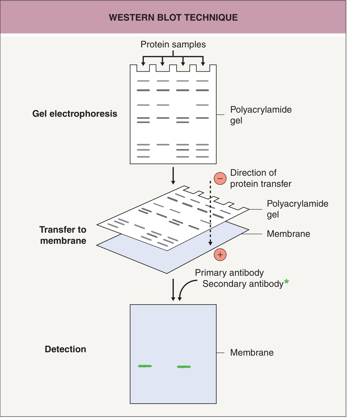
Definition
Western blotting (also called protein immunoblot) is a technique used to detect specific proteins in a sample - either the microbial proteins themselves, or patient antibodies directed against those proteins. It is a variation of the Southern blot (for DNA, developed by Edwin Southern) and the Northern blot (for RNA). The term "Western blot" was introduced by Burnette in 1981.
- Medical Microbiology 9e
Principle
The technique uses three core elements:
- Separation of proteins by molecular weight (size)
- Transfer of separated proteins to a solid membrane support
- Detection of the target protein using antibody probes
Step-by-Step Procedure
1. Sample Preparation
- Cells or organisms are mechanically or chemically disrupted to release proteins
- Proteins are denatured with a strong reducing agent (e.g., SDS - sodium dodecyl sulfate)
- SDS coats proteins with a uniform negative charge, so separation is purely by size
2. Gel Electrophoresis (SDS-PAGE)
- The protein mixture is loaded into a polyacrylamide gel
- An electric current drives proteins through the gel - smaller proteins migrate faster and travel further
- This separates proteins into distinct bands by molecular weight
3. Transfer (Blotting)
- The separated protein bands are transferred electrophoretically from the gel onto a nitrocellulose or nylon membrane
- Transfer occurs by passing an electric current through a "sandwich" (gel + membrane) submerged in transfer buffer - proteins migrate toward the positive electrode onto the membrane
- The membrane is then blocked (usually with milk proteins) to prevent nonspecific antibody binding
4. Primary Antibody Incubation
- The membrane is incubated with the patient's serum or a specific primary antibody
- The primary antibody binds to its target protein on the membrane
- Polyclonal antibodies recognize multiple epitopes; monoclonal antibodies recognize a single epitope
5. Secondary Antibody Detection
-
A labeled secondary antibody (anti-human IgG) is added - it binds to the primary antibody
-
The secondary antibody carries a detection label: enzyme, fluorescent probe, or radioactive isotope
-
Enzyme-labeled antibodies convert a substrate to produce a colorimetric or chemiluminescent signal
-
The pattern of bands reveals which proteins the antibodies bind to
-
Medical Microbiology 9e; Dermatology 2-Volume Set 5e; Jawetz Melnick & Adelbergs Medical Microbiology 28E
The Antibody System
| Antibody | Source | Target |
|---|---|---|
| Primary Ab | Polyclonal (animal serum) or monoclonal (hybridoma) | Specific protein of interest |
| Secondary Ab | Labeled anti-species IgG | Primary antibody |
Detection labels used on the secondary antibody:
- Enzyme (e.g., HRP) - produces color or chemiluminescence on substrate
- Fluorescent probe - produces light signal
- Radioactive isotope (e.g., I-125, older method)
Purpose / Uses
| Use | Examples |
|---|---|
| Detect patient antibodies to a pathogen | HIV confirmatory test, Lyme disease |
| Identify specific proteins in a sample | Research, diagnostic microbiology |
| Determine molecular weight of a protein | Research |
| Quantify protein expression | Research, biomarker studies |
| Confirm pathogen identity | Prion proteins (CJD) |
Clinical Applications
-
HIV - historically used as confirmatory test after positive ELISA/EIA. A positive Western blot in the right context confirms HIV infection. The CDC no longer recommends Western blot for routine HIV diagnosis (replaced by newer supplemental assays), but it remains conceptually relevant.
-
Lyme disease - a two-tier approach: ELISA first, then Western blot to confirm a positive/equivocal ELISA result. The IgM Western blot has a high false-positive rate when pretest probability is low. - Textbook of Family Medicine 9e
-
Prion diseases - used in Creutzfeldt-Jakob disease (CJD) diagnosis
-
HIV-2 - indeterminate HIV-1 Western blot results may indicate HIV-2 infection, requiring a separate HIV-2 Western blot - Jawetz 28E
Benefits
- Simple and sensitive method to detect and quantify proteins
- Can determine the molecular weight of a specific protein relative to standard molecular weight markers
- Can differentiate specific reactivity from nonspecific (negative) reactions
Limitations / Sources of Error
-
Requires a specific antibody that recognizes the target protein (unavailable for some antigens)
-
May produce high background of nonspecific staining
-
Proteins may degrade during extraction
-
IgM Western blot results can give false positives when pretest probability is low (e.g., Lyme disease)
-
Indeterminate results occur (e.g., HIV-2 cross-reactivity with HIV-1 test)
-
Dermatology 2-Volume Set 5e
Related / Alternative Techniques
| Technique | Key difference |
|---|---|
| Immunoprecipitation (IP) | Antibody added to protein mixture first; proteins not denatured - detects native conformation |
| IP-Western | Combines IP + Western blot to study protein-protein interactions |
| ELISA | Quantifies protein amount; protein captured on antibody-coated plate; no gel separation |
| Immunohistochemistry | Visualizes protein localization within cells/tissue sections |
| Southern blot | Same principle but for DNA |
| Northern blot | Same principle but for RNA |
- Dermatology 2-Volume Set 5e
Blot Family Summary
| Blot | Detects | Separation Method |
|---|---|---|
| Southern | DNA | Agarose gel |
| Northern | RNA | Agarose gel |
| Western | Protein | SDS-PAGE |
Southern blotting
Southern blotting is a technique for detecting specific DNA sequences in a sample, developed by Edwin Southern in 1975. It involves DNA restriction digestion, gel electrophoresis, transfer to a membrane, and hybridization with a labeled probe.
Searching sources...
Finding Sources
Searching the Web
southern blotting technique principle steps procedure applications
Reading File
Finding Sources
Reading File
Reading File
Reading File
Loading Image
Loading Image
Reading File
Here is a complete overview of Southern blotting, drawn from multiple medical and biochemistry textbooks:
Southern Blotting
Definition
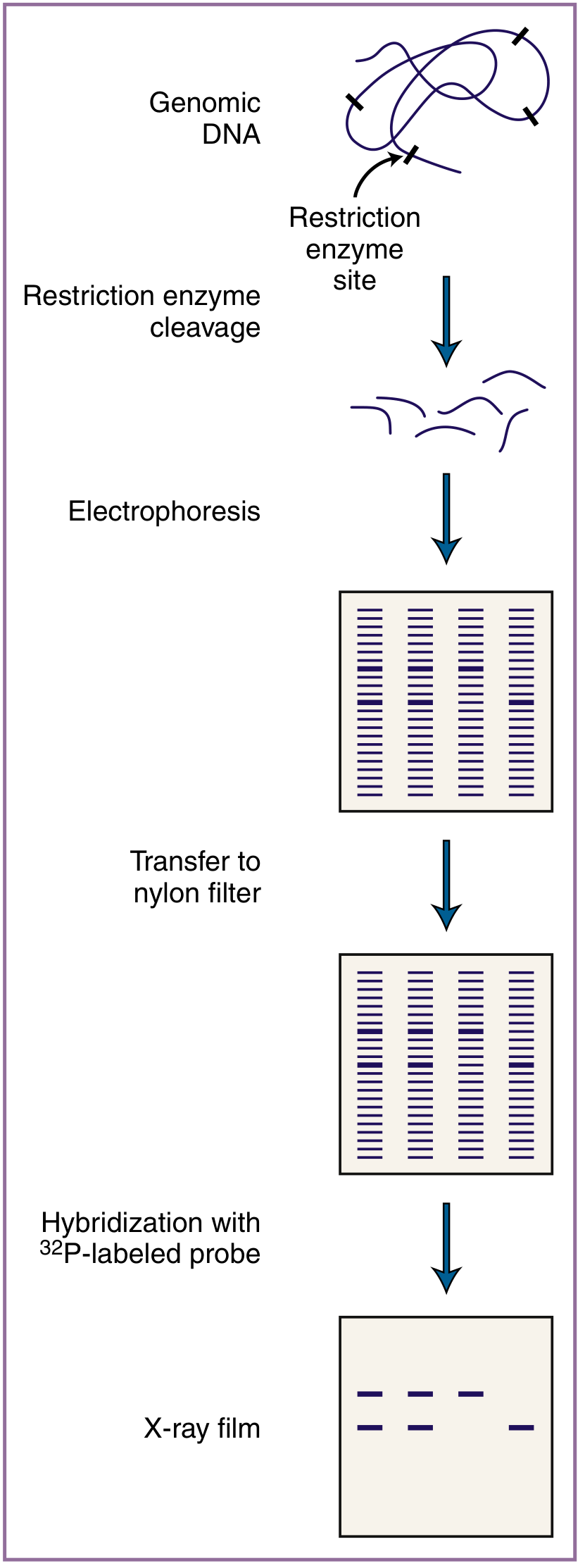
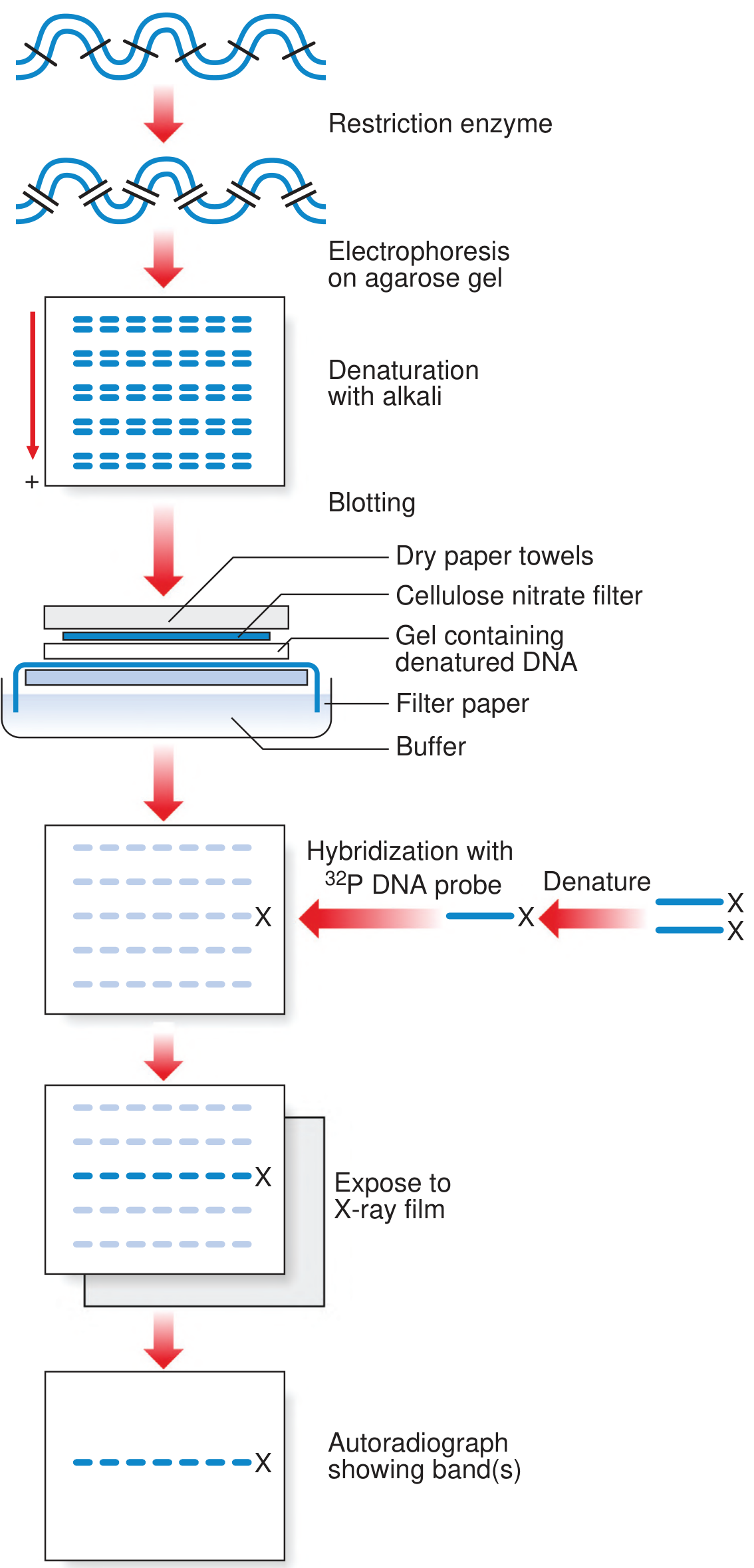
Southern blotting is a molecular technique that combines restriction enzyme digestion, agarose gel electrophoresis, membrane transfer, and nucleic acid hybridization to detect and analyze specific DNA sequences in a complex sample. It was developed by Edwin Mellor Southern in 1975 and is named after him.
- Emery's Elements of Medical Genetics and Genomics; Schwartz's Principles of Surgery 11e
Core Principle
The technique allows discrimination of DNA at three levels simultaneously:
- At the level of restriction enzyme recognition (where cuts are made)
- By the size of the resulting DNA fragment
- By hybridization of a DNA probe to a specific locus (sequence identity)
- Jawetz Melnick & Adelbergs Medical Microbiology 28E
Step-by-Step Procedure
Step 1: DNA Extraction
- DNA is extracted from the sample (e.g., patient's white blood cells, bacterial colony)
- The entire genomic digest may yield ≥10⁶ fragments
Step 2: Restriction Enzyme Digestion
- DNA is cleaved with a restriction endonuclease - a highly specific bacterial enzyme that recognizes a unique palindromic sequence (usually 4-8 bp, e.g., GAATTC)
- The pattern of fragments produced is unique to each gene sequence
- Wild-type and mutant DNA are discriminated by the size of resulting fragments
Step 3: Gel Electrophoresis
- The DNA fragments (all negatively charged) are loaded onto an agarose gel
- An electric current drives them through the gel - smaller fragments migrate faster and travel farther
- A DNA ladder (standard fragments of known size) runs alongside to allow sizing of unknown bands
- The gel is stained with ethidium bromide and photographed with a ruler alongside
Step 4: Denaturation
- The double-stranded DNA in the gel is denatured with alkali (e.g., NaOH), converting the fragments to single-stranded DNA
- This is necessary for probe hybridization in the next steps
Step 5: Transfer (Blotting) to Membrane
- The single-stranded DNA fragments are transferred from the gel to a nitrocellulose or nylon membrane, preserving their relative positions exactly
- Transfer occurs either by:
- Capillary diffusion - buffer wicks upward through the gel, carrying DNA fragments onto the membrane above (via a "sandwich" of filter paper, gel, membrane, and paper towels)
- Electrophoretic transfer - electric current drives DNA onto the membrane
- The membrane permanently binds the single-stranded DNA
Step 6: Probe Hybridization
- A single-stranded DNA probe - with a known sequence complementary to the target DNA - is added to the membrane
- The probe is labeled with:
- ³²P radioactive isotope (traditional method) - detected by autoradiography/X-ray film
- Digoxigenin (non-radioactive) - detected by chemiluminescence (safer, faster)
- The probe hybridizes only to fragments with a complementary sequence
Step 7: Detection
-
Radioactive method: Membrane is exposed to X-ray film (autoradiography) - the probe-bound bands appear as dark bands on the film
-
Chemiluminescent method: Generates a light signal detected on film or digitally
-
Lippincott Biochemistry 8e; Emery's Elements of Medical Genetics and Genomics; Creasy & Resnik's Maternal-Fetal Medicine; Schwartz's Principles of Surgery 11e
What Southern Blotting Can Detect
| Type of Mutation | Mechanism of Detection |
|---|---|
| Large insertions or deletions | Changes the size of the restriction fragment (band shifts up or down) |
| Trinucleotide repeat expansions | Produces a larger fragment (e.g., Fragile X, Huntington's) |
| Point mutations at restriction sites | Loss of a cut site → larger fragment; gain of a cut site → smaller fragment |
| Gene rearrangements | Altered banding pattern |
| Large deletions in multi-exon genes | Missing bands (e.g., dystrophin gene in DMD/BMD) |
- Lippincott Biochemistry 8e
Clinical Applications
| Application | Details |
|---|---|
| Fragile X syndrome | Detects large trinucleotide (CGG) repeat expansions - still one of the primary uses |
| Duchenne/Becker muscular dystrophy | Identifies large deletions in the dystrophin gene |
| Prenatal genetic diagnosis | Used to identify disease-associated alleles for single-gene disorders |
| Genetic fingerprinting | Restriction fragment length polymorphism (RFLP) analysis for identity testing |
| Microbial ribotyping | Subtyping bacterial isolates (e.g., outbreak investigation); probes the 16S/23S rRNA genes |
| HIV confirmatory (historical) | Detection of proviral DNA integration patterns |
- Creasy & Resnik's Maternal-Fetal Medicine; Jawetz 28E
Restriction Fragment Length Polymorphism (RFLP)
A major application of Southern blotting is RFLP analysis. Because any two unrelated individuals' genomes differ at many nucleotide positions, restriction enzyme digestion produces fragments of different lengths in different people. Southern blotting reveals these differences as different band patterns, which can be used for:
- Linkage analysis and gene mapping
- Prenatal diagnosis of hereditary conditions
- Forensic DNA profiling
Limitations
-
Requires large amounts of DNA (relatively)
-
Labor-intensive and difficult to automate → expensive
-
Low throughput compared to PCR-based methods
-
For point mutations not at restriction sites, Southern blotting alone cannot detect them
-
Has been largely replaced by PCR for most applications, though it remains the gold standard for detecting large repeat expansions (e.g., Fragile X)
-
Creasy & Resnik's Maternal-Fetal Medicine
Blot Family Comparison
| Blot | Named After | Target Molecule | Gel Used | Probe Type |
|---|---|---|---|---|
| Southern | Edwin Southern (person) | DNA | Agarose | Labeled DNA |
| Northern | Analogy to Southern | RNA (mRNA) | Agarose | Labeled DNA |
| Western | Analogy to Southern | Protein | Polyacrylamide (SDS-PAGE) | Antibody |
Note from Lippincott Biochemistry: "Northern" and "Western" are facetious extensions of the name - they do not refer to any person's name, nor to points of the compass.
This is a shared conversation. Sign in to Orris to start your own chat.