I now have everything needed. Here is the complete structured long answer based on Guyton and Hall Textbook of Medical Physiology (4th South Asia Edition):
Cerebellum - Structured Long Answer (15 Marks)
PART 1: Diagram - Functional Divisions of the Cerebellum (3 Marks)
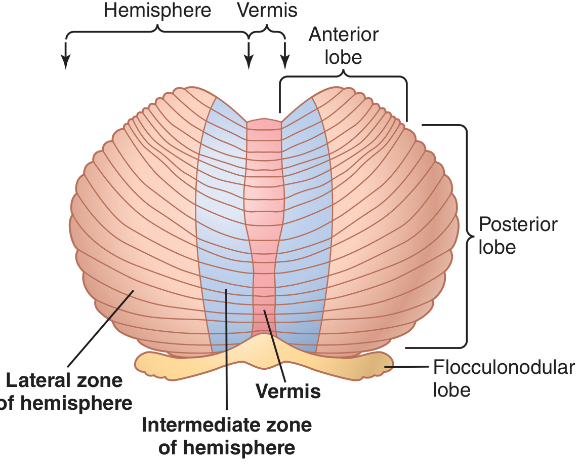
Fig. 57.2 from Guyton & Hall - Functional parts of the cerebellum (posteroinferior view):
From Guyton & Hall Textbook of Medical Physiology - Fig. 57.2: Functional parts of the cerebellum (posteroinferior view, inferiormost portion rolled outward)
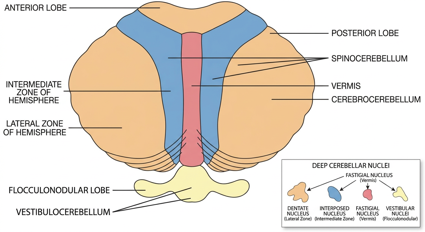
The cerebellum is divided into three functional zones along the longitudinal axis:
┌──────────────────────────────────────────────────────────┐
│            CEREBELLUM - Functional Divisions              │
│                                                          │
│  ANTERIOR LOBE          POSTERIOR LOBE                   │
│ ┌──────────────────────────────────────┐                 │
│ │    Lateral  │ Intermed │   Vermis  │ Intermed │ Lateral│
│ │    Zone     │  Zone    │  (midline)│  Zone    │ Zone   │
│ │             │          │           │          │        │
│ │ CEREBRO-    │ SPINO-   │  SPINO-   │  SPINO-  │CEREBRO-│
│ │ CEREBELLUM  │CEREBELLUM│CEREBELLUM │CEREBELLUM│CEREBEL.│
│ │             │          │           │          │        │
│ │ Deep Nuc.:  │ Deep Nuc.│ Deep Nuc. │ Deep Nuc.│ Deep   │
│ │ Dentate     │ Interposed│ Fastigial │ Interpos.│ Dentate│
└──────────────────────────────────────────────────────────┘
│              FLOCCULONODULAR LOBE                        │
│            (Vestibulocerebellum)                         │
│    Vestibular nuclei / Fastigial nucleus                 │
└──────────────────────────────────────────────────────────┘
| Functional Division | Anatomical Parts | Deep Nucleus |
|---|
| Vestibulocerebellum (Archicerebellum) | Flocculonodular lobe + adjacent vermis | Fastigial + Vestibular nuclei |
| Spinocerebellum (Paleocerebellum) | Vermis + Intermediate zones of hemispheres | Fastigial (vermis) + Interposed (intermediate) |
| Cerebrocerebellum (Neocerebellum) | Lateral zones of hemispheres | Dentate nucleus |
PART 2: Functions of Each Division (2 Marks)
1. Vestibulocerebellum (Archicerebellum)
- Controls equilibrium and postural movements in association with the vestibular apparatus
- Coordinates balance between agonist and antagonist muscles of the spine, hips, and shoulders during rapid changes in body position
- Calculates in advance where body parts will be during the next few milliseconds using signals from the vestibular apparatus and body periphery - this is predictive control of equilibrium
- Loss causes severe disturbance of equilibrium, especially during rapid motion
2. Spinocerebellum (Paleocerebellum)
The spinocerebellum has two sub-zones:
- Vermis: Receives information from the axial body (neck, shoulders, hips). Controls postural and locomotor movements. Sends output via fastigial nucleus to reticulospinal and vestibulospinal tracts.
- Intermediate zone of hemisphere: Receives information from the distal limbs. Compares intended movements (from cerebral motor cortex and red nucleus) with actual movements (from peripheral proprioceptors). Sends corrective signals via the interposed nucleus back to the motor cortex (through thalamus) and to the rubrospinal tract, which controls the distal limb muscles. Also prevents overshoot and provides damping of pendular movements.
3. Cerebrocerebellum (Neocerebellum)
- Controls planning and sequencing of complex voluntary movements
- Receives input exclusively from the cerebral motor, premotor, and somatosensory cortices (via corticopontocerebellar pathway)
- Plans the next sequential movement a fraction of a second in advance while the current movement is still being executed
- Provides timing for sequential movements
- Also has extramotor predictive functions - helps predict rates of progression of visual and auditory phenomena
- Works via the dentate nucleus - cerebellothalamocortical pathway back to the premotor and supplementary motor cortex
- Destruction causes incoordination of complex movements (hands, fingers, speech) - failure of smooth progression
PART 3: Afferent and Efferent Pathways (7 Marks)
AFFERENT (INPUT) PATHWAYS
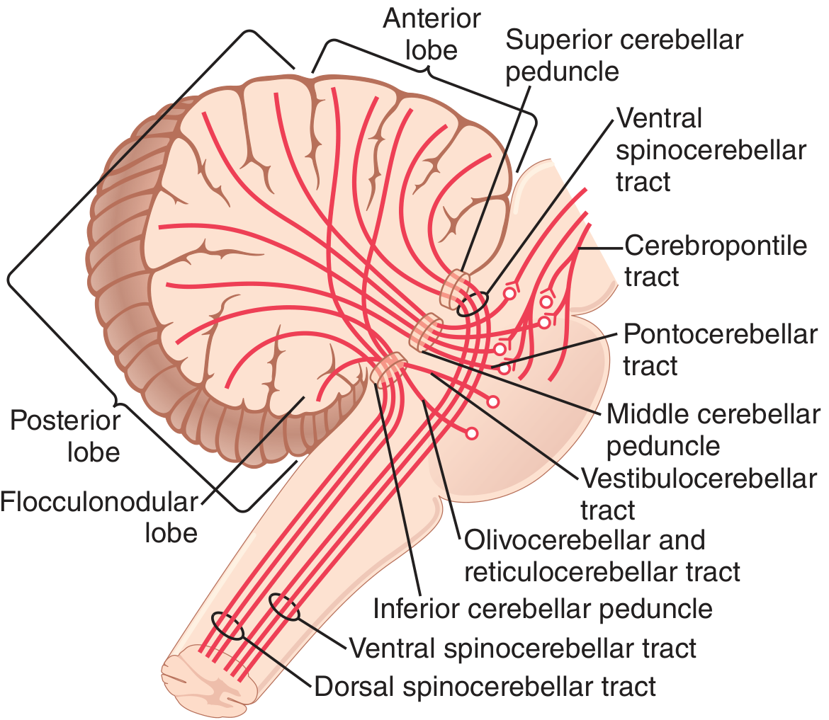
Fig. 57.4 from Guyton & Hall - Principal afferent tracts to the cerebellum
A. From Higher Brain Centers (Brain - to - Cerebellum)
| Tract | Origin | Pathway | Termination |
|---|
| Corticopontocerebellar | Cerebral motor, premotor, somatosensory cortex | Pontile nuclei → pontocerebellar fibers via middle cerebellar peduncle | Lateral zones of opposite cerebellar hemisphere |
| Olivocerebellar | Inferior olive (excited by cerebral cortex, basal ganglia, reticular formation, spinal cord) | Via inferior cerebellar peduncle | All parts of cerebellum |
| Vestibulocerebellar | Vestibular apparatus (directly) and vestibular nuclei of brain stem | Via inferior cerebellar peduncle | Flocculonodular lobe + fastigial nucleus |
| Reticulocerebellar | Brain stem reticular formation | Via inferior cerebellar peduncle | Midline cerebellar areas (vermis) |
B. From the Periphery (Spinal Cord - to - Cerebellum)
Four tracts transmit peripheral information (two dorsal, two ventral):
| Tract | Origin | Carries | Peduncle | Terminates |
|---|
| Dorsal spinocerebellar (most important) | Clark's cells (nucleus dorsalis, T1-L2) | Muscle spindle, Golgi tendon organ, joint receptor, large tactile receptor signals (proprioception) | Inferior cerebellar peduncle | Vermis + intermediate zone, same side (ipsilateral) |
| Ventral spinocerebellar | Anterior horn cells | Efference copy - which motor signals reached the anterior horn (from corticospinal + rubrospinal tracts + intrinsic cord generators) | Superior cerebellar peduncle (crosses twice - once at spinal cord level, once in cerebellum - net result = ipsilateral) | Both sides of cerebellum |
| Cuneocerebellar | Accessory cuneate nucleus (medulla) | Upper limb proprioception | Inferior cerebellar peduncle | Ipsilateral cerebellum |
| Spinoreticulocerebellar + Spinoolivocerebellar | Via reticular formation and inferior olive | Indirect spinal signals | Various | Cerebellum |
The spinocerebellar pathways conduct impulses at up to 120 m/sec - the fastest conduction velocity in the CNS - to allow instantaneous appraisal of changes in peripheral muscle actions.
EFFERENT (OUTPUT) PATHWAYS
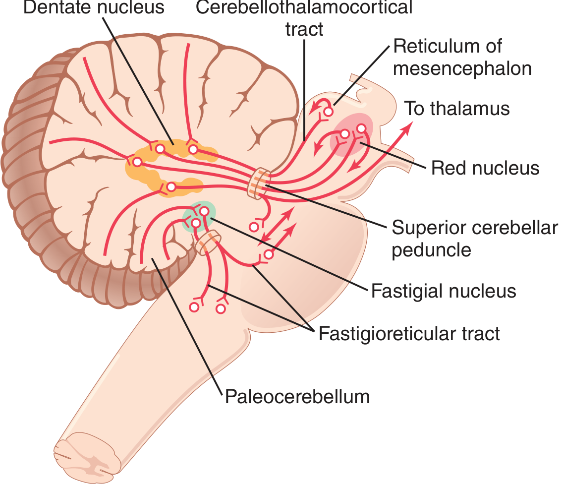
Fig. 57.6 from Guyton & Hall - Principal efferent tracts from the cerebellum
All output signals leave via the deep cerebellar nuclei and exit through the superior cerebellar peduncle (mainly) or the inferior cerebellar peduncle (fastigial):
| Pathway | Origin | Route | Destination | Function |
|---|
| 1. Fastigioreticular / Fastigiovestibular | Vermis → Fastigial nucleus | Via inferior cerebellar peduncle | Medullary and pontile reticular formation + vestibular nuclei | Controls equilibrium and body posture via reticulospinal and vestibulospinal tracts |
| 2. Interpositorubral / Interpositoreticulocortical | Intermediate zone → Interposed nucleus | Via superior cerebellar peduncle → crosses in midbrain | (a) Ventrolateral thalamus → cerebral motor cortex; (b) Magnocellular red nucleus → rubrospinal tract | Feedback control of distal limb movements; prevents overshoot; damps movements |
| 3. Dentatorubrothalamocortical | Lateral zone → Dentate nucleus | Via superior cerebellar peduncle → crosses in midbrain → ventrolateral + ventroanterior thalamus | Cerebral cortex (premotor, supplementary motor area) | Planning and sequencing of voluntary movements |
Summary of peduncle usage:
- Superior cerebellar peduncle: Main EFFERENT pathway (dentate and interposed nuclei outputs)
- Middle cerebellar peduncle: Main AFFERENT (corticopontocerebellar)
- Inferior cerebellar peduncle: Afferent (spinocerebellar dorsal, vestibulocerebellar, olivocerebellar) AND efferent (fastigial nucleus)
PART 4: Ipsilateral Control by the Cerebellum (1.5 Marks)
The cerebellum controls the ipsilateral (same) side of the body. This is explained by the presence of two decussations that cancel each other out:
Step-by-step explanation:
-
The cerebral motor cortex (right side) controls the left side of the body via the crossed corticospinal tract.
-
The cortical motor signals are sent to the contralateral (opposite side) cerebellar hemisphere via the corticopontocerebellar pathway (crosses in the pons). Thus, the right cerebral cortex projects to the left cerebellar hemisphere.
-
The cerebellar output (from the dentate/interposed nucleus of the left cerebellum) travels via the superior cerebellar peduncle and crosses again in the midbrain (decussates in the brachium conjunctivum) to reach the right thalamus and then back to the right cerebral cortex.
-
The right cerebral cortex then controls the left side of the body.
Net result: Right cerebellar lesion → affects right cerebellar hemisphere → signals return to right motor cortex (via double-cross) → but the right motor cortex controls the LEFT body. Yet cerebellar lesions produce ipsilateral deficits.
The explanation is that the peripheral sensory feedback (from the dorsal spinocerebellar tract) goes ipsilaterally to the same cerebellar hemisphere - i.e., sensory signals from the left hand go to the left cerebellum. The cerebellum on the left thus monitors and corrects movements of the left side. The double-crossing of efferent pathways ensures that the cerebellar hemisphere always feeds back to the same cerebral cortex that originated the command, and this motor cortex then corrects its ipsilateral side's movement.
In summary: A lesion of the RIGHT cerebellum produces defects on the RIGHT (ipsilateral) side of the body. This occurs because the pathway crosses TWICE - once at the pons (afferent corticopontocerebellar) and once at the midbrain (efferent superior cerebellar peduncle decussation) - resulting in the cerebellum correcting movements ipsilateral to itself.
PART 5: Clinical Signs of Cerebellar Lesion (1.5 Marks)
Serious and continuing cerebellar dysfunction usually involves lesions of the deep cerebellar nuclei (dentate, interposed, or fastigial). The signs are ipsilateral to the side of the lesion.
| Sign | Description |
|---|
| Dysmetria | Inability to judge distances/extent of movement accurately; movements overshoot or undershoot the intended target (the fundamental defect) |
| Ataxia | General loss of coordination of voluntary movements - uncoordinated gait, balance problems, incoordinate limb movements |
| Past-pointing | Moving a limb considerably beyond the intended point (manifestation of dysmetria - cerebellum cannot initiate the "stop" signal in time) |
| Intention tremor (Action tremor) | Oscillating overshoot in pendular movements due to failure of the cerebellar damping function; worsens as the limb approaches the target |
| Dysdiadochokinesia | Inability to perform rapid alternating movements (e.g., pronation/supination); the system "loses" the position of the limb during rapid motion |
| Dysarthria | Slurred, scanning, or explosive speech - failure of progression in the rapid sequential muscular actions of the larynx, mouth, and respiratory system |
| Nystagmus | Tremulous movements of the eyes on lateral gaze; failure of cerebellar damping; particularly with flocculonodular lobe lesions |
| Hypotonia | Decreased muscle tone on the side of the lesion (from loss of tonic facilitatory signals from dentate and interposed nuclei to motor cortex and brain stem motor nuclei) |
| Rebound phenomenon (Holmes sign) | Inability to check movements; excessive rebound when resistance is suddenly released |
Note: Cerebellar lesions do NOT cause paralysis. The cerebellum has no direct pathway to cause muscle contraction.
All content from: Guyton and Hall Textbook of Medical Physiology (Chapter 57 - Cerebellum and Basal Ganglia), pp. 699-709