
create a diagram for arterial supply for radius
Clean anatomical diagram showing the arterial supply of the radius bone, anterior (volar) view. The radius bone is shown in the center with labeled arteries: 1. BRACHIAL ARTERY at the top splitting into two branches at the neck of the radius 2. RADIAL ARTERY on the lateral (right) side traveling down the length of the radius, with branches labeled: - Radial recurrent artery (branching proximally upward toward elbow) - Nutrient artery entering the volar aspect of the shaft at 9 cm distal to the radial head - Palmar carpal branch (distal) - Superficial palmar branch (most distal) 3. ULNAR ARTERY on the medial (left) side with: - Common interosseous artery branching off - Anterior interosseous artery running along the interosseous membrane, giving perforating branches to the radius - Posterior interosseous artery (branching posteriorly) 4. The interosseous membrane between radius and ulna shown schematically 5. Color coding: arteries in red, bone in beige/ivory, interosseous membrane in blue-gray 6. Title at top: "ARTERIAL SUPPLY OF THE RADIUS" 7. Clean white background, medical textbook style, clearly labeled with arrows pointing to each structure
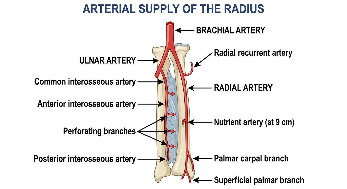
Arterial Supply of the Radius
Main Source
Radial Artery (lateral supply)
- Originates at the neck of the radius
- Travels along the lateral aspect of the forearm, deep to brachioradialis proximally
- Key branches to the radius:
- Radial recurrent artery - runs proximally, anastomoses around the elbow joint
- Nutrient artery to the radius - enters the volar (anterior) shaft approximately 9 cm distal to the radial head (range 6-12 cm); the primary endosteal blood supply
- Palmar carpal branch - supplies distal radius and carpal bones
- Superficial palmar branch - enters the thenar region distally
Anterior Interosseous Artery (medial/posterior supply)
- Arises from the common interosseous artery (a branch of the ulnar artery)
- Runs along the anterior surface of the interosseous membrane
- Gives perforating branches that supply the anterior surface and periosteum of the radius throughout its shaft
- Divides distally into anterior and posterior branches supplying the distal metaphysis
Posterior Interosseous Artery
- Also from the common interosseous artery
- Supplies the posterior periosteum of the proximal radius
Clinical Relevance
- The nutrient artery enters at the volar midshaft - this is why plating on the volar surface at mid-radius risks its injury
- The proximal metaphysis receives cancellous supply over ~4 cm; the distal metaphysis over ~5 cm
- Rich periosteal anastomoses between radial and interosseous arteries ensure collateral supply after fracture