
Recent classification and morphology of giant cell tumours of bone and tumor like lesions
giant cell tumor of bone histology microscopy
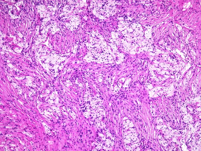
This histopathology image depicts a giant cell tumor of bone observed under light microscopy following Hematoxylin and Eosin staining. The cellular stroma consists of uniform spindle-shaped mononuclear cells interspersed with numerous osteoclast-like multinucleated giant cells. Notably, foam cells—lipid-laden macrophages—may populate the interstitium, contributing foamy cytoplasm in parts of the lesion. Giant cells contain multiple nuclei with even distribution and lack pronounced cytologic atypia; mitotic activity, if present, is typically limited. The architectural pattern can be variable, and in some cases the classic giant cell-rich areas are inconspicuous, complicating diagnosis without radiographic context. Clinically, GCTs arise in the epiphyseal/metaphyseal region of long bones in young to middle-aged adults and demonstrate local aggressiveness comparable to other bone tumors. Radiographs often reveal a soap-bubble lytic lesion with cortical thinning and expansion, providing a critical diagnostic adjunct. Differential considerations include fibrous histiocytoma and other giant cell-containing lesions. Definitive diagnosis relies on correlating histology with imaging and tumor location; immunohistochemical markers such as RANKL expression in stromal cells and tartrate-resistant acid phosphatase in giant cells can support confirmation.
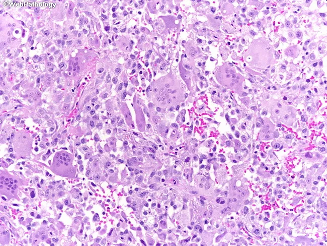
Imaging modality: Light microscopy of Hematoxylin and Eosin stained bladder tumor tissue. Primary subject: Osteoclast-type giant cell-rich urothelial carcinoma, a rare aggressive variant of urothelial carcinoma of the urinary bladder. Specimen: Bladder wall biopsy / surgical resection specimen. Perspective: Histopathology micrograph at high magnification showing cellular detail. In this image the osteoclast-type giant cells (multinucleated, large, eosinophilic cytoplasm) are interspersed with mononuclear tumor cells in a fibromuscular stromal background. Areas of conventional urothelial carcinoma are present in parts of the slide; the giant cells resemble those seen in giant cell tumors of bone. The tumor demonstrates marked cytologic atypia in the mononuclear component, frequent mitotic figures, and occasional necrosis; the stromal milieu may be inflammatory with capillary-rich vasculature. The combination signifies an aggressive biological behavior with poor prognosis. Clinically, this variant may be associated with early invasion and metastasis, and can mimic benign giant cell lesions histologically, necessitating careful differential diagnosis. Immunophenotyping typically shows urothelial markers in carcinomatous cells (e.g., GATA3, CK7/CK20) and histiocytic markers (CD68) in the giant cells. This image is relevant for educational diagnosis, differential diagnosis, and research on tumor heterogeneity within urothelial neoplasms and osteoclast-like giant cell-rich carcinomas. Potential use cases include pathology teaching, biopsy interpretation, and correlating histology with prognosis and therapy.
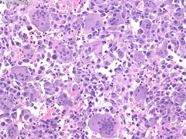
Modality: Light microscopy of a formalin-fixed bladder biopsy, single hematoxylin and eosin stained section. Comprehensive description: The tumor shows an admixture of osteoclast-type giant cells and mononuclear stromal cells with scant intervening necrosis and a fibrous to myxoid background. Osteoclast-like multinucleated giant cells are prominent, with abundant pale cytoplasm and variably sized nuclei, often showing conspicuous nucleoli. Interspersed mononuclear cells are pleomorphic and can exhibit atypia and mitotic activity. In some regions, conventional urothelial carcinoma components are present, reinforcing urothelial origin, while other areas may lack overt epithelial differentiation, recapitulating a giant cell tumor of bone-like morphology. The histology can mimic true giant cell tumors of bone, particularly when giant cells predominate and the stroma is relatively undifferentiated. This pattern represents an aggressive variant of urothelial carcinoma with poor outcome, higher propensity for invasion and metastasis, and increased risk of recurrence after resection. Immunophenotype typically supports urothelial lineage in carcinoma areas (e.g., cytokeratins, GATA3); osteoclast-like giant cells often express CD68. Clinically, this entity requires accurate diagnosis to guide management, given its dual components and potential therapeutic implications, including radical surgery, adjuvant therapy, and vigilant follow-up. This description emphasizes diagnostic nuances, therapeutic considerations, and prognostic implications for urothelial-origin neoplasms with giant cell components.
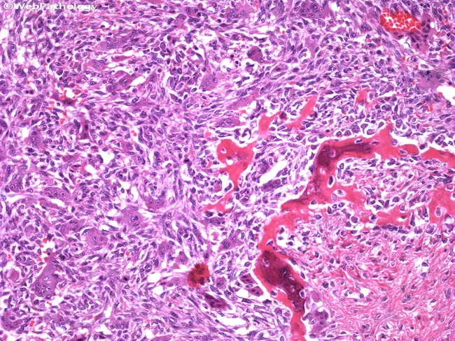
Histopathology of a giant cell tumor of bone. This light-m microscopy image uses Hematoxylin and Eosin staining to reveal the characteristic architecture of a GCT rather than an osteoid-producing sarcoma. The tissue section demonstrates a cellular stroma composed of mononuclear ovoid to spindle-shaped cells with evenly distributed, numerous osteoclast-like multinucleated giant cells. The giant cells display uniformly stained cores with multiple nuclei per cell, set within a vascular, often hemorrhagic background. The mononuclear component tends to be relatively bland, with minimal nuclear atypia and occasional mitotic activity, supporting a benign but locally aggressive neoplasm. Reactive bone formation can be present at the periphery in rare cases, potentially confounding the diagnosis by suggesting osteosarcoma; however, malignant osteoid is typically absent or scant, and the overall pattern is a mosaic of giant cells interspersed with mononuclear stromal cells rather than malignant osteoid production. Clinically, this histology correlates with an epiphyseal/metaphyseal bone lesion in adults, presenting with pain and swelling. The diagnostic significance lies in distinguishing GCT from osteosarcoma and other giant cell-rich lesions, guiding surgical management such as intralesional curettage with adjuvant therapy or resection, and informing prognosis given the risk of local recurrence. Correlation with imaging and clinical data improves accuracy.
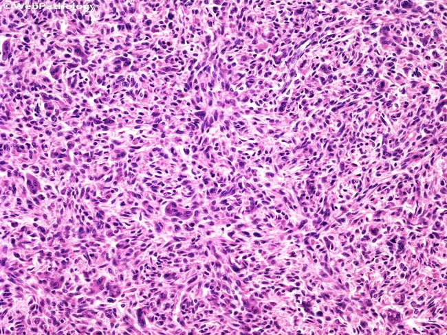
Histopathology of giant cell tumor of bone demonstrated on Hematoxylin and Eosin stained sections, visualized by bright-field light microscopy. The specimen represents a skeletally derived tumor bed with abundant spindle-shaped stromal cells interspersed with numerous multinucleated giant cells resembling osteoclasts. The mononuclear cells are typically evenly distributed, with minimal cytologic atypia and rare mitotic activity, providing a cellular yet non-atypical background. The giant cells are conspicuous but evenly dispersed, with numerous nuclei per cell in a peripherally oriented pattern. The matrix is typically collagenous with a vascular stroma, and hemorrhagic or foamy macrophages may be present. In this illustrative image, sheets of spindle cells exhibit mild pleomorphism but lack overt nuclear atypia, supporting a benign/low-grade neoplasm rather than high-grade sarcoma. Diagnostic significance lies in recognizing the characteristic osteoclast-like giant cells coexisting with a uniform mononuclear stromal component. Knowledge of this histology helps differentiate GCT from other sarcomas, guides management (curettage versus en bloc resection), and correlates with radiographic epiphyseal lesions in long bones. Additional differential considerations include fibrosarcoma and other spindle-cell sarcomas, but the uniform stromal population with evenly distributed osteoclast-like giant cells and absence of nuclear atypia aids accurate classification. This histologic profile informs surgical planning, biopsy interpretation, adjuvant decisions, and radiologic correlation in long-bone epiphyseal lesions.
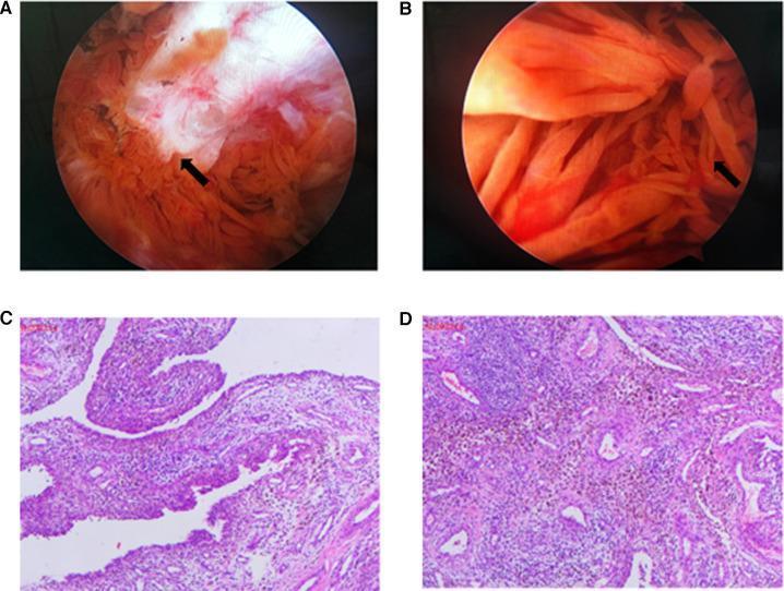
A multi-panel figure containing intraoperative arthroscopic photographs and histopathology slides related to Tenosynovial Giant Cell Tumor, localized type (formerly Pigmented Villonodular Synovitis - PVNS). Panel A shows an arthroscopic view of bone erosion on the medial femoral condyle, indicated by an arrow, characterized by irregular, white fibrillated tissue and exposed bone. Panel B demonstrates synovial proliferation with characteristic yellowish-orange, finger-like villous projections and nodules typical of PVNS. Panels C and D are light microscopy images of excised synovial tissue stained with Hematoxylin and Eosin (H&E) and CD68 immunohistochemistry. The histology reveals synovial hyperplasia with a dense infiltrate of mononuclear cells, foamy macrophages, multinucleated giant cells, and prominent brownish hemosiderin deposits. These panels illustrate the diagnostic features used in orthopedic surgery and pathology to identify proliferative synovial disorders in pediatric patients.
"giant cell tumor of bone"[MeSH] AND classification
WHO classification 2020 2024 giant cell tumour of bone tumor-like lesions bone tumors update
https://www.pathologyoutlines.com/topic/bonewhoclass.html
Giant Cell Tumour of Bone and Giant Cell-Rich Tumour-Like Lesions: Current Classification and Morphology
1. Current classification framework (WHO Classification of Tumours, 5th edition, 2020 - Soft Tissue and Bone Tumours)
| Entity | Biological behaviour |
|---|---|
| Non-ossifying fibroma (NOF) | Benign |
| Aneurysmal bone cyst (ABC) | Benign |
| Giant cell tumour of bone, NOS | Intermediate (locally aggressive, rarely metastasizing) |
| Giant cell tumour of bone, malignant | Malignant |
- ABC and NOF have been moved into this group with GCT, reflecting shared giant cell-rich morphology, rather than being classified as isolated "cystic" or "fibrous" lesions.
- "Benign fibrous histiocytoma of bone" has been eliminated as a distinct entity - it is now regarded as a variant/synonym overlapping with NOF.
- "Giant cell lesion of small bones" and "giant cell reparative granuloma" (older terms for lesions in hands/feet/jaw) are now considered obsolete terminology; these lesions are reclassified as a solid variant of aneurysmal bone cyst.
- ABC is now subdivided by pathogenesis into primary (de novo, often with USP6 gene rearrangement) and secondary ABC (arising within a pre-existing lesion such as GCT, chondroblastoma, fibrous dysplasia, or NOF) - distinguishing the two matters because secondary ABC-like change can mask an underlying neoplasm requiring different management.
- GCT of bone is graded as intermediate (locally aggressive) rather than purely benign, because of its capacity for local destruction and rare (about 1-4%) benign-appearing pulmonary metastases; a separate malignant GCT category exists for sarcomatous transformation.
2. Morphology of conventional GCT of bone
- Neoplastic mononuclear stromal cells - the true tumour cell, oval to spindle-shaped with round-to-oval nuclei, inconspicuous nucleoli, and indistinct cell borders. These cells harbor the H3.3 G34W mutation and drive tumour biology by overexpressing RANKL, which recruits and activates osteoclast precursors.
- Osteoclast-like multinucleated giant cells - large cells with 20-100+ evenly-dispersed, bland nuclei per cell, uniformly distributed throughout the lesion (not focal). These are non-neoplastic, reactive cells (derived from monocyte/macrophage precursors fused via RANK-RANKL signaling) and stain positively for TRAP (tartrate-resistant acid phosphatase) and CD68.
- Mononuclear histiocyte-like/round cells - a variably prominent third population.
- Minimal cytologic atypia and low mitotic activity in the mononuclear cells in conventional GCT; significant nuclear atypia or atypical mitoses should raise suspicion for malignant transformation.
- Vascular, sometimes hemorrhagic stroma; foam cells (lipid-laden macrophages) and reactive woven bone at the periphery may be seen, occasionally causing confusion with osteosarcoma.
- Radiographically corresponds to an eccentric, lytic, "soap-bubble" epiphyseal/metaphyseal lesion in a skeletally mature patient (growth plate closed), most often distal femur, proximal tibia, or distal radius - - Robbins & Kumar Basic Pathology; Miller's Review of Orthopaedics.
3. Malignant GCT of bone
- Primary malignant GCT - sarcomatous component identified concurrently with conventional GCT at first presentation.
- Secondary malignant GCT - develops within a previously treated GCT, often years later, frequently linked to prior radiotherapy.
- Osteosarcoma is the most common sarcomatous subtype in both categories. A large 2025 clinicopathologic/genetic series (Papke et al., Am J Surg Pathol, PMID 40077813) found malignant GCT and metastasizing "conventional" GCT carry distinct genetic alterations (e.g., TP53 abnormalities) compared with ordinary GCT, supporting molecular distinction between these entities - relevant since morphology alone is often indistinguishable from aggressive conventional GCT - Campbell's Operative Orthopaedics, 15th ed.
4. Tumor-like lesions now grouped with GCT (per current classification)
- Aneurysmal bone cyst (ABC) - multiloculated blood-filled cystic spaces separated by fibrous septa containing spindle cells, scattered osteoclast-type giant cells, and reactive woven bone; primary ABC often shows USP6 rearrangement.
- Non-ossifying fibroma (NOF) - storiform arrangement of bland spindle (fibrohistiocytic) cells admixed with scattered giant cells and hemosiderin-laden macrophages; now the preferred term over "benign fibrous histiocytoma of bone."
- Solid variant of ABC (formerly "giant cell reparative granuloma"/"giant cell lesion of small bones") - seen typically in small bones of hands/feet and craniofacial skeleton, histologically resembling ABC without cystic spaces.